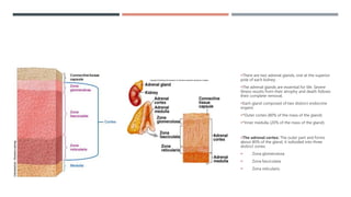
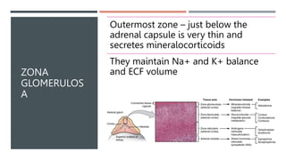
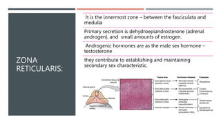
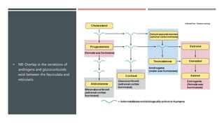
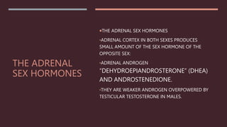
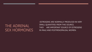
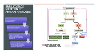
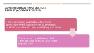
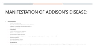
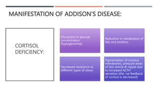
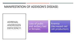
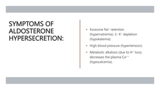
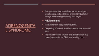
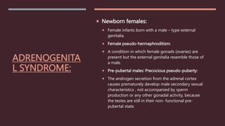
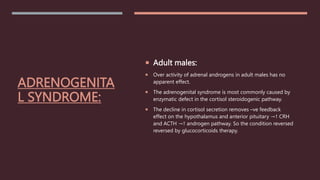
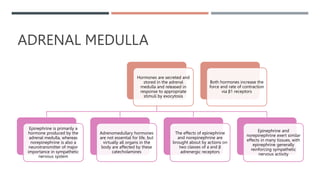
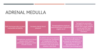
Endocrine
system
Physiology
Presented by/
Dr/ dina hamdy merzeban
ADRENAL
GLAND
There are two adrenal glands, one at the superior
pole of each kidney.
The adrenal glands are essential for life. Severe
illness results from their atrophy and death follows
their complete removal.
Each gland composed of two distinct endocrine
organs:
*Outer cortex (80% of the mass of the gland).
*Inner medulla (20% of the mass of the gland).
The adrenal cortex: The outer part and forms
about 80% of the gland, it isdivided into three
distinct zones.
· Zona glomerulosa
· Zona fasciculata
· Zona reticularis.
Hormones produced by the adrenal cortex are
steroids derived from the common precursor
cholesterol
These comprise mineralocorticoids, glucocorticoids
and sex hormones
The three categories of adrenal steroids are
produced in anatomically distinct portions of the
adrenal cortex as a result of differential distribution
of the enzymes required to catalyze the different
biosynthetic pathways leading to the formation of
each of these steroids
ZONA
GLOMERULOS
A
Outermost zone – just below the
adrenal capsule is very thin and
secretes mineralocorticoids
They maintain Na+ and K+ balance
and ECF volume
ZONA
FASCICULATA
It is the middle widest zone
Primary secretion is glucocorticoids (cortisol).
Glucocorticoids play a major role in glucose
metabolism, as well as protein and lipid metabolism
ZONA
RETICULARIS:
It is the innermost zone – between the fasciculata and
medulla
Primary secretion is dehydroepiandrosterone (adrenal
androgen), and small amounts of estrogen.
Androgenic hormones are as the male sex hormone –
testosterone
they contribute to establishing and maintaining
secondary sex characteristic.
 NB: Overlap in the secretions of
androgens and glucocorticoids
exist between the fasciculata and
reticularis
TRANSPORT OF
ADRENOCORTICAL
HORMONES:
 Being lipophilic, the adrenocortical
hormones are all carried in the
blood extensively bound to plasma
proteins
 Cortisol is bound mostly to a
plasma protein specific for it called
corticosteroid binding globulin ,
about 15% is bound to albumin,
only 10% is free
MECHANISM OF
ACTION OF
STEROID HORMONES:
 ¯ Steroid hormones are made
from cholesterol (lipophilic) so
can enter target cell. They are
slower acting and have longer
half-life than peptide
hormones.
MINERALOCORTICOID
S
¯ IT REGULATES THE ELECTROLYTE CONCENTRATIONS OF
EXTRACELLULAR FLUIDS.
¯ MINERALOCORTICOIDS INCLUDE MAINLY ALDOSTERONE AND
DEOXY-CORTICOSTERONE.
¯ MINERALOCORTICOIDS ARE ESSENTIAL FOR LIFE, WITHOUT
ALDOSTERONE, A PERSON RAPIDLY DIES FROM CIRCULATORY
SHOCK (MARKED FALL IN PLASMA VOLUME CAUSED BY
EXCESSIVE LOSSES OF H2O HOLDING NA).
• ACTION OF
ALDOSTERONE:
 ¯ The site of aldosterone action is on the principal cells of the late
distal tubules and collecting duct of the kidney. It affects Na+ and K+
balance.
 - It promotes Na+ reabsorption and K+ excretion .
 ¯ More aldosterone → more Na+ retention, so more water retention in
the body (osmotic retention). Expanding of the ECF volume, which is
important in the long term regulation of blood pressure.
 NB: Aldosterone also increases Na+ absorption from other body fluid
(sweat and saliva)
 Aldosterone also causes H+ to be secreted by renal tubules in exchange
with Na+. Thus, aldosterone excess causes mild alkalosis and an
increase in urine acidity.
CONTROL OF
ALDOSTERO
NE
SECRETION:
 Angiotensin II stimulates conversion of corticosterone to aldosterone in the
zona glomerulosa cells and secretion of aldosterone from these
cells Angiotensin II promotes growth of the zona glomerulosa (tropic effect).
 Direct stimulation of adrenal cortex by a rise in plasma K+ concentration
 N.B. The regulation of aldosterone secretion is largely independent of anterior
pituitary control as the ACTH from anterior pituitary primarily promotes the
secretion of cortisol, not aldosterone.
GLUCOCORTICOIDS
¯Glucocorticoids include
mainly cortisol and
corticosterone
Cortisol account 95% of all
glucocorticoid activity
¯Cortisol is essential for life; it
plays an important role in
carbohydrate, protein and fat
metabolism, helps people
resist stress and has a
significant permissive action
for other hormonal activities
PHYSIOLOGICAL
ACTIONS OF
GLUCOCORTICOI
DS
 EFFECT ON METABOLISM:
THE OVERALL EFFECT OF CORTISOL'S METABOLIC ACTIONS IS TO
INCREASE THE CONCENTRATION OF GLUCOSE AT THE EXPENSE OF
PROTEIN AND FAT STORES.
ON
CARBOHYDRAT
E METABOLISM:
Stimulation of gluconeogenesis by the liver.
Decrease the utilization of glucose by muscle
and adipose tissue (not the brain) and lowers
their sensitivity to insulin (anti- insulin action).
ON PROTEIN
METABOLISM
Increase protein degradation
(catabolism) in many tissue especially
muscle, increases the blood amino acid
concentration, thus providing more
amino acids to liver or for tissue repair.
Decreased protein synthesis.
ON FAT
METABOLISM
Increase lipolysis (the mobilized fatty
acids are available as an alternative
metabolic fuel for tissues that can use
this energy source as an alternative to
glucose, conserving glucose for the brain.
In diabetics, it increases ketone body
formation (ketogenic).
PERMISSIVE ACTION:
Mean that in small amounts it allows certain processes to occur,
although glucocorticoids by themselves do not initiate these
reactions
Cortisol must be present in adequate amounts to permit the
catecholamines to induce vasoconstriction and glucagon to
produce glycogenolysis.
Cortisol is necessary for catecholamines and growth
hormone to exert their lipolytic effect.
ROLE IN
ADAPTATION
TO STRESS
ROLE IN ADAPTATION TO
STRESS
Cortisol plays a key role in adaptation to stress. This rise is essential for survival in
these conditions.
Cortisol provokes:
 Gluconeogenesis (formation of glucose from non -carbohydrates).
 Rises in blood glucose, fatty acids, and amino acids.
 Physical or mental stresses include: Trauma, pain, infection, surgery, anxiety,
shock, intense heat or cold. Stressful stimuli are defined as the noxious stimuli
that increase secretion of ACTH through CRH secretion.
 An increased pool of glucose, amino acids and fatty acids is available for use as
needed.
• Other effects:
Cortisol has a very
slight
mineralocorticoid
activity
During fetal life,
cortisol accelerates
the maturation of
surfactant in the lung
PHARMACOLOGICAL ACTION:
When cortisol or synthetic cortisol like compounds are administered to yield higher
than physiologic concentrations of glucocorticoids during treatment of certain
diseases; or in case of its hypersecretion by adrenal cortex
Corticosteroids are anti-inflammatory and immunosuppressive
It suppress the inflammatory reaction by reducing phagocytic action of white
blood cells , inhibiting release of the lysosomal enzymes and decreasing capillary
permeability
Suppresses allergic reactions by preventing release of histamine from the mast
cells
PHARMACOLOGICAL ACTIONS
OTHER UNDESIRABLE EFFECTS MAY BE OBSERVED WITH PROLONGED EXPOSURE
TO HIGHER THAN NORMAL CONCENTRATIONS OF GLUCOCORTICOIDS
The administration of large doses
of cortisol causes significant
atrophy of all the lymphoid tissue
throughout the body, which in
turn decreases the output of both
T cells and antibodies from the
lymphoid tissue
NB: This occasionally can lead to
fulminating infection and death
from diseases that would
otherwise not be lethal, such as
fulminating tuberculosis in a
person whose disease had
previously been arrested.
Conversely, this ability of cortisol
and other glucocorticoids to
suppress immunity makes them
useful drugs in preventing
immunological rejection of
transplanted hearts, kidneys, and
other tissues.
PHARMACOLOGICAL ACTIONS
OTHER UNDESIRABLE EFFECTS MAY BE OBSERVED WITH PROLONGED EXPOSURE
TO HIGHER THAN NORMAL CONCENTRATIONS OF GLUCOCORTICOIDS
Cortisol increases the production
of red blood cells by mechanisms
that are unclear. When excess
cortisol is secreted by the adrenal
glands, polycythemia often
results, and conversely, when the
adrenal glands secrete no
cortisol, anemia often results.
Prolonged suppression of this
axis can lead to irreversible
atrophy of cortisol secreting
cells of the adrenal gland and
thus to permanent inability of
the bodyto produce its own
cortisol.
CONTROL OF
COTRICOSTEROID
S
Hypothalamic control is via CRH
CRH is secreted into the hypothalamic-
hypophyseal portal blood and sent to the
anterior pituitary
CRH binds to receptors causing synthesis of
POMC a precursor of ACTH
POMC is a large precursor of MSH, and β-
endorphin
ACTH being tropic to zona fasciculata and
zona reticularis
CONTROL OF COTRICOSTEROIDS
Diurnal rhythm: The plasma
cortisol concentration
display a characteristic
diurnal rhythm, with the
highest level occurring in
the morning and lowest
level at mid night
Stress: The magnitude of the
increase in plasma cortisol
concentration is
proportional to intensity of
the stressful stimuli
THE ADRENAL
SEX HORMONES
THE ADRENAL SEX HORMONES
•ADRENAL CORTEX IN BOTH SEXES PRODUCES
SMALL AMOUNT OF THE SEX HORMONE OF THE
OPPOSITE SEX:
•ADRENAL ANDROGEN
“DEHYDROEPIANDROSTERONE” (DHEA)
AND ANDROSTENEDIONE.
•THEY ARE WEAKER ANDROGEN OVERPOWERED BY
TESTICULAR TESTOSTERONE IN MALES.
THE ADRENAL
SEX HORMONES
• FUNCTION OF DHEA IN FEMALE:
•1- ENHANCEMENT OF THE PUBERTAL GROWTH
SPURT
• 2- GROWTH OF PUBIC AND AXILLARY HAIR.
•3 - DEVELOPMENT AND MAINTENANCE OF FEMALE
SEX DRIVE.
•4- HAVE NO MASCULINIZING EFFECT IN THEIR
NORMAL AMOUNT.
THE ADRENAL
SEX HORMONES
•ESTROGENS ARE NORMALLY PRODUCED IN VERY
SMALL QUANTITIES FROM THIS SOURCE;
THEY ARE IMPORTANT SOURCE OF ESTROGENS
IN MALE AND POSTMENOPAUSAL WOMEN.
REGULATION OF
SECRETION OF
ADRENAL ANDROGEN:
ACTH controls adrenal
androgen secretion
Adrenal androgens feedback
outside the hypothalamus
pituitary adrenal cortex loop
Instead of inhibiting CRH, it
inhibits gonadotropin releasing
hormone GRH, just as testicular
androgen do
Adrenal androgen secretion
undergoes a marked surge, at
the time of puberty, and peaks
between the ages 25 and
ADRENOCORTICAL HYPOFUNCTION:
PRIMARY (ADDISON'S DISEASE):
Is most commonly caused by autoimmune
destruction of the adrenal cortex by erroneous
production of adrenal cortex – attacking antibodies
Characterized by deficiency of all
adrenocortical hormones and hyper-
pigmentation
SECONDARY
Pigmentation of mucous
membranes, pressure
areas of skin areola &
nipple due to increased
ACTH secretion
Loss of pubic and axillary
hair in females
Anemia
MANIFESTATION OF ADDISON’S DISEASE:
 Aldosterone deficiency:
 Decreased sodium (hyponatremia).
 Decrease ECF volume (dehydration, hypotension and decrease cardiac output).
 Hyperkalemia → disturbs cardiac rhythm and metabolic acidosis (↑ H+).
 Patient dies in shock if untreated.
 Cortisol deficiency:
 Disruption in glucose concentration (hypoglycemia).
 Reduction in metabolism of fats and proteins.
 Decreased resistance to different types of stress.
 Pigmentation of mucous membranes, pressure areas of skin areola & nipple due to increased ACTH secretion (the –ve feedback of cortisol is decreased).
 Adrenal androgen deficiency:
 Loss of pubic and axillary hair in females.
 Anemia (decreased red cell production).
 NB: Addisonian crisis:
 Addissonian’s patients fail to respond to stress by an increase in glucocorticoids, so an exposure to stress, the body systems respond by collapse, shock, hyperkalemia and hypoglycemia. Emergency treatment: I.V. cortisol and isotonic NaCl infusion.
MANIFESTATION OF ADDISON’S DISEASE:
Decreased sodium
(hyponatremia).
Decrease ECF volume
(dehydration,
hypotension and
decrease cardiac
output).
Hyperkalemia →
disturbs cardiac
rhythm and
metabolic acidosis (↑
H+).
Patient dies in shock
if untreated.
ALDOSTERON
E DEFICIENCY:
MANIFESTATION OF ADDISON’S DISEASE:
Disruption in glucose
concentration
(hypoglycemia).
Reduction in metabolism of
fats and proteins.
Decreased resistance to
different types of stress.
Pigmentation of mucous
membranes, pressure areas
of skin areola & nipple due
to increased ACTH
secretion (the –ve feedback
of cortisol is decreased).
CORTISOL
DEFICIENCY:
MANIFESTATION OF ADDISON’S DISEASE:
Loss of pubic
and axillary hair
in females.
Anemia
(decreased red
cell production).
ADRENAL
ANDROGEN
DEFICIENCY:
ADDISONIAN
CRISIS:
 Addissonian’s patients fail to respond to stress
by an increase in glucocorticoids, so an
exposure to stress, the body systems respond
by collapse, shock, hyperkalemia and
hypoglycemia. Emergency treatment: I.V.
cortisol and isotonic NaCl infusion.
ADRENOCORTICAL
HYPER-FUNCTION:
 THIS INCLUDES THREE SYNDROMES,
EACH OF WHICH IS CHARACTERIZED
BY OVER ACTIVITY OF A PARTICULAR
ZONE OF THE GLAND.
ALDOSTERONE
HYPERSECRETION:
 Primary hyperaldosteronism (Conn's syndrome):
 Caused by over activity of the zona glomerulosa as a result of
hypersecreting adrenal tumor.
 Secondary hyperaldosteronism:
 Caused by inappropriately high activity of the renin – angiotensin
system (atherosclerotic narrowing of renal arteries).
 The symptoms of both are related to exaggerated effects of
aldosterone.
SYMPTOMS OF
ALDOSTERONE
HYPERSECRETION:
 Excessive Na+ retention
(hypernatremia). 2- K+ depletion
(hypokalemia).
 High blood pressure (hypertension).
 Metabolic alkalosis (due to H+ loss),
decreases the plasma Ca++
(hypocalcemia).
EXCESSIVE
CORTISOL
SECRETION
(CUSHING’S
SYNDROME)
:
 Causes:
 Overstimulation of the adrenal cortex by excessive amount of CRH
or ACTH.
 Adrenal tumors that uncontrollably secrete cortisol independent of
ACTH.
 ACTH secreting tumors located in places other than the pituitary,
most commonly in the lung.
 Administration of pharmacological doses of glucocorticoids.
 The prominent characteristic of this syndrome are related to the
exaggerated effects of glucocorticoids. The main symptoms being
reflections of excessive gluconeogenesis (combined glucose
excess and protein shortage).
CUSHING'S
SYNDROME IS
CHARACTERIZE
D BY:
 ↑ Cortisol and androgen levels.
 ↑ ACTH (if caused by overproduction of ACTH), ↓ ACTH (if
caused by primary adrenal hyperplasia).
CUSHING'S
SYNDROME IS
CHARACTERIZE
D BY:
 ↑ Cortisol and androgen levels.
 ↑ ACTH (if caused by overproduction of ACTH), ↓ ACTH (if
caused by primary adrenal hyperplasia).
 Hyperglycemia, glucosuria (adrenal diabetes).
 Central obesity (abdominal), round face (moon face)
supraclavicular fat (buffalo hump).
 ↑ Protein catabolism leads to muscle wasting and fatigue.
 Poor wound healing and easy bruisability.
 Osteoporosis (↑ bone resorption).
 The protein poor thin skin of the abdomen becomes over
overstretched by the excessive underlying fat deposits forming
irregular reddish purple linear streaks (striae).
CUSHING'S
SYNDROME IS
CHARACTERIZE
D BY:
 Hyperglycemia, glucosuria (adrenal diabetes).
 Central obesity (abdominal), round face (moon face)
supraclavicular fat (buffalo hump).
 ↑ Protein catabolism leads to muscle wasting and fatigue.
 Poor wound healing and easy bruisability.
 Osteoporosis (↑ bone resorption).
 The protein poor thin skin of the abdomen becomes over
overstretched by the excessive underlying fat deposits forming
irregular reddish purple linear streaks (striae).
CUSHING'S
SYNDROME IS
CHARACTERIZED BY:
 Hypertension.
 Virilization of women
(caused by elevated levels
of adrenal androgens).
CUSHING SYNDROME
↑ Cortisol and androgen
levels
↑ ACTH , ↓ ACTH Hyperglycemia, glucosuria
Central obesity , round face
supraclavicular fat
↑ Protein catabolism leads to
muscle wasting and fatigue
Poor wound healing and
easy bruisability
Hypertension Osteoporosis
The protein poor thin skin of
the abdomen becomes over
overstretched by the
excessive underlying fat
deposits forming irregular
reddish purple linear streaks
Virilization of women
ADRENOGENITA
L SYNDROME:
 The symptoms that result from excess androgen
secretion depend on the sex of the individual and
the age when the hyperactivity first begins.
 Adult females:
 Male pattern of body hair (hirsutism).
 Deepening of the voice and more muscular arms and
legs.
 The breast become smaller, and menstruation may
cease (suppression of GRH), and sterility occur.
ADRENOGENITAL SYNDROME
Male pattern of
body hair
Deepening of the
voice and more
muscular arms and
legs
The breast become
smaller, and
menstruation may
cease , and sterility
occur
Female infants born
with a male – type
external genitalia
ADRENOGENITA
L SYNDROME:
 Newborn females:
 Female infants born with a male – type external
genitalia.
 Female pseudo-hermaphroditism:
 A condition in which female gonads (ovaries) are
present but the external genitalia resemble those of
a male.
 Pre-pubertal males: Precocious pseudo-puberty:
 The androgen secretion from the adrenal cortex
causes prematurely develop male secondary sexual
characteristics , not accompanied by sperm
production or any other gonadal activity, because
the testes are still in their non- functional pre-
pubertal state.
PRE-PUBERTAL
MALES:
PRECOCIOUS
PSEUDO-PUBERTY
ADRENOGENITAL SYNDROME
PRE-PUBERTAL MALES: PRECOCIOUS PSEUDO-PUBERTY
Over activity of adrenal androgens in adult males has no
apparent effect
The adrenogenital syndrome is most commonly caused by
enzymatic defect in the cortisol steroidogenic pathway
The decline in cortisol secretion removes –ve feedback effect
on the hypothalamus and anterior pituitary →↑ CRH and
ACTH →↑ androgen pathway
ADRENOGENITA
L SYNDROME:
 Adult males:
 Over activity of adrenal androgens in adult males has no
apparent effect.
 The adrenogenital syndrome is most commonly caused by
enzymatic defect in the cortisol steroidogenic pathway.
 The decline in cortisol secretion removes –ve feedback
effect on the hypothalamus and anterior pituitary →↑ CRH
and ACTH →↑ androgen pathway. So the condition reversed
reversed by glucocorticoids therapy.
DRENAL MEDULLA
ADRENAL MEDULLA
The adrenal medulla
forms about 20% of the
adrenal gland
It is a modified
postganglionic
sympathetic neuron
where the neurons have
lost their axons and
become secretory cells
Controlled by
preganglionic
sympathetic innervation
Secretes epinephrine
and norepinephrine
ADRENAL MEDULLA
Hormones are secreted and
stored in the adrenal
medulla and released in
response to appropriate
stimuli by exocytosis
Epinephrine is primarily a
hormone produced by the
adrenal medulla, whereas
norepinephrine is also a
neurotransmitter of major
importance in sympathetic
nervous system
Adrenomedullary hormones
are not essential for life, but
virtually all organs in the
body are affected by these
catecholamines
The effects of epinephrine
and norepinephrine are
brought about by actions on
two classes of a and β
adrenergic receptors
Epinephrine and
norepinephrine exert similar
effects in many tissues, with
epinephrine generally
reinforcing sympathetic
nervous activity
Both hormones increase the
force and rate of contraction
via β1 receptors
ADRENAL MEDULLA
Both hormones also increase
myocardial excitability
Increase arterial blood
pressure
Norepinephrine produces
vasoconstriction in almost all
organs via a1
Epinephrine promotes
vasodilation of the blood
vessels that supply skeletal
muscle and the heart
through β2 receptor
activation
Epinephrine constricts blood
vessels which have α-
adrenergic receptors in their
smooth muscle
A central role of epinephrine
is to increase the availability
of metabolites for the
intensive physical activity
involved in the acute stress
situation described
The release of glucose from
the liver to the blood is
increased by epinephrine in
several ways: it increases
glycogenolysis, and
stimulates gluconeogenesis
ADRENAL MEDULLA
Epinephrine stimulates glycogenolysis in skeletal muscles, leading to the formation of lactic acid
In pancreatic beta cells, epinephrine inhibits the production of insulin, and stimulates glucagon
In adipose tissue, epinephrine stimulates the lipolysis
Epinephrine increases the overall metabolic rate
Catecholamines affect the central nervous system to promote a state of arousal and increased CNS
alertness

Endocrine Physiology ADRENAL CORTEX.pptx

  • 1.
  • 2.
    Presented by/ Dr/ dinahamdy merzeban
  • 3.
  • 4.
    There are twoadrenal glands, one at the superior pole of each kidney. The adrenal glands are essential for life. Severe illness results from their atrophy and death follows their complete removal. Each gland composed of two distinct endocrine organs: *Outer cortex (80% of the mass of the gland). *Inner medulla (20% of the mass of the gland). The adrenal cortex: The outer part and forms about 80% of the gland, it isdivided into three distinct zones. · Zona glomerulosa · Zona fasciculata · Zona reticularis.
  • 5.
    Hormones produced bythe adrenal cortex are steroids derived from the common precursor cholesterol These comprise mineralocorticoids, glucocorticoids and sex hormones The three categories of adrenal steroids are produced in anatomically distinct portions of the adrenal cortex as a result of differential distribution of the enzymes required to catalyze the different biosynthetic pathways leading to the formation of each of these steroids
  • 6.
    ZONA GLOMERULOS A Outermost zone –just below the adrenal capsule is very thin and secretes mineralocorticoids They maintain Na+ and K+ balance and ECF volume
  • 7.
    ZONA FASCICULATA It is themiddle widest zone Primary secretion is glucocorticoids (cortisol). Glucocorticoids play a major role in glucose metabolism, as well as protein and lipid metabolism
  • 8.
    ZONA RETICULARIS: It is theinnermost zone – between the fasciculata and medulla Primary secretion is dehydroepiandrosterone (adrenal androgen), and small amounts of estrogen. Androgenic hormones are as the male sex hormone – testosterone they contribute to establishing and maintaining secondary sex characteristic.
  • 9.
     NB: Overlapin the secretions of androgens and glucocorticoids exist between the fasciculata and reticularis
  • 10.
    TRANSPORT OF ADRENOCORTICAL HORMONES:  Beinglipophilic, the adrenocortical hormones are all carried in the blood extensively bound to plasma proteins  Cortisol is bound mostly to a plasma protein specific for it called corticosteroid binding globulin , about 15% is bound to albumin, only 10% is free
  • 11.
    MECHANISM OF ACTION OF STEROIDHORMONES:  ¯ Steroid hormones are made from cholesterol (lipophilic) so can enter target cell. They are slower acting and have longer half-life than peptide hormones.
  • 12.
    MINERALOCORTICOID S ¯ IT REGULATESTHE ELECTROLYTE CONCENTRATIONS OF EXTRACELLULAR FLUIDS. ¯ MINERALOCORTICOIDS INCLUDE MAINLY ALDOSTERONE AND DEOXY-CORTICOSTERONE. ¯ MINERALOCORTICOIDS ARE ESSENTIAL FOR LIFE, WITHOUT ALDOSTERONE, A PERSON RAPIDLY DIES FROM CIRCULATORY SHOCK (MARKED FALL IN PLASMA VOLUME CAUSED BY EXCESSIVE LOSSES OF H2O HOLDING NA).
  • 13.
    • ACTION OF ALDOSTERONE: ¯ The site of aldosterone action is on the principal cells of the late distal tubules and collecting duct of the kidney. It affects Na+ and K+ balance.  - It promotes Na+ reabsorption and K+ excretion .  ¯ More aldosterone → more Na+ retention, so more water retention in the body (osmotic retention). Expanding of the ECF volume, which is important in the long term regulation of blood pressure.  NB: Aldosterone also increases Na+ absorption from other body fluid (sweat and saliva)  Aldosterone also causes H+ to be secreted by renal tubules in exchange with Na+. Thus, aldosterone excess causes mild alkalosis and an increase in urine acidity.
  • 14.
    CONTROL OF ALDOSTERO NE SECRETION:  AngiotensinII stimulates conversion of corticosterone to aldosterone in the zona glomerulosa cells and secretion of aldosterone from these cells Angiotensin II promotes growth of the zona glomerulosa (tropic effect).  Direct stimulation of adrenal cortex by a rise in plasma K+ concentration  N.B. The regulation of aldosterone secretion is largely independent of anterior pituitary control as the ACTH from anterior pituitary primarily promotes the secretion of cortisol, not aldosterone.
  • 15.
    GLUCOCORTICOIDS ¯Glucocorticoids include mainly cortisoland corticosterone Cortisol account 95% of all glucocorticoid activity ¯Cortisol is essential for life; it plays an important role in carbohydrate, protein and fat metabolism, helps people resist stress and has a significant permissive action for other hormonal activities
  • 16.
    PHYSIOLOGICAL ACTIONS OF GLUCOCORTICOI DS  EFFECTON METABOLISM: THE OVERALL EFFECT OF CORTISOL'S METABOLIC ACTIONS IS TO INCREASE THE CONCENTRATION OF GLUCOSE AT THE EXPENSE OF PROTEIN AND FAT STORES.
  • 17.
    ON CARBOHYDRAT E METABOLISM: Stimulation ofgluconeogenesis by the liver. Decrease the utilization of glucose by muscle and adipose tissue (not the brain) and lowers their sensitivity to insulin (anti- insulin action).
  • 18.
    ON PROTEIN METABOLISM Increase proteindegradation (catabolism) in many tissue especially muscle, increases the blood amino acid concentration, thus providing more amino acids to liver or for tissue repair. Decreased protein synthesis.
  • 19.
    ON FAT METABOLISM Increase lipolysis(the mobilized fatty acids are available as an alternative metabolic fuel for tissues that can use this energy source as an alternative to glucose, conserving glucose for the brain. In diabetics, it increases ketone body formation (ketogenic).
  • 20.
    PERMISSIVE ACTION: Mean thatin small amounts it allows certain processes to occur, although glucocorticoids by themselves do not initiate these reactions Cortisol must be present in adequate amounts to permit the catecholamines to induce vasoconstriction and glucagon to produce glycogenolysis. Cortisol is necessary for catecholamines and growth hormone to exert their lipolytic effect.
  • 21.
  • 22.
    ROLE IN ADAPTATIONTO STRESS Cortisol plays a key role in adaptation to stress. This rise is essential for survival in these conditions. Cortisol provokes:  Gluconeogenesis (formation of glucose from non -carbohydrates).  Rises in blood glucose, fatty acids, and amino acids.  Physical or mental stresses include: Trauma, pain, infection, surgery, anxiety, shock, intense heat or cold. Stressful stimuli are defined as the noxious stimuli that increase secretion of ACTH through CRH secretion.  An increased pool of glucose, amino acids and fatty acids is available for use as needed.
  • 23.
    • Other effects: Cortisolhas a very slight mineralocorticoid activity During fetal life, cortisol accelerates the maturation of surfactant in the lung
  • 24.
    PHARMACOLOGICAL ACTION: When cortisolor synthetic cortisol like compounds are administered to yield higher than physiologic concentrations of glucocorticoids during treatment of certain diseases; or in case of its hypersecretion by adrenal cortex Corticosteroids are anti-inflammatory and immunosuppressive It suppress the inflammatory reaction by reducing phagocytic action of white blood cells , inhibiting release of the lysosomal enzymes and decreasing capillary permeability Suppresses allergic reactions by preventing release of histamine from the mast cells
  • 25.
    PHARMACOLOGICAL ACTIONS OTHER UNDESIRABLEEFFECTS MAY BE OBSERVED WITH PROLONGED EXPOSURE TO HIGHER THAN NORMAL CONCENTRATIONS OF GLUCOCORTICOIDS The administration of large doses of cortisol causes significant atrophy of all the lymphoid tissue throughout the body, which in turn decreases the output of both T cells and antibodies from the lymphoid tissue NB: This occasionally can lead to fulminating infection and death from diseases that would otherwise not be lethal, such as fulminating tuberculosis in a person whose disease had previously been arrested. Conversely, this ability of cortisol and other glucocorticoids to suppress immunity makes them useful drugs in preventing immunological rejection of transplanted hearts, kidneys, and other tissues.
  • 26.
    PHARMACOLOGICAL ACTIONS OTHER UNDESIRABLEEFFECTS MAY BE OBSERVED WITH PROLONGED EXPOSURE TO HIGHER THAN NORMAL CONCENTRATIONS OF GLUCOCORTICOIDS Cortisol increases the production of red blood cells by mechanisms that are unclear. When excess cortisol is secreted by the adrenal glands, polycythemia often results, and conversely, when the adrenal glands secrete no cortisol, anemia often results.
  • 27.
    Prolonged suppression ofthis axis can lead to irreversible atrophy of cortisol secreting cells of the adrenal gland and thus to permanent inability of the bodyto produce its own cortisol.
  • 28.
    CONTROL OF COTRICOSTEROID S Hypothalamic controlis via CRH CRH is secreted into the hypothalamic- hypophyseal portal blood and sent to the anterior pituitary CRH binds to receptors causing synthesis of POMC a precursor of ACTH POMC is a large precursor of MSH, and β- endorphin ACTH being tropic to zona fasciculata and zona reticularis
  • 29.
    CONTROL OF COTRICOSTEROIDS Diurnalrhythm: The plasma cortisol concentration display a characteristic diurnal rhythm, with the highest level occurring in the morning and lowest level at mid night Stress: The magnitude of the increase in plasma cortisol concentration is proportional to intensity of the stressful stimuli
  • 30.
    THE ADRENAL SEX HORMONES THEADRENAL SEX HORMONES •ADRENAL CORTEX IN BOTH SEXES PRODUCES SMALL AMOUNT OF THE SEX HORMONE OF THE OPPOSITE SEX: •ADRENAL ANDROGEN “DEHYDROEPIANDROSTERONE” (DHEA) AND ANDROSTENEDIONE. •THEY ARE WEAKER ANDROGEN OVERPOWERED BY TESTICULAR TESTOSTERONE IN MALES.
  • 31.
    THE ADRENAL SEX HORMONES •FUNCTION OF DHEA IN FEMALE: •1- ENHANCEMENT OF THE PUBERTAL GROWTH SPURT • 2- GROWTH OF PUBIC AND AXILLARY HAIR. •3 - DEVELOPMENT AND MAINTENANCE OF FEMALE SEX DRIVE. •4- HAVE NO MASCULINIZING EFFECT IN THEIR NORMAL AMOUNT.
  • 32.
    THE ADRENAL SEX HORMONES •ESTROGENSARE NORMALLY PRODUCED IN VERY SMALL QUANTITIES FROM THIS SOURCE; THEY ARE IMPORTANT SOURCE OF ESTROGENS IN MALE AND POSTMENOPAUSAL WOMEN.
  • 33.
    REGULATION OF SECRETION OF ADRENALANDROGEN: ACTH controls adrenal androgen secretion Adrenal androgens feedback outside the hypothalamus pituitary adrenal cortex loop Instead of inhibiting CRH, it inhibits gonadotropin releasing hormone GRH, just as testicular androgen do Adrenal androgen secretion undergoes a marked surge, at the time of puberty, and peaks between the ages 25 and
  • 35.
    ADRENOCORTICAL HYPOFUNCTION: PRIMARY (ADDISON'SDISEASE): Is most commonly caused by autoimmune destruction of the adrenal cortex by erroneous production of adrenal cortex – attacking antibodies Characterized by deficiency of all adrenocortical hormones and hyper- pigmentation
  • 36.
    SECONDARY Pigmentation of mucous membranes,pressure areas of skin areola & nipple due to increased ACTH secretion Loss of pubic and axillary hair in females Anemia
  • 37.
    MANIFESTATION OF ADDISON’SDISEASE:  Aldosterone deficiency:  Decreased sodium (hyponatremia).  Decrease ECF volume (dehydration, hypotension and decrease cardiac output).  Hyperkalemia → disturbs cardiac rhythm and metabolic acidosis (↑ H+).  Patient dies in shock if untreated.  Cortisol deficiency:  Disruption in glucose concentration (hypoglycemia).  Reduction in metabolism of fats and proteins.  Decreased resistance to different types of stress.  Pigmentation of mucous membranes, pressure areas of skin areola & nipple due to increased ACTH secretion (the –ve feedback of cortisol is decreased).  Adrenal androgen deficiency:  Loss of pubic and axillary hair in females.  Anemia (decreased red cell production).  NB: Addisonian crisis:  Addissonian’s patients fail to respond to stress by an increase in glucocorticoids, so an exposure to stress, the body systems respond by collapse, shock, hyperkalemia and hypoglycemia. Emergency treatment: I.V. cortisol and isotonic NaCl infusion.
  • 38.
    MANIFESTATION OF ADDISON’SDISEASE: Decreased sodium (hyponatremia). Decrease ECF volume (dehydration, hypotension and decrease cardiac output). Hyperkalemia → disturbs cardiac rhythm and metabolic acidosis (↑ H+). Patient dies in shock if untreated. ALDOSTERON E DEFICIENCY:
  • 39.
    MANIFESTATION OF ADDISON’SDISEASE: Disruption in glucose concentration (hypoglycemia). Reduction in metabolism of fats and proteins. Decreased resistance to different types of stress. Pigmentation of mucous membranes, pressure areas of skin areola & nipple due to increased ACTH secretion (the –ve feedback of cortisol is decreased). CORTISOL DEFICIENCY:
  • 40.
    MANIFESTATION OF ADDISON’SDISEASE: Loss of pubic and axillary hair in females. Anemia (decreased red cell production). ADRENAL ANDROGEN DEFICIENCY:
  • 41.
    ADDISONIAN CRISIS:  Addissonian’s patientsfail to respond to stress by an increase in glucocorticoids, so an exposure to stress, the body systems respond by collapse, shock, hyperkalemia and hypoglycemia. Emergency treatment: I.V. cortisol and isotonic NaCl infusion.
  • 42.
    ADRENOCORTICAL HYPER-FUNCTION:  THIS INCLUDESTHREE SYNDROMES, EACH OF WHICH IS CHARACTERIZED BY OVER ACTIVITY OF A PARTICULAR ZONE OF THE GLAND.
  • 43.
    ALDOSTERONE HYPERSECRETION:  Primary hyperaldosteronism(Conn's syndrome):  Caused by over activity of the zona glomerulosa as a result of hypersecreting adrenal tumor.  Secondary hyperaldosteronism:  Caused by inappropriately high activity of the renin – angiotensin system (atherosclerotic narrowing of renal arteries).  The symptoms of both are related to exaggerated effects of aldosterone.
  • 44.
    SYMPTOMS OF ALDOSTERONE HYPERSECRETION:  ExcessiveNa+ retention (hypernatremia). 2- K+ depletion (hypokalemia).  High blood pressure (hypertension).  Metabolic alkalosis (due to H+ loss), decreases the plasma Ca++ (hypocalcemia).
  • 45.
    EXCESSIVE CORTISOL SECRETION (CUSHING’S SYNDROME) :  Causes:  Overstimulationof the adrenal cortex by excessive amount of CRH or ACTH.  Adrenal tumors that uncontrollably secrete cortisol independent of ACTH.  ACTH secreting tumors located in places other than the pituitary, most commonly in the lung.  Administration of pharmacological doses of glucocorticoids.  The prominent characteristic of this syndrome are related to the exaggerated effects of glucocorticoids. The main symptoms being reflections of excessive gluconeogenesis (combined glucose excess and protein shortage).
  • 46.
    CUSHING'S SYNDROME IS CHARACTERIZE D BY: ↑ Cortisol and androgen levels.  ↑ ACTH (if caused by overproduction of ACTH), ↓ ACTH (if caused by primary adrenal hyperplasia).
  • 47.
    CUSHING'S SYNDROME IS CHARACTERIZE D BY: ↑ Cortisol and androgen levels.  ↑ ACTH (if caused by overproduction of ACTH), ↓ ACTH (if caused by primary adrenal hyperplasia).  Hyperglycemia, glucosuria (adrenal diabetes).  Central obesity (abdominal), round face (moon face) supraclavicular fat (buffalo hump).  ↑ Protein catabolism leads to muscle wasting and fatigue.  Poor wound healing and easy bruisability.  Osteoporosis (↑ bone resorption).  The protein poor thin skin of the abdomen becomes over overstretched by the excessive underlying fat deposits forming irregular reddish purple linear streaks (striae).
  • 48.
    CUSHING'S SYNDROME IS CHARACTERIZE D BY: Hyperglycemia, glucosuria (adrenal diabetes).  Central obesity (abdominal), round face (moon face) supraclavicular fat (buffalo hump).  ↑ Protein catabolism leads to muscle wasting and fatigue.  Poor wound healing and easy bruisability.  Osteoporosis (↑ bone resorption).  The protein poor thin skin of the abdomen becomes over overstretched by the excessive underlying fat deposits forming irregular reddish purple linear streaks (striae).
  • 49.
    CUSHING'S SYNDROME IS CHARACTERIZED BY: Hypertension.  Virilization of women (caused by elevated levels of adrenal androgens).
  • 50.
    CUSHING SYNDROME ↑ Cortisoland androgen levels ↑ ACTH , ↓ ACTH Hyperglycemia, glucosuria Central obesity , round face supraclavicular fat ↑ Protein catabolism leads to muscle wasting and fatigue Poor wound healing and easy bruisability Hypertension Osteoporosis The protein poor thin skin of the abdomen becomes over overstretched by the excessive underlying fat deposits forming irregular reddish purple linear streaks Virilization of women
  • 51.
    ADRENOGENITA L SYNDROME:  Thesymptoms that result from excess androgen secretion depend on the sex of the individual and the age when the hyperactivity first begins.  Adult females:  Male pattern of body hair (hirsutism).  Deepening of the voice and more muscular arms and legs.  The breast become smaller, and menstruation may cease (suppression of GRH), and sterility occur.
  • 52.
    ADRENOGENITAL SYNDROME Male patternof body hair Deepening of the voice and more muscular arms and legs The breast become smaller, and menstruation may cease , and sterility occur Female infants born with a male – type external genitalia
  • 53.
    ADRENOGENITA L SYNDROME:  Newbornfemales:  Female infants born with a male – type external genitalia.  Female pseudo-hermaphroditism:  A condition in which female gonads (ovaries) are present but the external genitalia resemble those of a male.  Pre-pubertal males: Precocious pseudo-puberty:  The androgen secretion from the adrenal cortex causes prematurely develop male secondary sexual characteristics , not accompanied by sperm production or any other gonadal activity, because the testes are still in their non- functional pre- pubertal state.
  • 54.
  • 55.
    ADRENOGENITAL SYNDROME PRE-PUBERTAL MALES:PRECOCIOUS PSEUDO-PUBERTY Over activity of adrenal androgens in adult males has no apparent effect The adrenogenital syndrome is most commonly caused by enzymatic defect in the cortisol steroidogenic pathway The decline in cortisol secretion removes –ve feedback effect on the hypothalamus and anterior pituitary →↑ CRH and ACTH →↑ androgen pathway
  • 56.
    ADRENOGENITA L SYNDROME:  Adultmales:  Over activity of adrenal androgens in adult males has no apparent effect.  The adrenogenital syndrome is most commonly caused by enzymatic defect in the cortisol steroidogenic pathway.  The decline in cortisol secretion removes –ve feedback effect on the hypothalamus and anterior pituitary →↑ CRH and ACTH →↑ androgen pathway. So the condition reversed reversed by glucocorticoids therapy.
  • 57.
  • 58.
    ADRENAL MEDULLA The adrenalmedulla forms about 20% of the adrenal gland It is a modified postganglionic sympathetic neuron where the neurons have lost their axons and become secretory cells Controlled by preganglionic sympathetic innervation Secretes epinephrine and norepinephrine
  • 59.
    ADRENAL MEDULLA Hormones aresecreted and stored in the adrenal medulla and released in response to appropriate stimuli by exocytosis Epinephrine is primarily a hormone produced by the adrenal medulla, whereas norepinephrine is also a neurotransmitter of major importance in sympathetic nervous system Adrenomedullary hormones are not essential for life, but virtually all organs in the body are affected by these catecholamines The effects of epinephrine and norepinephrine are brought about by actions on two classes of a and β adrenergic receptors Epinephrine and norepinephrine exert similar effects in many tissues, with epinephrine generally reinforcing sympathetic nervous activity Both hormones increase the force and rate of contraction via β1 receptors
  • 60.
    ADRENAL MEDULLA Both hormonesalso increase myocardial excitability Increase arterial blood pressure Norepinephrine produces vasoconstriction in almost all organs via a1 Epinephrine promotes vasodilation of the blood vessels that supply skeletal muscle and the heart through β2 receptor activation Epinephrine constricts blood vessels which have α- adrenergic receptors in their smooth muscle A central role of epinephrine is to increase the availability of metabolites for the intensive physical activity involved in the acute stress situation described The release of glucose from the liver to the blood is increased by epinephrine in several ways: it increases glycogenolysis, and stimulates gluconeogenesis
  • 61.
    ADRENAL MEDULLA Epinephrine stimulatesglycogenolysis in skeletal muscles, leading to the formation of lactic acid In pancreatic beta cells, epinephrine inhibits the production of insulin, and stimulates glucagon In adipose tissue, epinephrine stimulates the lipolysis Epinephrine increases the overall metabolic rate Catecholamines affect the central nervous system to promote a state of arousal and increased CNS alertness