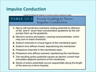
Membrane Potential
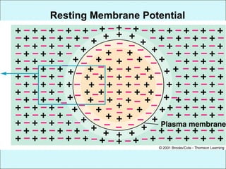
Membrane potential
ļ‚—Separation of charges across the membrane
Or
ļ‚—Difference in relative number of cations and anions
in the ECF
ļ‚—Separated charges create the
ability to do work
ļ‚—Membrane potential is measured
in millivolts
ļ‚—1mv = 1/1000 volts
Chapter 3 The Plasma Membrane and
Membrane Potential
Human Physiology by Lauralee Sherwood ©2007
Brooks/Cole-Thomson Learning
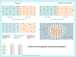
Which has the greatest membrane potential?
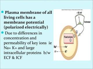
ļ‚—Plasma membrane of all
living cells has a
membrane potential
(polarized electrically)
ļ‚—Due to differences in
concentration and
permeability of key ions ie
Na+ K+ and large
intracellular proteins b/w
ECF & ICF
ļ‚—Nerve and muscle cells
ļ‚—Excitable cells
ļ‚—Have ability to produce rapid,
transient changes in their membrane
potential when excited which serves as
electric signals
ļ‚—Resting membrane potential (RMP)
Constant membrane potential present in cells of
non excitable tissues and those of excitable
tissues when they are at rest
Generation and maintenance of
RMP
ļ‚—The unequal distribution of a few key ions b/w the
ICF and ECF and their selective movement through
the plasma membrane are responsible for the
electrical properties of the membrane
ļ‚—In body electric charges are carried by ions .
ļ‚—So the ions primarily responsible for the generation of
resting membrane potential are Na+
, K+
, and A-
Resting Membrane Potential
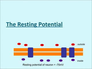
The Resting Potential
inside
outside
Resting potential of neuron = -70mV
+
-
+
-
+
-
+
-
+
-
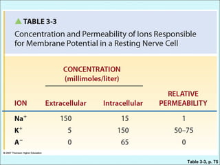
Table 3-3, p. 75
ļ‚—The concentration difference of Na+
and K+ are maintained by the Na+ K+
pump.
ļ‚—Since the plasma membrane is
impermeable to proteins so A- are
inside the membrane
More permeability of K+ as
compared to Na+ in resting state
ļ‚—The plasma membrane is more
permeable to K+ in resting state than
Na+ because the membrane has got
more leak channels for K+ than for
Na+
ļ‚—Moreover the hydrated form of K+ is
smaller than the hydrated form of Na+
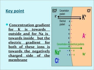
Key point
ļ‚—Concentration gradient
for K is towards
outside and for Na is
towards inside but the
electric gradient for
both of these ions is
towards the negatively
charged side of the
membrane
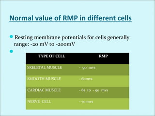
Normal value of RMP in different cells
ļ‚—Resting membrane potentials for cells generally
range: -20 mV to -200mV
ļ‚—
TYPE OF CELL RMP
SKELETAL MUSCLE - 90 mvs
SMOOTH MUSCLE - 60mvs
CARDIAC MUSCLE - 85 to - 90 mvs
NERVE CELL - 70 mvs
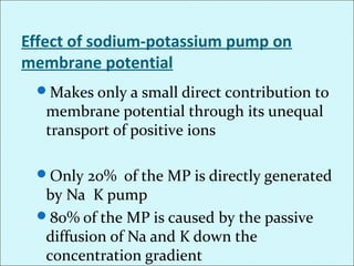
Effect of sodium-potassium pump on
membrane potential
ļ‚—Makes only a small direct contribution to
membrane potential through its unequal
transport of positive ions
ļ‚—Only 20% of the MP is directly generated
by Na K pump
ļ‚—80% of the MP is caused by the passive
diffusion of Na and K down the
concentration gradient
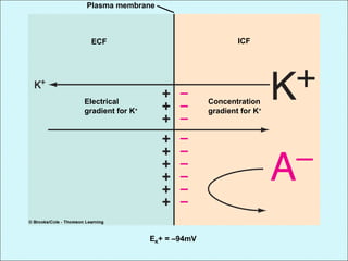
Effect of movement of K+
alone on MP (K+ equilibrium
potential)
Plasma membrane
ECF ICF
Concentration
gradient for K+
Electrical
gradient for K+
EK+ = –94mV
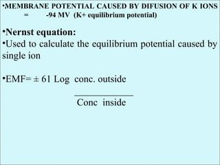
•.•MEMBRANE POTENTIAL CAUSED BY DIFUSION OF K IONS
= -94 MV (K+ equilibrium potential)
•Nernst equation:
•Used to calculate the equilibrium potential caused by
single ion
•EMF= ± 61 Log conc. outside
____________
Conc inside
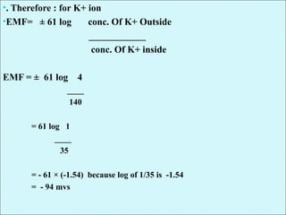
•. Therefore : for K+ ion
•EMF= ± 61 log conc. Of K+ Outside
____________
conc. Of K+ inside
EMF = ± 61 log 4
____
140
= 61 log 1
____
35
= - 61 Ɨ (-1.54) because log of 1/35 is -1.54
= - 94 mvs
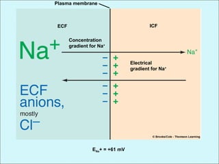
Effect of movement of Na+ alone on
MP (Na+ equilibrium potential)
Plasma membrane
ECF ICF
Concentration
gradient for Na+
Electrical
gradient for Na+
ENa+ = +61 mV
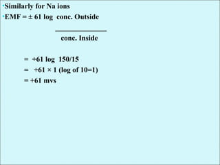
•Similarly for Na ions
•EMF = ± 61 log conc. Outside
______________
conc. Inside
= +61 log 150/15
= +61 Ɨ 1 (log of 10=1)
= +61 mvs
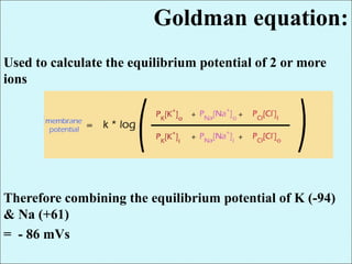
Goldman equation:
Used to calculate the equilibrium potential of 2 or more
ions
Therefore combining the equilibrium potential of K (-94)
& Na (+61)
= - 86 mVs
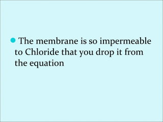
ļ‚—The membrane is so impermeable
to Chloride that you drop it from
the equation
Maintaining the Resting Potential by
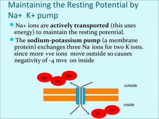
Na+ K+ pump
ļ‚—Na+ ions are actively transported (this uses
energy) to maintain the resting potential.
ļ‚—The sodium-potassium pump (a membrane
protein) exchanges three Na+
ions for two K+
ions.
since more +ve ions move outside so causes
negativity of -4 mvs on inside
inside
outside
Na+
Na+
K+
K+
Na+
NET RMP
ļ‚—Net RMP = - 86 - 4 = -90mvs
ļ‚—This means when the cell is at rest it has negativity of
-90 mvs inside i.e. inside the cell there is 90 mvs more
negative as compared to outside the cell
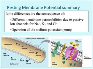
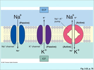
Resting Membrane Potential summary
Ionic differences are the consequence of:
•Different membrane permeabilities due to passive
ion channels for Na+
, K+
,and Cl-
•Operation of the sodium-potassium pump
Fig. 3-23, p. 79
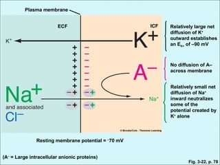
Plasma membrane
ECF ICF Relatively large net
diffusion of K+
outward establishes
an EK+ of –90 mV
No diffusion of A–
across membrane
Relatively small net
diffusion of Na+
inward neutralizes
some of the
potential created by
K+
alone
Resting membrane potential = –
70 mV
(A–
= Large intracellular anionic proteins)
Fig. 3-22, p. 78
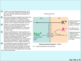
Fig. 3-22, p. 78
Usefulness?
ļ‚—Neurons and muscle fibers can alter membrane
potential to send signals and create motion.
ļ‚— So called excitable tissues because when they are
excited by appropriate stimulation they can rapidly
and transiently alter membrane permeabilities to the
involved ions so bringing fluctuations in membrane
potential which bring about nerve impulse in nerve
cells and triggering contraction in muscle cells
ļ‚—Changes in ion movement
in turn are brought about by
changes in membrane
permeability in response to
a triggering agent or a
stimuli
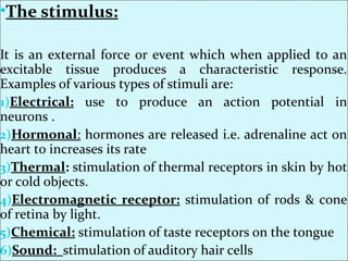
•The stimulus:
It is an external force or event which when applied to an
excitable tissue produces a characteristic response.
Examples of various types of stimuli are:
1)Electrical: use to produce an action potential in
neurons .
2)Hormonal: hormones are released i.e. adrenaline act on
heart to increases its rate
3)Thermal: stimulation of thermal receptors in skin by hot
or cold objects.
4)Electromagnetic receptor: stimulation of rods & cone
of retina by light.
5)Chemical: stimulation of taste receptors on the tongue
6)Sound: stimulation of auditory hair cells
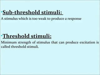
•Sub-threshold stimuli:
A stimulus which is too weak to produce a response
•Threshold stimuli:
Minimum strength of stimulus that can produce excitation is
called threshold stimuli.
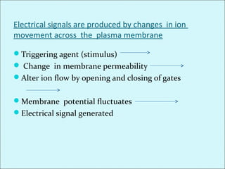
Electrical signals are produced by changes in ion
movement across the plasma membrane
ļ‚—Triggering agent (stimulus)
ļ‚— Change in membrane permeability
ļ‚—Alter ion flow by opening and closing of gates
ļ‚—Membrane potential fluctuates
ļ‚—Electrical signal generated
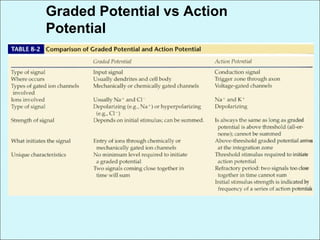
Types of electrical signals
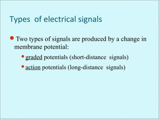
ļ‚—Two types of signals are produced by a change in
membrane potential:
ļ‚—graded potentials (short-distance signals)
ļ‚—action potentials (long-distance signals)
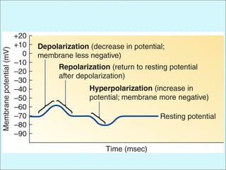
Terminology Associated with Changes in
Membrane PotentialF8-7, F8-8
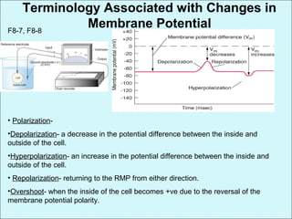
• Polarization-
•Depolarization- a decrease in the potential difference between the inside and
outside of the cell.
•Hyperpolarization- an increase in the potential difference between the inside and
outside of the cell.
• Repolarization- returning to the RMP from either direction.
•Overshoot- when the inside of the cell becomes +ve due to the reversal of the
membrane potential polarity.
Terminologies
ļ‚—Polarization
ļ‚—Depolarization
ļ‚—Repolarization
ļ‚—Hyperpolarization
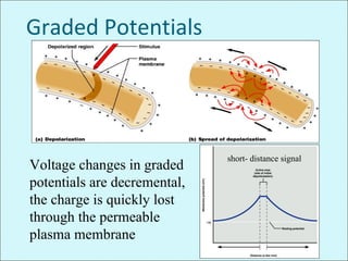
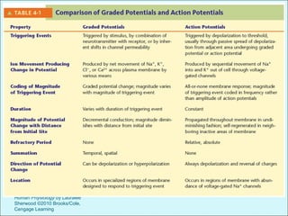
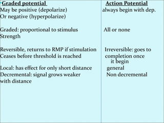
Graded Potentials
Short-lived, local changes in membrane potential
(either depolarizations or hyperpolarizations)
Cause passive current flow that decreases in
magnitude with distance so serve as short distance
signals
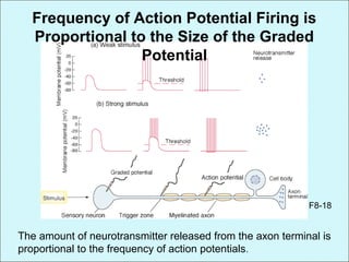
Their magnitude varies directly with the strength of
the stimulus – the stronger the stimulus the more the
voltage changes and the farther the current goes
Sufficiently strong graded potentials can initiate
action potentials
Graded Potentials
ļ‚—The Stronger a triggering event, the larger the
resultant graded potential
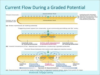
ļ‚—Graded Potential spread by passive Current flow.
ļ‚—Graded potentials die over short distances
ļ‚—K+ leaks out of the membrane
ļ‚—Decremental: gradually decreases
ļ‚—If strong enough, graded potentials trigger action
potentials
ļ‚—The wave of depolarization or
hyperpolarization which moves
through the cell with a graded
potential is known as local current
flow.
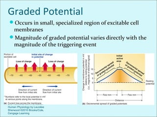
Current Flow During a Graded Potential
Chapter 4 Principles of Neural and Hormonal
Communication
Human Physiology by Lauralee Sherwood ©2010
Brooks/Cole, Cengage Learning
Chapter 4 Principles of Neural
and Hormonal Communication
Human Physiology by Lauralee
Sherwood ©2010 Brooks/Cole,
Cengage Learning
Graded Potential
ļ‚—Occurs in small, specialized region of excitable cell
membranes
ļ‚—Magnitude of graded potential varies directly with the
magnitude of the triggering event
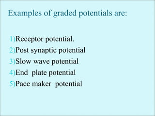
Examples of graded potentials are:
1)Receptor potential.
2)Post synaptic potential
3)Slow wave potential
4)End plate potential
5)Pace maker potential
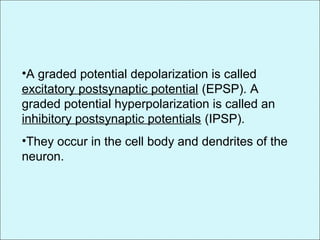
•A graded potential depolarization is called
excitatory postsynaptic potential (EPSP). A
graded potential hyperpolarization is called an
inhibitory postsynaptic potentials (IPSP).
•They occur in the cell body and dendrites of the
neuron.
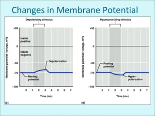
Changes in Membrane Potential
Graded Potentials
Voltage changes in graded
potentials are decremental,
the charge is quickly lost
through the permeable
plasma membrane
short- distance signal
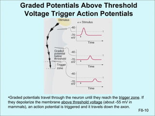
•Graded potentials travel through the neuron until they reach the trigger zone. If
they depolarize the membrane above threshold voltage (about -55 mV in
mammals), an action potential is triggered and it travels down the axon.
F8-10
Graded Potentials Above Threshold
Voltage Trigger Action Potentials
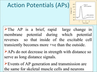
Action Potentials (APs)
The AP is a brief, rapid large change in
membrane potential during which potential
reverses so that inside of the excitable cell
transiently becomes more +ve than the outside.
APs do not decrease in strength with distance so
serve as long distance signals.
Events of AP generation and transmission are
the same for skeletal muscle cells and neurons
Course of the Action Potential
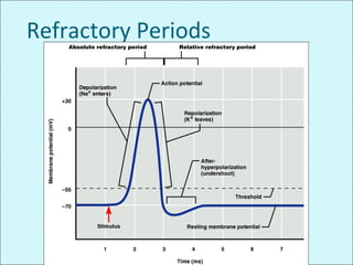
ļ‚—The action potential begins with a partial
depolarization [A].
ļ‚—When the excitation threshold is reached there is a
sudden large depolarization [B].
ļ‚—This is followed rapidly by repolarization [C] and a
brief hyperpolarization [D].
Membrane
potential
(mV)
[A]
[B] [C]
[D] excitation threshold
Time (msec)
-70
+40
0
0 1 2 3
Marked changes in membrane
permeability and ion movement lead to
an action potential (AP)
ļ‚—Passive diffusion of K+ makes greatest contribution to
the RMP due to more permeability of plasma
membrane to K+ through leak channels at rest.
ļ‚—During an AP marked changes in membrane
permeability to Na+ and K+ take place permitting
rapid fluxes down their electrochemical gradient
ļ‚—These ions carry the current responsible for the
potential changes that occur during an AP
ļ‚—Action potential takes place as
a result of the triggered
opening and subsequent closing
of 2 specific types of channels
Voltage gated Na+ channels
Voltage gated K+ channels
ROLE OF VOLTAGE GATED Na+
CHANNEL & VOLTAGE GATED K+
CHANNELS IN ACTION POTENTIAL
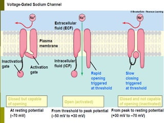
Voltage gated Na+ channels
ļ‚— Most important channels during AP
ļ‚—It has two gates:
ACTIVATION GATES: At RMP activation gates are
closed so no Na+ influx at RMP thru these channels
These activation gates open when membrane
potential become less negative than during resting
state then the activation gates of these voltage gated
channels open so increasing Na+ permeability to 500-
5000 fold.
Inactivation of Na+ channels
ļ‚—The same increase in voltage that open the activation
gates also closes the inactivation gates but closing of
gates is a slower process than opening so large
amount of Na+ influx has occurred
ļ‚—Another important feature of Na+ channels
inactivation is that the inactivation gate will not
reopen until the membrane potential returns to or
near the original RMP.
•.
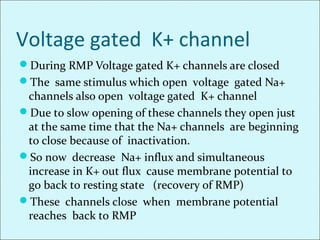
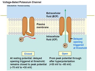
Voltage gated K+ channel
ļ‚—During RMP Voltage gated K+ channels are closed
ļ‚—The same stimulus which open voltage gated Na+
channels also open voltage gated K+ channel
ļ‚—Due to slow opening of these channels they open just
at the same time that the Na+ channels are beginning
to close because of inactivation.
ļ‚—So now decrease Na+ influx and simultaneous
increase in K+ out flux cause membrane potential to
go back to resting state (recovery of RMP)
ļ‚—These channels close when membrane potential
reaches back to RMP
•.
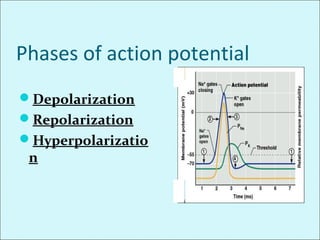
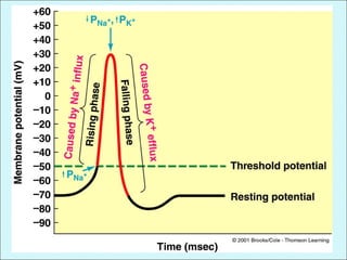
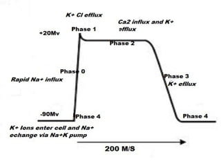
Phases of action potential
ļ‚—Depolarization
ļ‚—Repolarization
ļ‚—Hyperpolarizatio
n
Initiation of action potential
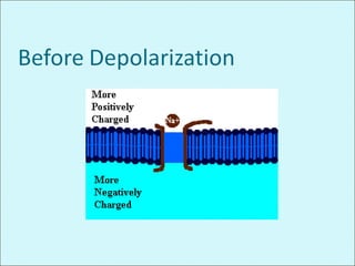
ļ‚—To initiate an AP a triggering event causes the
membrane to depolarize from the resting potential of
-90 mvs.
ļ‚—Depolarization proceeds slowly at first until it reaches
a critical level known as threshold potential. i.e.
-65 mvs . At threshold explosive depolarization occurs.
ļ‚§ An AP will not occur until the initial rise in membrane
potential reaches a threshold level.
ļ‚§ This occurs when no. of Na+ entering the cell
becomes greater than the no. of K+ leaving the
cell.
Threshold and Action Potentials
Threshold Voltage– membrane is depolarized by
15 to 20 mV
Subthreshold stimuli produce subthreshold
depolarizations and are not translated into APs
Stronger threshold stimuli produce depolarizing
currents that are translated into action potentials
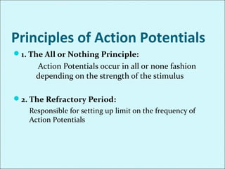
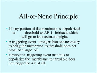
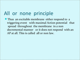
All-or-None phenomenon – action potentials
either happen completely, or not at all
depending on threshold
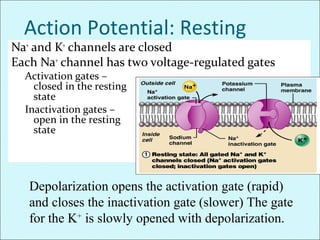
Action Potential: Resting
StateNa+
and K+
channels are closed
Each Na+
channel has two voltage-regulated gates
Activation gates –
closed in the resting
state
Inactivation gates –
open in the resting
state
Depolarization opens the activation gate (rapid)
and closes the inactivation gate (slower) The gate
for the K+
is slowly opened with depolarization.
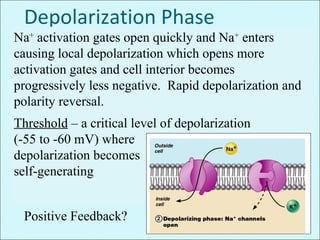
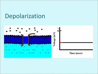
Depolarization Phase
Na+
activation gates open quickly and Na+
enters
causing local depolarization which opens more
activation gates and cell interior becomes
progressively less negative. Rapid depolarization and
polarity reversal.
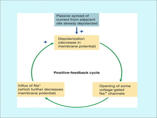
Threshold – a critical level of depolarization
(-55 to -60 mV) where
depolarization becomes
self-generating
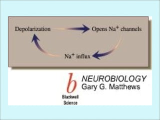
Positive Feedback?
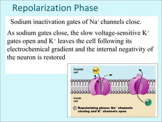
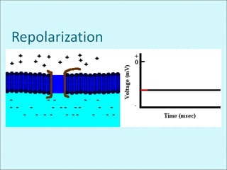
Repolarization Phase
Sodium inactivation gates of Na+
channels close.
As sodium gates close, the slow voltage-sensitive K+
gates open and K+
leaves the cell following its
electrochemical gradient and the internal negativity of
the neuron is restored
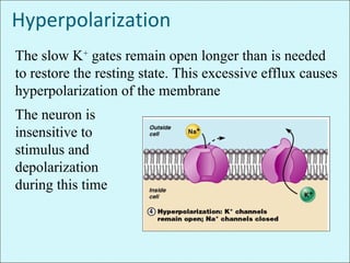
Hyperpolarization
The slow K+
gates remain open longer than is needed
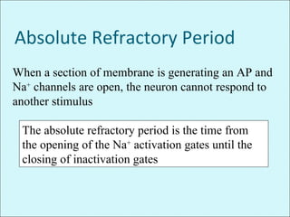
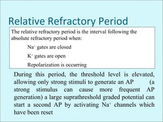
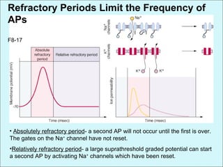
to restore the resting state. This excessive efflux causes
hyperpolarization of the membrane
The neuron is
insensitive to
stimulus and
depolarization
during this time
Depolarization increases the probability of
producing nerve impulses. Hyperpolarization
reduces the probability of producing nerve
impulses.
Role of the Sodium-Potassium Pump
Repolarization restores the resting electrical
conditions of the neuron, but does not restore the
resting ionic conditions
Ionic redistribution is accomplished by the
sodium-potassium pump following
repolarization
Importance of Action Potential
Generation
ļ‚—Nerve traffic, muscle contraction, hormone
release, G.I. secretions, Cognitive thought,
etc.
ļ‚—Action Potentials are required for the
senses - Sight, hearing, and touch are all
dependent on action potentials for
transmission of information to the brain
ļ‚—Threshold stimuli (Graded Potential) cause
the.generation of an action potential
Role of Calcium ions in Action
potential
ļ‚—Calcium pump in almost all cells of the body
maintain the calcium gradient with high Ca in ECF
as compared to ICF.
ļ‚—In addition to Ca pumps there are voltage gated Ca
channels which are slightly permeable to Na+ as well
as to Ca++ ions.
ļ‚—So when they open both Na and Ca flow to the
interior of the fiber. So called Ca Na channels.
ļ‚—They are slow to open requiring 20 times as long for
activation as Na channels so called slow channels in
contrast to Na channels which are fast channels.
ļ‚—Ca++ channels are numerous in smooth muscles and
cardiac muscle. In some smooth muscles the fast
Na+ channels are hardly present so that the AP are
caused almost entirely by activation of slow Ca++
channels.
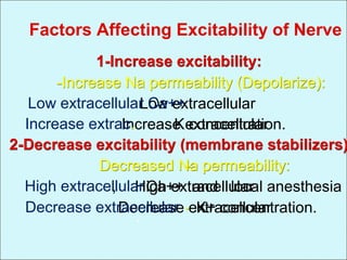
Increased permeability of Na channels
when there is deficit of Ca ions
ļ‚—The conc. Of Ca ions in ECF has profound effect on
the voltage level at which the Na channels become
activated.
ļ‚—So when there is a deficit of Calcium ions in the
ECF the voltage gated Na channels open by very little
increase of MP from its normal very negative level.
so nerve fiber become highly excitable .
ļ‚—When Ca levels fall 50% below normal spontaneous
discharge occurs in some peripheral nerves causing
tetany. Its lethal when respiratory muscles are
involved.
Cause:
ļ‚—Ca bind to the exterior surface of the voltage gated
Na channels protein molecule.
ļ‚—The +ve charge of Ca ions in turn alter the electrical
state of the channel protein itself.
ļ‚—So altering the voltage level required to open the
sodium gates.
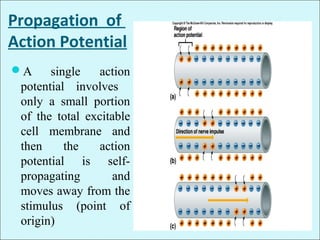
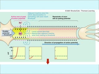
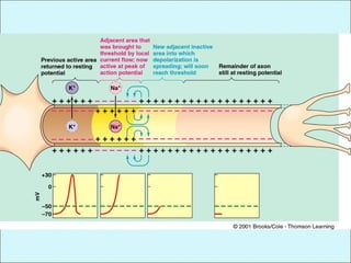
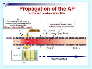
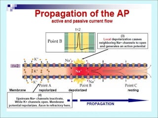
Propagation of
Action Potential
ļ‚—A single action
potential involves
only a small portion
of the total excitable
cell membrane and
then the action
potential is self-
propagating and
moves away from the
stimulus (point of
origin)
Direction of Action potential
ļ‚—AP travels in all directions away from the stimulus
until the entire membrane is depolarized
Conduction of Action Potentials
ļ‚—Two types of propagation
ļ‚—Contiguous conduction
ļ‚— Conduction in unmyelinated fibers
ļ‚— Action potential spreads along every portion of the
membrane
ļ‚—Saltatory conduction
ļ‚— Rapid conduction in myelinated fibers
ļ‚— Impulse jumps over sections of the fiber covered with
insulating myelin
Nerve or muscle impulse
ļ‚—The transmission of the depolarization process along
a nerve or muscle fibre is called impulse
ļ‚—An action potential in the axon of a neuron is called a nerve
impulse and is the way neurons communicate.
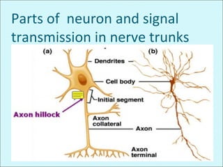
Parts of neuron and signal
transmission in nerve trunks
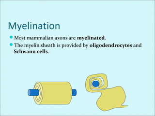
Myelination
ļ‚—Most mammalian axons are myelinated.
ļ‚—The myelin sheath is provided by oligodendrocytes and
Schwann cells.
MYELIN
ļ‚—Myelin
ļ‚—Primarily composed of lipids sphingomyelin
ļ‚—Formed by oligodendrocytes in CNS
ļ‚—Formed by Schwann cells in PNS
• Myelin is insulating, preventing passage of ions
over the membrane as it is made up of lipids so
water soluble ions cannot permeate so current
cannot leak out in the ECF
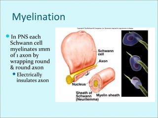
Myelination
ļ‚—In PNS each
Schwann cell
myelinates 1mm
of 1 axon by
wrapping round
& round axon
ļ‚—Electrically
insulates axon
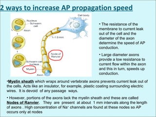
• The resistance of the
membrane to current leak
out of the cell and the
diameter of the axon
determine the speed of AP
conduction.
• Large diameter axons
provide a low resistance to
current flow within the axon
and this in turn, speeds up
conduction.
•Myelin sheath which wraps around vertebrate axons prevents current leak out of
the cells. Acts like an insulator, for example, plastic coating surrounding electric
wires. It is devoid of any passage ways.
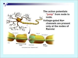
• However, portions of the axons lack the myelin sheath and these are called
Nodes of Ranvier. They are present at about 1 mm intervals along the length
of axons . High concentration of Na+
channels are found at these nodes so AP
occurs only at nodes
2 ways to increase AP propagation speed
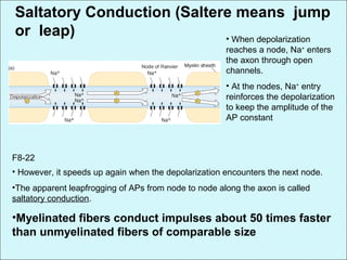
Saltatory Conduction (Saltere means jump
or leap) • When depolarization
reaches a node, Na+
enters
the axon through open
channels.
• At the nodes, Na+
entry
reinforces the depolarization
to keep the amplitude of the
AP constant
• However, it speeds up again when the depolarization encounters the next node.
•The apparent leapfrogging of APs from node to node along the axon is called
saltatory conduction.
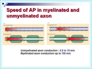
•Myelinated fibers conduct impulses about 50 times faster
than unmyelinated fibers of comparable size
F8-22
•Saltatory conduction in myelinated fibers
from node to node
•As no ions can flow through myelin sheath they can
flow with ease through node of ranvier.
•Therefore, action potential or flow of electrical
currents occurs from node to node in a jumping
manner known as saltatory conduction
Importance of saltatory
conduction
•Increases the conduction velocity through myelinated
nerve fiber.
•Conserves energy for the axon
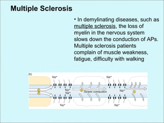
Multiple Sclerosis
• In demylinating diseases, such as
multiple sclerosis, the loss of
myelin in the nervous system
slows down the conduction of APs.
Multiple sclerosis patients
complain of muscle weakness,
fatigue, difficulty with walking