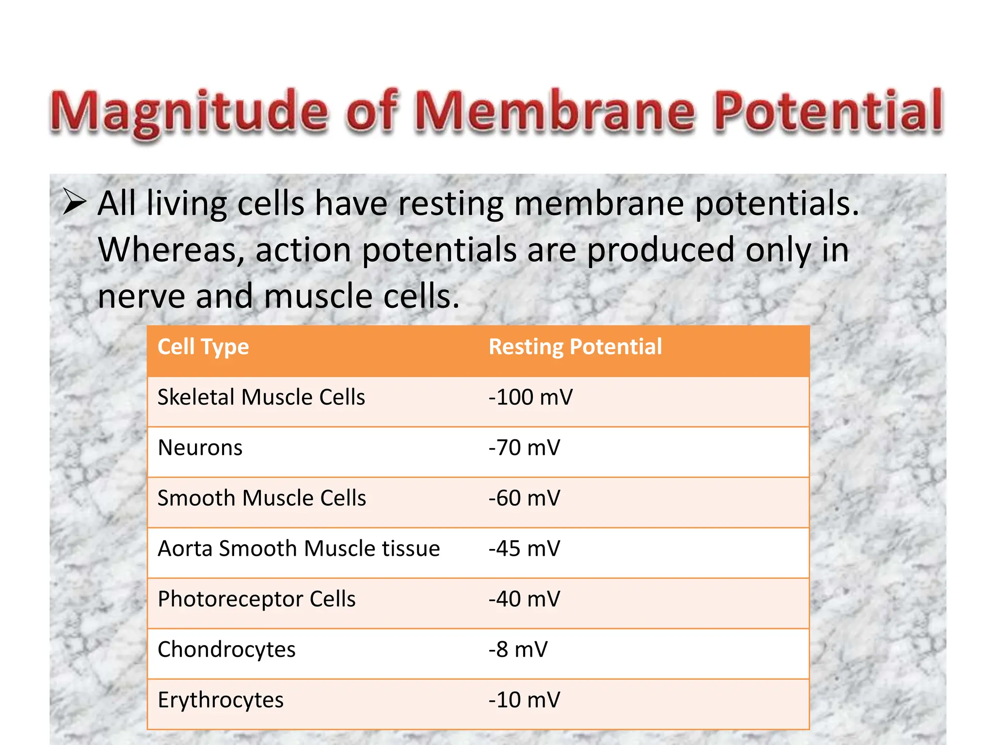
1. Presented By
(M.Sc. PG Student)
Department Of Physiology
Dr. Balasaheb Vikhe Patil Rural Medical College, Loni
2. Membrane potential is a difference in
electrical potential across the cell membrane.
There exists a potential difference across the
membrane of all living cells with inside being
negative in relation to the outside, i.e. the
cations and anions arrange themselves along
the outer and inner surfaces of the cell
membrane.
3. The build up of charges occurs only very close
to cell membrane. The ECF and ICF are else
where contains equal number of +ve and –ve
charges and is electrically neutral.
Thus, the number of ions responsible
for membrane potential is minute fraction of
the total number present across the cell
membrane in each respective compartment.
The greater the difference across the
membrane, the larger the membrane
potential.
4. The membrane potential at rest is called
resting membrane potential. It is written
with minus sign, signifying that inside is
negative relative to exterior.
The membrane potential measured
during the excited state of cell is called
action potential. It is written with plus
sign, signifying that outside is negative
relative to interior.
5. All living cells have resting membrane potentials.
Whereas, action potentials are produced only in
nerve and muscle cells.
Cell Type Resting Potential
Skeletal Muscle Cells -100 mV
Neurons -70 mV
Smooth Muscle Cells -60 mV
Aorta Smooth Muscle tissue -45 mV
Photoreceptor Cells -40 mV
Chondrocytes -8 mV
Erythrocytes -10 mV
6. In a particular cell also membrane
potential varies according to its
functional status. For example, a nerve
cell has a membrane potential of -70 mV
(inside negative) at rest , but when it gets
excited, the membrane potentials
becomes +30 mV (inside positive).
7. The essential instruments used in recording
the activity of an excitable tissue are:
1. Microelectrode
2. Electronic amplifiers
3. Cathode ray oscilloscope (CRO i.e. voltage
amplifier recorder system)
8. Measurement of the membrane potential of
the nerve fiber using a microelectrode.
9. • When one electrode is placed on the surface
of the cell and another electrode is inserted
into interior of the cell, both are connected
through a suitable amplifier to a cathode ray
oscilloscope (CRO), a constant (or steady)
potential difference is observed between the
inside and outside of the cell at rest, called as
the resting membrane potential.
10. • Factors involved in genesis of membrane
potential:
1. Selective permeability of the membrane
2. Gibbs–Donnan effect
3. Nernst equation
4. Constant field Goldman equation
5. Sodium-potassium ATPase Pump
11. The cell membrane is selectively permeable, i.e. to
some ions it is freely permeable, to others
impermeable to some others, it has variable
permeability.
12.
13. 1. Ions like Na+, K+, Cl- and HCO3
- are diffusible ions. The
cell membrane is freely permeable to K+ and Cl- and
moderately permeable to Na+. The permeability of K+
is 50-100 times greater than that of Na+.
2. The cell membrane is practically impermeable to
intracellular proteins and organic phosphate, which
are negatively charged ions.
3. The presence of gated channels in the cell membrane
is responsible for the variable permeability of certain
ions in different circumstances.
14. According to Gibbs-Donnan membrane
equilibrium, when two ionized solutions are
separated by a semi-permeable membrane then
at equilibrium:
1. Each solution shall be electrically neural, i.e.
15. 2. The product of diffusible ions on each side of
the membrane will be equal, i.e.
16. From the above the ratio of diffusible ions will
be as below:
Thus, there will be symmetrical distribution
of ions at equilibrium.
17. But if one or more non-diffusible ions ‘X’ are
present on one side (A side) of the membrane,
then according to to Gibbs-Donnan membrane
equilibrium the distribution of diffusible ions
will be as follows:
1. Each solution shall be electrically neural, i.e.
(Na+)A = (Cl-)A + (X-)A
(Na+)B = (Cl-)B
18. 2. The product of diffusible ions on two sides will be
equal, i.e.
From the relationship of (1) and (2), it is found
that:
• (Na+)A > (Na+)B , and
• (Cl−)A < (Cl−)B
19. The Gibbs–Donnan effect is named after the
American physicist Josiah Willard Gibbs who
proposed it in 1878 and the British
British chemist Frederick George
Donnan who studied it experimentally in
1911.
22. Membrane Potentials Caused by
Diffusion
A diffusion potential is the potential
difference generated across a membrane
when a cation or anion diffuses down its
concentration gradient.
A diffusion potential can be generated only if
the membrane is permeable to the ion.
23. The size of the diffusion potential depends on
the size of concentration gradient
The sign of the diffusion potential depends
on whether the diffusing ion is positively or
negatively charged.
Diffusion potentials are created by diffusion of
very few ions and, therefore, do not result in
changes in concentration of diffusing ions.
24. 2. The Nernst Potential— Relation of the Diffusion
Potential to the Concentration Difference.
The diffusion potential level across a
membrane that exactly opposes the net
diffusion of a particular ion through the
membrane is called the Nernst potential for
that ion.
The magnitude of this Nernst potential is
determined by the ratio of the concentrations
of that specific ion on the two sides of the
membrane.
25. The greater this ratio, the greater the tendency
for the ion to diffuse in one direction, and
therefore the greater the Nernst potential
required to prevent additional net diffusion.
The following equation, called the Nernst
equation, can be used to calculate the Nernst
potential for any univalent ion at normal body
temperature of 98.6°F (37°C):
27. The Nernst equation helps in calculating
the equilibrium potential for each ion
individually.
However, the magnitude of the
membrane potential at any given time
depends on the distribution of Na+, K+
and Cl- and the permeability of each of
these ions.
28. The integrated role of different ions in
generation of membrane potential can
be describe accurately by the
Goldman’s constant field equation or
the so called Goldman-Hodgkin-Katz
Eqaution:
29. • Interference of Goldman constant field equation
The following important inferences can be
drawn from gold-man constant field equation:
1. Most important ions for development of
membrane potentials in nerve and muscle
fibres are Na+, K+ and Cl-. The voltage of
membrane potential is determined by
concentration gradient of each of these ions.
30. 2.
• Degree of importance of each of the ions in
determining the voltage depends upon the membrane
permeability of the individual ion.
• Each permeable ion attempts to drive the membrane
potential towards its equilibrium potential.
• Ions with highest permeability will make greatest
contribution to the membrane potential and those with
lowest permeability will make very little or no
contribution.
• For example, if the membrane is impermeable to K+
and Cl-, then the membrane potential will be
determined by the Na+ gradient alone and the resulting
potential will be equal to the Nernst potential for
sodium.
31. 3. Higher concentration of cations in
the intracellular fluid as compared to
extracellular fluid is responsible for
electronegativity inside the
membrane. This is because of the fact
that due to concentration gradient,
the cations diffuse out outside leaving
the non-diffusible anion inside the
cell.
32. 4. Signal transmission in the
nerves is primarily due to change
in the sodium and potassium
permeability because their
channel undergo rapid change
during conduction of the nerve
impulse and not much change is
seen in chloride channels.
33. The discoverers of Goldman
Equation are David E.
Goldman of Columbia University,
and the Medicine Nobel
laureates Alan Lloyd
Hodgkin and Bernard Katz.
37. • Na+-K+ pump provides an additional contribution to the
resting potential.
• There is continuous pumping of three sodium ions to
the outside for each two potassium ions pumped to
the inside of the membrane.
• The fact that more sodium ions are being pumped to
the outside than potassium to the inside causes
continual loss of positive charges from inside the
membrane; this creates an additional degree of
negativity (about −4 millivolts additional) on the inside
beyond that which can be accounted for by diffusion
alone.