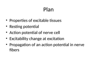
Plan
• Properties ofexcitable tissues
• Resting potential
• Action potential of nerve cell
• Excitability change at excitation
• Propagation of an action potential in nerve
fibers
3.
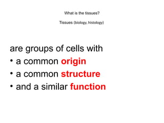
What is thetissues?
Tissues (biology, histology)
are groups of cells with
• a common origin
• a common structure
• and a similar function
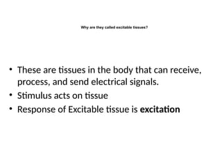
Why are theycalled excitable tissues?
• These are tissues in the body that can receive,
process, and send electrical signals.
• Stimulus acts on tissue
• Response of Excitable tissue is excitation
6.
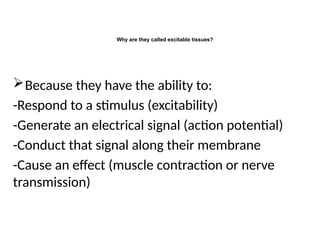
Why are theycalled excitable tissues?
Because they have the ability to:
-Respond to a stimulus (excitability)
-Generate an electrical signal (action potential)
-Conduct that signal along their membrane
-Cause an effect (muscle contraction or nerve
transmission)
7.
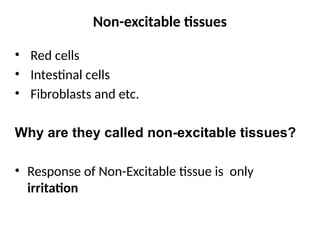
Non-excitable tissues
• Redcells
• Intestinal cells
• Fibroblasts and etc.
Why are they called non-excitable tissues?
• Response of Non-Excitable tissue is only
irritation
8.
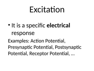
Excitation
• It isa specific electrical
response
Examples: Action Potential,
Presynaptic Potential, Postsynaptic
Potential, Receptor Potential, …
9.
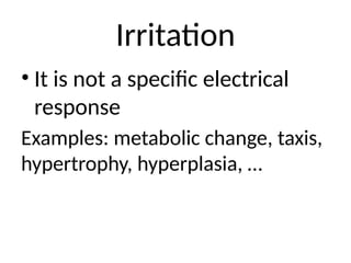
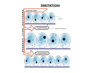
Irritation
• It isnot a specific electrical
response
Examples: metabolic change, taxis,
hypertrophy, hyperplasia, …
Classification of stimuli:
•By the nature
- the external: physical, chemical, biological
- the internal: physiologically active substances
(e.g neurotransmitters)
• By force
-The Threshold is the minimal stimulus capable
to cause tissue response.
13.
THE GENERAL PROPERTIESOF EXCITABLE
TISSUES
•
1. EXCITABILITY
-Ability to respond when stimulated.
-Ability to detect stimulus.
-Ability to change electrical state.
Example: Pinch your skin → nerves respond.
2. CONDUCTIVITY
- Ability to transmit electrical signals
-Nerves: Conduct impulses from one cell to another
-Muscles: Conduct signals across muscle fibers to coordinate contraction
3. COMMUNICATION (Neurons)
- Neurons transmit impulses to: Other neurons, Muscles, Glands
- Achieved through action potentials and neurotransmitters
14.
THE GENERAL PROPERTIESOF EXCITABLE
TISSUES
•
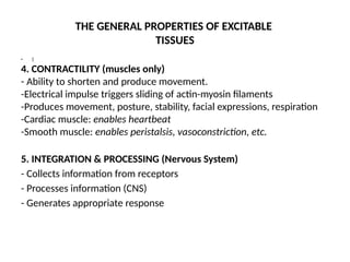
4. CONTRACTILITY (muscles only)
- Ability to shorten and produce movement.
-Electrical impulse triggers sliding of actin-myosin filaments
-Produces movement, posture, stability, facial expressions, respiration
-Cardiac muscle: enables heartbeat
-Smooth muscle: enables peristalsis, vasoconstriction, etc.
5. INTEGRATION & PROCESSING (Nervous System)
- Collects information from receptors
- Processes information (CNS)
- Generates appropriate response
15.
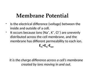
Membrane Potential
- Isthe electrical difference (voltage) between the
inside and outside of a cell.
- It occurs because ions (Na⁺, K⁺, Cl⁻) are unevenly
distributed across the cell membrane, and the
membrane has different permeability to each ion.
Em=Ein-Eout
It is the charge difference across a cell’s membrane
created by ions moving in and out.
16.
VARIATIONS IN MEMBRANE
POTENTIAL
Thevalue of the membrane potential changes
depending on:
- Ion concentrations (Na⁺, K⁺, Cl⁻) inside vs outside the
cell
- Membrane permeability to each ion
- How easily each ion can cross the membrane
17.
Goldman-Hodgkin-Katz (GHK)
Equation
The valueof the membrane potential changes
depending on:
- Ion concentrations (Na⁺, K⁺, Cl⁻) inside vs outside the
cell
- Membrane permeability to each ion
- How easily each ion can cross the membrane
18.
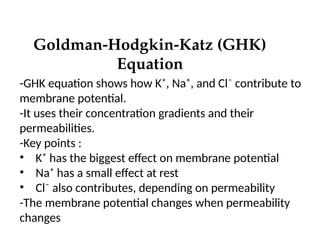
Goldman-Hodgkin-Katz (GHK)
Equation
-GHK equationshows how K⁺, Na⁺, and Cl⁻ contribute to
membrane potential.
-It uses their concentration gradients and their
permeabilities.
-Key points :
• K⁺ has the biggest effect on membrane potential
• Na⁺ has a small effect at rest
• Cl⁻ also contributes, depending on permeability
-The membrane potential changes when permeability
changes
19.
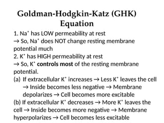
Goldman-Hodgkin-Katz (GHK)
Equation
1. Na⁺has LOW permeability at rest
→ So, Na⁺ does NOT change resting membrane
potential much
2. K⁺ has HIGH permeability at rest
→ So, K⁺ controls most of the resting membrane
potential.
(a) If extracellular K⁺ increases → Less K⁺ leaves the cell
→ Inside becomes less negative → Membrane
depolarizes → Cell becomes more excitable
(b) If extracellular K⁺ decreases → More K⁺ leaves the
cell → Inside becomes more negative → Membrane
hyperpolarizes → Cell becomes less excitable
20.
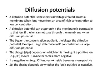
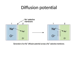
Diffusion potentials
• Adiffusion potential is the electrical voltage created across a
membrane when ions move from an area of high concentration to
low concentration.
• A diffusion potential can occur only if the membrane is permeable
to that ion. If the ion cannot pass through the membrane → no
diffusion potential.
• The bigger the concentration gradient, the bigger the diffusion
potential. Example: Large difference in K⁺ concentration → large
diffusion potential.
• The charge (sign) depends on which ion is moving: If a positive ion
(e.g., K⁺) moves → inside becomes more negative
• If a negative ion (e.g., Cl⁻) moves → inside becomes more positive
• So, the charge depends on whether the ion is positive or negative.
21.
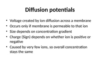
Diffusion potentials
• Voltagecreated by ion diffusion across a membrane
• Occurs only if membrane is permeable to that ion
• Size depends on concentration gradient
• Charge (Sign) depends on whether ion is positive or
negative
• Caused by very few ions, so overall concentration
stays the same
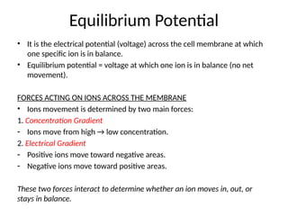
Equilibrium Potential
• Itis the electrical potential (voltage) across the cell membrane at which
one specific ion is in balance.
• Equilibrium potential = voltage at which one ion is in balance (no net
movement).
FORCES ACTING ON IONS ACROSS THE MEMBRANE
• Ions movement is determined by two main forces:
1. Concentration Gradient
- Ions move from high → low concentration.
2. Electrical Gradient
- Positive ions move toward negative areas.
- Negative ions move toward positive areas.
These two forces interact to determine whether an ion moves in, out, or
stays in balance.
24.
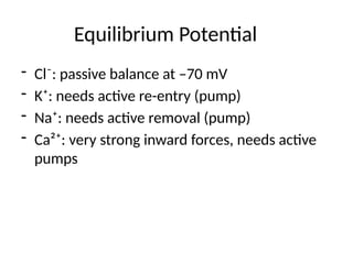
Equilibrium Potential
- Cl⁻:passive balance at –70 mV
- K⁺: needs active re-entry (pump)
- Na⁺: needs active removal (pump)
- Ca²⁺: very strong inward forces, needs active
pumps
25.
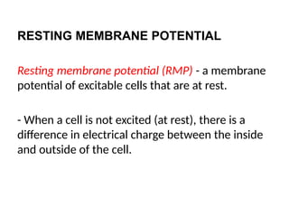
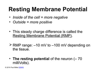
RESTING MEMBRANE POTENTIAL
Restingmembrane potential (RMP) - a membrane
potential of excitable cells that are at rest.
- When a cell is not excited (at rest), there is a
difference in electrical charge between the inside
and outside of the cell.
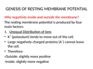
GENESIS OF RESTINGMEMBRANE POTENTIAL
Why negativity inside and outside the membrane?
The resting membrane potential is produced by four
main factors:
1. Unequal Distribution of Ions
• K⁺ (potassium) tends to move out of the cell.
• Large negatively charged proteins (A⁻) cannot leave
the cell.
• Therefore:
-Outside: slightly more positive
-Inside: slightly more negative
28.
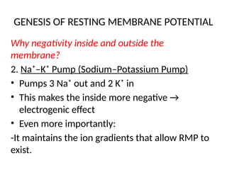
GENESIS OF RESTINGMEMBRANE POTENTIAL
Why negativity inside and outside the
membrane?
2. Na⁺–K⁺ Pump (Sodium–Potassium Pump)
• Pumps 3 Na⁺ out and 2 K⁺ in
• This makes the inside more negative →
electrogenic effect
• Even more importantly:
-It maintains the ion gradients that allow RMP to
exist.
29.
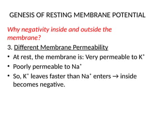
GENESIS OF RESTINGMEMBRANE POTENTIAL
Why negativity inside and outside the
membrane?
3. Different Membrane Permeability
• At rest, the membrane is: Very permeable to K⁺
• Poorly permeable to Na⁺
• So, K⁺ leaves faster than Na⁺ enters → inside
becomes negative.
30.
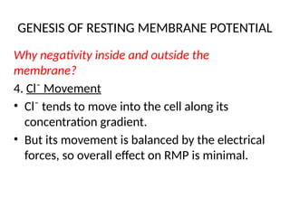
GENESIS OF RESTINGMEMBRANE POTENTIAL
Why negativity inside and outside the
membrane?
4. Cl⁻ Movement
• Cl⁻ tends to move into the cell along its
concentration gradient.
• But its movement is balanced by the electrical
forces, so overall effect on RMP is minimal.
31.
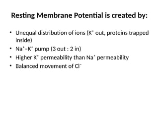
Resting Membrane Potentialis created by:
• Unequal distribution of ions (K⁺ out, proteins trapped
inside)
• Na⁺–K⁺ pump (3 out : 2 in)
• Higher K⁺ permeability than Na⁺ permeability
• Balanced movement of Cl⁻
32.
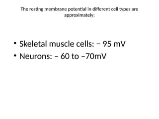
The resting membranepotential in different cell types are
approximately:
• Skeletal muscle cells: − 95 mV
• Neurons: – 60 to –70mV
33.
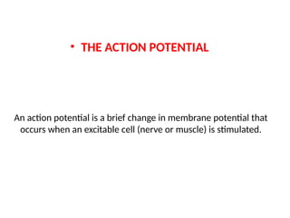
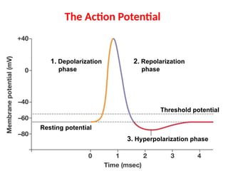
An action potentialis a brief change in membrane potential that
occurs when an excitable cell (nerve or muscle) is stimulated.
• THE ACTION POTENTIAL
1. Resting state(RMP): At rest, inside of the membrane
is negative and outside is positive. Since K+
permeability is greater than Na+ permeability,
therefore, K+ channels maintain the RMP and the
inside is negative.
2. Depolarization (during activation of the membrane)
- Na⁺ channels open → Na⁺ rushes in → inside becomes
positive.
3. Repolarization: K⁺ channels open → K⁺ exits → inside
returns negative.
4. After-hyperpolarization: K⁺ channels slowly close;
Na⁺/K⁺ pump restores full ionic balance.
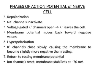
37.
PHASES OF ACTIONPOTENTIAL of NERVE
CELL
1. Resting membrane potential
• The neuron is at rest, typically around –70 mV.
2. Initial depolarization
• Some voltage-gated Na⁺ channels open.
Na⁺ enters the cell → membrane becomes less negative.
3. Rapid depolarization (upstroke)
• More Na⁺ channels open → rapid influx of Na⁺.
• Membrane potential rises quickly toward positive
values.
4. Peak of action potential
• Membrane reaches maximum positive voltage.
38.
PHASES OF ACTIONPOTENTIAL of NERVE
CELL
5. Repolarization
• Na⁺ channels inactivate.
• Voltage-gated K⁺ channels open → K⁺ leaves the cell.
• Membrane potential moves back toward negative
values.
6. Hyperpolarization
• K⁺ channels close slowly, causing the membrane to
become slightly more negative than resting.
7. Return to resting membrane potential
• Ion channels reset, membrane stabilizes at –70 mV.
39.
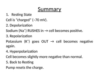
Summary
1. Resting State
Cellis “charged” (–70 mV).
2. Depolarization
Sodium (Na⁺) RUSHES in → cell becomes positive.
3. Repolarization
Potassium (K⁺) goes OUT → cell becomes negative
again.
4. Hyperpolarization
Cell becomes slightly more negative than normal.
5. Back to Resting
Pump resets the charge.
40.
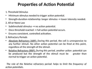
Properties of ActionPotential
1. Threshold Stimulus
• Minimum stimulus needed to trigger action potential.
• Strength-duration relationship: longer stimulus → lower intensity needed.
2. All-or-None Law
• Subthreshold stimulus → no action potential.
• Once threshold reached → full action potential occurs.
• Ensures consistent, controlled activation.
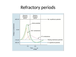
3. Refractory Periods
• Absolute Refractory (ARP): During this period, the cell is unresponsive to
any further stimuli. No other action potential can be fired at this point,
regardless of the strength of the stimuli.
• Relative Refractory (RRP): During this period, another action potential can
be produced but the strength of the stimuli must be greater than
normal to trigger an action potential.
The role of the Relative refractory period: helps to limit the frequency of
action potentials.
41.
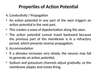
Properties of ActionPotential
4. Conductivity / Propagation
• An action potential in one part of the axon triggers an
action potential in the next part.
• This creates a wave of depolarization along the axon.
• The action potential cannot travel backward because
the previous part of the membrane is in a refractory
period, which prevents reverse propagation.
5. Accommodation
• If a stimulus increases very slowly, the neuron may fail
to generate an action potential.
• Sodium and potassium channels adjust gradually, so the
membrane adapts and resists firing.
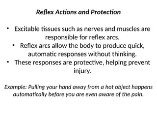
Reflex Actions andProtection
• Excitable tissues such as nerves and muscles are
responsible for reflex arcs.
• Reflex arcs allow the body to produce quick,
automatic responses without thinking.
• These responses are protective, helping prevent
injury.
Example: Pulling your hand away from a hot object happens
automatically before you are even aware of the pain.
44.
The main physiologicalcharacteristics of the AP
1. Obeys the law of "all or nothing." This means that:
• AP occurs when the stimulus, the power which is no less
than certain thresholds;
• Physical characteristics of the AP (amplitude, duration,
shape) does not depend on the power of stimulus.
2. Ability to auto spread along the cell membrane without damping, i.e.
without changing their physical characteristics.
3. AP accompanied with refractory.
4. AP is no capable to summation.
#32 The resting membrane potential in different cell types are approximately:
Skeletal muscle cells: −95 mV[3]
Smooth muscle cells: –60mV
Astroglia: –80 to –90mV
Neurons: –60 to –70mV
Erythrocytes: –9mV