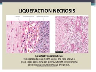
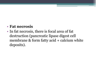
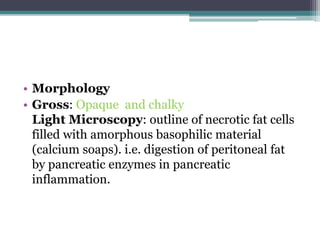
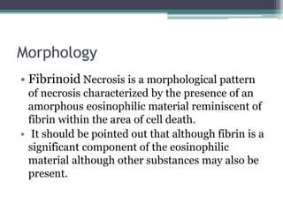
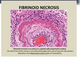
NECROSIS
Dr. Tulika Chaudhary
NECROSIS
Necrosis was long regarded as an
unprogrammed death of cells and living tissues,
which does not follow a highly regulated
intracellular program such as the apoptotic
signal transduction pathway.
Various agents ; Hypoxia, chemical and physical
agents , microbial agents .
TYPES
Coagulative
 Liquifactive
 Caseous
 Fat necrosis
 Fibrinoid necrosis.
• Coagulative necrosis is the commonest type and
is ischemic. It may occur in heart, kidney, or
adrenal glands and is firm in texture.
• It may occur due to denaturation of proteins
including enzymes.
• Morphology
• Gross features:
• The necrosis area is swollen, firm and pale.
• Light Microscopy:
• Cell detail is lost, but architecture preserved. The
dead cells retain their outline but only indistinctly.
This type of necrosis is frequently caused by lack of
blood supply and is exemplified well in infarcts of
solid organs, e. g. heart, spleen, kidney.
LIQUEFACTIVE NECROSIS
• Necrosis of big tissue with black, foul-smelling
appearance is known as liquefactive necrosis (black
or green color is due to breakdown of haemoglobin).
• In liquefactive necrosis, digestion of dead cells leads
to liquid mass. Autolysis predominates resulting in
liquified mass. Examples include cerebral infarction
and abscess. Brain cells secrete increased
hydrolases. These make neural tissue soft & liquid.
Abscess hyrolases from neutrophils liquefy tissue.
• Morphology
• Gross:
• Soft and liquid
• Microscopy:
• Enzymes digest the cell and convert it to a
formless proteinaceous mass. Ultimately,
discharge of the contents forms a cystic space
• Caseous Necrosis
• It is cheese-like, as in tuberculosis.
• Morphology
• Gross features: soft, granular, and friable as
cream -cheesy appearance.
Light Microscopy: Granular and eosinophilic.
Architecture is completely destroyed.
Examples: Tuberculosis, syphilis, some fungal
infections.
• Fat necrosis
• In fat necrosis, there is focal area of fat
destruction (pancreatic lipase digest cell
membrane & form fatty acid + calcium white
deposits).
• Morphology
• Gross: Opaque and chalky
Light Microscopy: outline of necrotic fat cells
filled with amorphous basophilic material
(calcium soaps). i.e. digestion of peritoneal fat
by pancreatic enzymes in pancreatic
inflammation.
• Types
• a. Truamatic fat necrosis –Foreign body giant
cell + foamy histiocytes form calcifications
producing hard lump.
b. Acute pancreatits -released enzymes digest
fat
c. Adipose tissue -TG + FFA lead to
saponification + calcification.
• Fibrinoid necrosis
• This is not a true degeneration but a strongly
eosinophilic stain like fibrin.
Location: interstitial collagen and blood vessels
(small artery and arteriole)
Nature: one kind of necrosis.
Morphology
• Fibrinoid Necrosis is a morphological pattern
of necrosis characterized by the presence of an
amorphous eosinophilic material reminiscent of
fibrin within the area of cell death.
• It should be pointed out that although fibrin is a
significant component of the eosinophilic
material although other substances may also be
present.
Necrosis
Necrosis

Necrosis

  • 1.
  • 2.
    NECROSIS Necrosis was longregarded as an unprogrammed death of cells and living tissues, which does not follow a highly regulated intracellular program such as the apoptotic signal transduction pathway. Various agents ; Hypoxia, chemical and physical agents , microbial agents .
  • 3.
    TYPES Coagulative  Liquifactive  Caseous Fat necrosis  Fibrinoid necrosis.
  • 4.
    • Coagulative necrosisis the commonest type and is ischemic. It may occur in heart, kidney, or adrenal glands and is firm in texture. • It may occur due to denaturation of proteins including enzymes.
  • 5.
    • Morphology • Grossfeatures: • The necrosis area is swollen, firm and pale. • Light Microscopy: • Cell detail is lost, but architecture preserved. The dead cells retain their outline but only indistinctly. This type of necrosis is frequently caused by lack of blood supply and is exemplified well in infarcts of solid organs, e. g. heart, spleen, kidney.
  • 7.
    LIQUEFACTIVE NECROSIS • Necrosisof big tissue with black, foul-smelling appearance is known as liquefactive necrosis (black or green color is due to breakdown of haemoglobin). • In liquefactive necrosis, digestion of dead cells leads to liquid mass. Autolysis predominates resulting in liquified mass. Examples include cerebral infarction and abscess. Brain cells secrete increased hydrolases. These make neural tissue soft & liquid. Abscess hyrolases from neutrophils liquefy tissue.
  • 8.
    • Morphology • Gross: •Soft and liquid • Microscopy: • Enzymes digest the cell and convert it to a formless proteinaceous mass. Ultimately, discharge of the contents forms a cystic space
  • 10.
    • Caseous Necrosis •It is cheese-like, as in tuberculosis.
  • 11.
    • Morphology • Grossfeatures: soft, granular, and friable as cream -cheesy appearance. Light Microscopy: Granular and eosinophilic. Architecture is completely destroyed. Examples: Tuberculosis, syphilis, some fungal infections.
  • 13.
    • Fat necrosis •In fat necrosis, there is focal area of fat destruction (pancreatic lipase digest cell membrane & form fatty acid + calcium white deposits).
  • 14.
    • Morphology • Gross:Opaque and chalky Light Microscopy: outline of necrotic fat cells filled with amorphous basophilic material (calcium soaps). i.e. digestion of peritoneal fat by pancreatic enzymes in pancreatic inflammation.
  • 16.
    • Types • a.Truamatic fat necrosis –Foreign body giant cell + foamy histiocytes form calcifications producing hard lump. b. Acute pancreatits -released enzymes digest fat c. Adipose tissue -TG + FFA lead to saponification + calcification.
  • 17.
    • Fibrinoid necrosis •This is not a true degeneration but a strongly eosinophilic stain like fibrin. Location: interstitial collagen and blood vessels (small artery and arteriole) Nature: one kind of necrosis.
  • 18.
    Morphology • Fibrinoid Necrosisis a morphological pattern of necrosis characterized by the presence of an amorphous eosinophilic material reminiscent of fibrin within the area of cell death. • It should be pointed out that although fibrin is a significant component of the eosinophilic material although other substances may also be present.