Downloaded 140 times


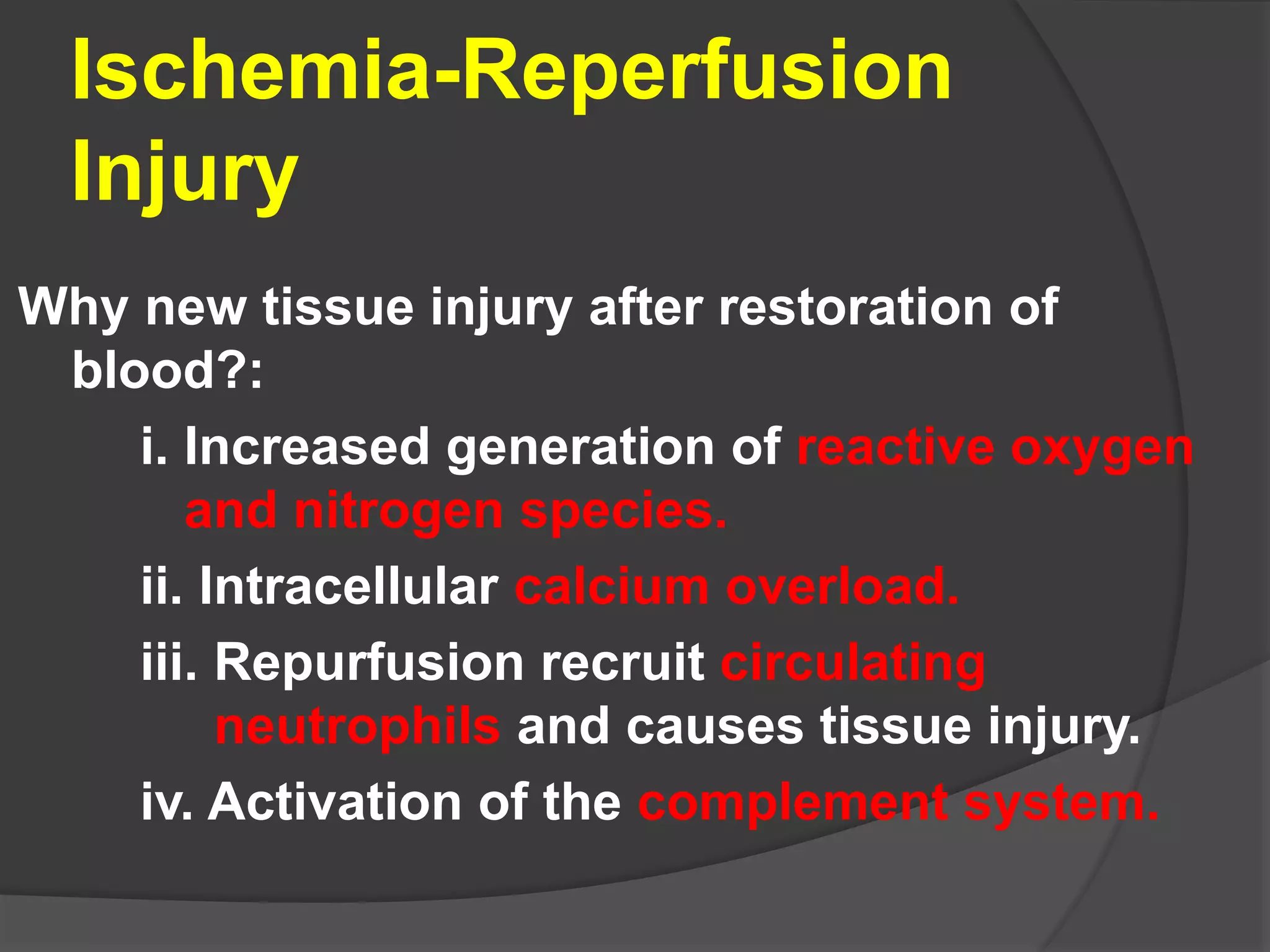


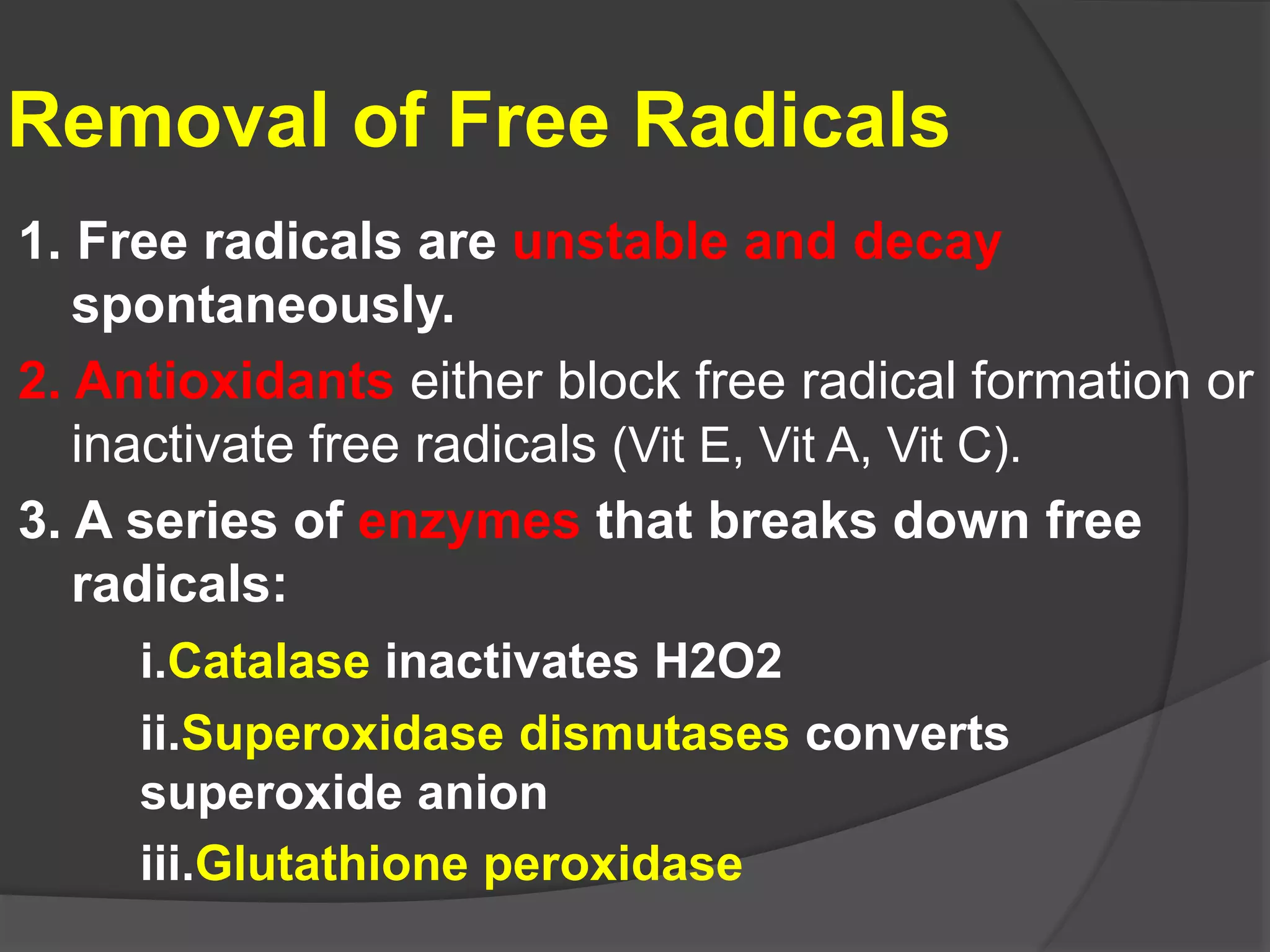


Irreversible cell injury can occur through necrosis or apoptosis. Necrosis is unprogrammed cell death due to intracellular protein denaturation and lysosomal enzyme digestion, resulting in cell membrane rupture and content leakage. Apoptosis is genetically programmed cell death where cells activate enzymes to degrade their own DNA and proteins. Key differences are that apoptotic cells shrink while necrotic cells swell, and apoptotic cells remain intact while necrotic cells lyse. Free radicals can also cause cell injury by attacking cell components and structures like membranes, proteins, and DNA, resulting in lipid peroxidation, protein damage, and DNA lesions.


