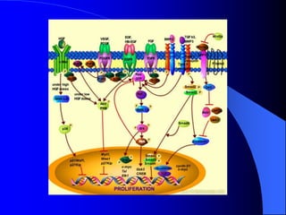
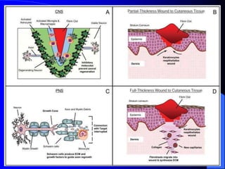
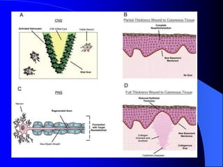
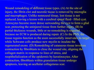
Healing & Repair
Tissue Repair
 may start early after tissue damage
 regeneration
– by parenchymal cells of the same type
 reparation
– replacement by connective tissue (fibrosis)
– result - scar
Regeneration and Reparation
 regeneration
– restoration of normal structure and function
– persistence of supportive “tissue skeleton“ necessary
 BM of epithelia
 Reticulin* frame in liver
 reparation
– restoration of normal shape x function is impaired or lost
– parenchyma replaced by fibrous tissue
– *Reticular fibers, reticular fibres or reticulin is a type of fiber in connective
tissue composed of type III collagen secreted by reticular cells. Reticular fibers
crosslink to form a fine meshwork (reticulin).
Tissue types
 permanent
– nonproliferative in postnatal life
– neurons, cardiomyocytes
 stable
– regeneration as response to injury
– parenchyma – liver, pancreas, renal tubules
– mesenchymal cells, endothelium
 labile
– continuous regeneration from stem cells (self-renewal)
– hematopoietic cells in bone marrow
– surface epithelia – skin, oral cavity, vagina, cervix
– duct epithelia – salivary glands, pancreas, biliary tract
– mucosas – GIT, uterus, fallopian tubes, urinary bladder
Cell-ECM interactions
 not only cells!
 EMC plays important role in healing
 interstitial matrix – by fibroblasts
 basement membrane – by fibroblast and epithelium
 components
– collagen (18 types) – I, III, IV, V; tensile strength*
– elastin (+ fibrillin) – return to normal structure after stress
– glycoproteins - adhesion, binding ECM to cells
(fibronectin, laminin)
– proteoglycans and hyalouronans – lubrication (gels)
*the resistance of a material to breaking under tension.
Cell-ECM interactions
 ECM function
– mechanical support
– determination of cell polarity
– control of cell growth
– maintenance of cell proliferation
– establishment of tissue micro-environments
– storage of regulatory molecules
Replacement of necrotic
tissue
 resorption by macrophages
 dissolution by enzymes
 replacement by granulation tissue
– uniform mechanism irrespective of initial trigger
– the same microscopic appearance
– angiogenesis
– migration and proliferation of fibroblasts
– deposition of ECM
– maturation and reorganization
Granulation tissue
 new-formed connective tissue, apparent from 3rd day
 thin-walled capillary vessels
 fibroblasts
 loose extracellular matrix
 stimulation
– PDGF, VEGF, FGF, TGF, TNF, EGF
 inhibition
– INFalfa, prostaglandins, angiostatins
 control
– cyclins, cyclin dependent kinases
Granulation tissue
 pink soft granular appearance
 richly vascularized
 highly cellular
 myxoid matrix
 inflammatory cells
 e.g. surface of wounds, bottom of ulcers
Angiogenesis
 neovascularization
 x vasculogenesis (embryonic process only)
 highly complex phenomenon
 angiogenic factors (FGF, VEGF)
 antiangiogenic factors
 healing, collateral circulation, tumors
Fibrosis and Remodeling
 scar formation
 fibroblasts
 myofibroblasts
– spindle cells of both fibroblastic and smooth
muscle phenotype
– production of collagen fibres
– wound contraction
Fibrosis and Remodeling
 abundant collagen fibres bridging the defect
 devoid of inflammatory cells
 reepithelization of surface defect
– from skin appendages and/or from periphery
 secondary changes
– calcification (dystrophic)
– ossification (metaplastic)
 remodeling
– synthesis and degradation of ECM
– metalloproteinases (MPs), tissue inhibitors of MPs
Pathological aspects
of healing
 proud flesh (caro luxurians)
– excessive amount of GT
 keloid
– excessive amount of collagen
 hyaline plaques
– serous membranes (spleen, pleura)
Summary of wound healing response in the brain versus nonneural tissue.
Hemostasis in damaged blood vessel. (a) An injury
to the vessel wall causes the release of blood-borne
cells, proteins, and platelets into the periphery.
Platelets are activated upon coming into contact
with the surrounding collagen outside of the blood
Wound repair of different tissue types. (A) In
the CNS, microglia and macrophages
migrate into the lesion, secreting various
cytokines and growth factors while also
removing necrotic tissue. Activated
astrocytes form a physical barrier around the
area
Primary versus secondary mineralized
tissue repair. (A) When a small fracture
gap occurs and the bone remains
stabilized, osteoblasts deposit bone
perpendicular to the bone’s long axis,
filling the gap and serving as a scaffold
for future remodeling.
(B) In primary contact repair, fractured ends are held in
direct apposition. No excess ECM is required. Cutting
cones are formed as osteoblasts absorb damaged bone,
followed by osteoblasts synthesizing osteoid, which
becomes mineralized to form lamellar bone. (C)
Unstabilized fractures are initially repaired through ECM
production by fibroblasts and chondrocytes arriving from
the periosteum to form a stabilizing soft callus. The
collagenous ECM becomes mineralized, forming woven
bone, also referred to as hard callus.
Histological analysis of
secondary fracture healing in
bone showing the progression
of repair on days 1, 3, 14, 21,
and 28. Fractured bone appears
denser than the surrounding
tissue. On day 7, extensive soft
callus is seen forming around
the injured bone. At day 14, the
soft callus becomes
mineralized to form new bone
and achieve union by day 21
and 28 (H&E stain, x40).
(Adapted from J. Bone Miner. Res., 16,
1004– 1014, 2001. With permission of the
American Society for Bone and Mineral
Research.)
Wound remodeling of different tissue types. (A) At the site of
injury, the fibrin clot and necrotic tissue is removed by microglia
and macrophages. Unlike nonneural tissue, lost tissue is not
replaced, leaving a lesion with a cerebral spinal fluid– filled cyst.
Astrocytes become more dense surrounding the cyst to form a glial
scar, protecting the uninjured tissue from further injury. (B) In
partial thickness wounds, little or no remodeling is required
because no ECM is produced during repair. (C) In the PNS, the
tissue regains function as the axon successfully innervates its target
while Schwann cells produce new myelin to insulate the
regenerated axons. (D) Remodeling of cutaneous tissue involves
contraction by fibroblasts to close the wound site, aligning the
collagen matrix in response to lines of stress, and
reepithelialization of the epidermis by keratinocytes. After
contraction, fibroblasts within granulation tissue undergo
apoptosis, leaving an acellular collagenous scar.
Primary versus secondary bone remodeling. (A)
Lamellar bone deposited perpendicular to the
bone’s long axis during primary repair is used as
a scaffold for cutting cones. Osteoclasts create
tunnels through which new blood vessels follow,
stimulating osteoblasts to produce new osteoid
that becomes mineralized to form lamellar bone.
A mature osteon forms when osteoclast and
osteoblast activity becomes quiescent. (B) In
primary contact remodeling the cutting cones
initiated during repair mature, forming “ mature
osteons.” The larger the number of osteons, the
greater the tissue strength of the repaired tissue.
(C) In secondary remodeling, the mineralized hard
callus near the periosteum not needed for structural
support is removed and remodeled by osteoclasts
arriving from the periosteum. Within the cortex,
cutting cones are formed as described in primary
remodeling. Less organized woven bone within the
wound gap is replaced with oriented lamellar bone.
The ultimate endpoint of both primary and
secondary bone healing is tissue with completely
restored function.
Time course of glial scar formation at four time points as
imaged by GFAP staining. At 2- and 4-week time points,
the astrocytic processes fall back into the void left by the
probe extraction before tissue processing. By 6 weeks, the
processes have interwoven to form a stronger, more dense
sheath surrounding the implant. Minimal changes between
the 6- and 12-week time points indicate the glial scar
completion within 6 weeks. (Reprinted from Exp. Neurol.,
156, 33–49, 1999. With permission from Elsevier.)

Pathology of Healing & repair

  • 1.
  • 2.
    Tissue Repair  maystart early after tissue damage  regeneration – by parenchymal cells of the same type  reparation – replacement by connective tissue (fibrosis) – result - scar
  • 3.
    Regeneration and Reparation regeneration – restoration of normal structure and function – persistence of supportive “tissue skeleton“ necessary  BM of epithelia  Reticulin* frame in liver  reparation – restoration of normal shape x function is impaired or lost – parenchyma replaced by fibrous tissue – *Reticular fibers, reticular fibres or reticulin is a type of fiber in connective tissue composed of type III collagen secreted by reticular cells. Reticular fibers crosslink to form a fine meshwork (reticulin).
  • 4.
    Tissue types  permanent –nonproliferative in postnatal life – neurons, cardiomyocytes  stable – regeneration as response to injury – parenchyma – liver, pancreas, renal tubules – mesenchymal cells, endothelium  labile – continuous regeneration from stem cells (self-renewal) – hematopoietic cells in bone marrow – surface epithelia – skin, oral cavity, vagina, cervix – duct epithelia – salivary glands, pancreas, biliary tract – mucosas – GIT, uterus, fallopian tubes, urinary bladder
  • 5.
    Cell-ECM interactions  notonly cells!  EMC plays important role in healing  interstitial matrix – by fibroblasts  basement membrane – by fibroblast and epithelium  components – collagen (18 types) – I, III, IV, V; tensile strength* – elastin (+ fibrillin) – return to normal structure after stress – glycoproteins - adhesion, binding ECM to cells (fibronectin, laminin) – proteoglycans and hyalouronans – lubrication (gels) *the resistance of a material to breaking under tension.
  • 6.
    Cell-ECM interactions  ECMfunction – mechanical support – determination of cell polarity – control of cell growth – maintenance of cell proliferation – establishment of tissue micro-environments – storage of regulatory molecules
  • 7.
    Replacement of necrotic tissue resorption by macrophages  dissolution by enzymes  replacement by granulation tissue – uniform mechanism irrespective of initial trigger – the same microscopic appearance – angiogenesis – migration and proliferation of fibroblasts – deposition of ECM – maturation and reorganization
  • 8.
    Granulation tissue  new-formedconnective tissue, apparent from 3rd day  thin-walled capillary vessels  fibroblasts  loose extracellular matrix  stimulation – PDGF, VEGF, FGF, TGF, TNF, EGF  inhibition – INFalfa, prostaglandins, angiostatins  control – cyclins, cyclin dependent kinases
  • 10.
    Granulation tissue  pinksoft granular appearance  richly vascularized  highly cellular  myxoid matrix  inflammatory cells  e.g. surface of wounds, bottom of ulcers
  • 11.
    Angiogenesis  neovascularization  xvasculogenesis (embryonic process only)  highly complex phenomenon  angiogenic factors (FGF, VEGF)  antiangiogenic factors  healing, collateral circulation, tumors
  • 12.
    Fibrosis and Remodeling scar formation  fibroblasts  myofibroblasts – spindle cells of both fibroblastic and smooth muscle phenotype – production of collagen fibres – wound contraction
  • 13.
    Fibrosis and Remodeling abundant collagen fibres bridging the defect  devoid of inflammatory cells  reepithelization of surface defect – from skin appendages and/or from periphery  secondary changes – calcification (dystrophic) – ossification (metaplastic)  remodeling – synthesis and degradation of ECM – metalloproteinases (MPs), tissue inhibitors of MPs
  • 14.
    Pathological aspects of healing proud flesh (caro luxurians) – excessive amount of GT  keloid – excessive amount of collagen  hyaline plaques – serous membranes (spleen, pleura)
  • 15.
    Summary of woundhealing response in the brain versus nonneural tissue.
  • 16.
    Hemostasis in damagedblood vessel. (a) An injury to the vessel wall causes the release of blood-borne cells, proteins, and platelets into the periphery. Platelets are activated upon coming into contact with the surrounding collagen outside of the blood
  • 18.
    Wound repair ofdifferent tissue types. (A) In the CNS, microglia and macrophages migrate into the lesion, secreting various cytokines and growth factors while also removing necrotic tissue. Activated astrocytes form a physical barrier around the area
  • 19.
    Primary versus secondarymineralized tissue repair. (A) When a small fracture gap occurs and the bone remains stabilized, osteoblasts deposit bone perpendicular to the bone’s long axis, filling the gap and serving as a scaffold for future remodeling.
  • 20.
    (B) In primarycontact repair, fractured ends are held in direct apposition. No excess ECM is required. Cutting cones are formed as osteoblasts absorb damaged bone, followed by osteoblasts synthesizing osteoid, which becomes mineralized to form lamellar bone. (C) Unstabilized fractures are initially repaired through ECM production by fibroblasts and chondrocytes arriving from the periosteum to form a stabilizing soft callus. The collagenous ECM becomes mineralized, forming woven bone, also referred to as hard callus.
  • 21.
    Histological analysis of secondaryfracture healing in bone showing the progression of repair on days 1, 3, 14, 21, and 28. Fractured bone appears denser than the surrounding tissue. On day 7, extensive soft callus is seen forming around the injured bone. At day 14, the soft callus becomes mineralized to form new bone and achieve union by day 21 and 28 (H&E stain, x40). (Adapted from J. Bone Miner. Res., 16, 1004– 1014, 2001. With permission of the American Society for Bone and Mineral Research.)
  • 23.
    Wound remodeling ofdifferent tissue types. (A) At the site of injury, the fibrin clot and necrotic tissue is removed by microglia and macrophages. Unlike nonneural tissue, lost tissue is not replaced, leaving a lesion with a cerebral spinal fluid– filled cyst. Astrocytes become more dense surrounding the cyst to form a glial scar, protecting the uninjured tissue from further injury. (B) In partial thickness wounds, little or no remodeling is required because no ECM is produced during repair. (C) In the PNS, the tissue regains function as the axon successfully innervates its target while Schwann cells produce new myelin to insulate the regenerated axons. (D) Remodeling of cutaneous tissue involves contraction by fibroblasts to close the wound site, aligning the collagen matrix in response to lines of stress, and reepithelialization of the epidermis by keratinocytes. After contraction, fibroblasts within granulation tissue undergo apoptosis, leaving an acellular collagenous scar.
  • 24.
    Primary versus secondarybone remodeling. (A) Lamellar bone deposited perpendicular to the bone’s long axis during primary repair is used as a scaffold for cutting cones. Osteoclasts create tunnels through which new blood vessels follow, stimulating osteoblasts to produce new osteoid that becomes mineralized to form lamellar bone. A mature osteon forms when osteoclast and osteoblast activity becomes quiescent. (B) In primary contact remodeling the cutting cones initiated during repair mature, forming “ mature osteons.” The larger the number of osteons, the greater the tissue strength of the repaired tissue.
  • 25.
    (C) In secondaryremodeling, the mineralized hard callus near the periosteum not needed for structural support is removed and remodeled by osteoclasts arriving from the periosteum. Within the cortex, cutting cones are formed as described in primary remodeling. Less organized woven bone within the wound gap is replaced with oriented lamellar bone. The ultimate endpoint of both primary and secondary bone healing is tissue with completely restored function.
  • 26.
    Time course ofglial scar formation at four time points as imaged by GFAP staining. At 2- and 4-week time points, the astrocytic processes fall back into the void left by the probe extraction before tissue processing. By 6 weeks, the processes have interwoven to form a stronger, more dense sheath surrounding the implant. Minimal changes between the 6- and 12-week time points indicate the glial scar completion within 6 weeks. (Reprinted from Exp. Neurol., 156, 33–49, 1999. With permission from Elsevier.)