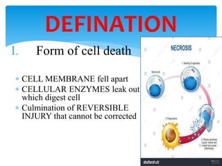
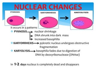
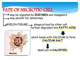
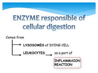
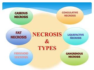
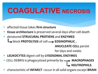
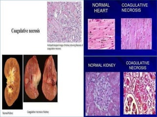
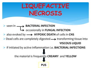
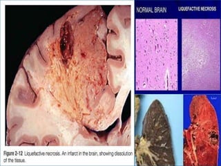
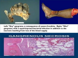
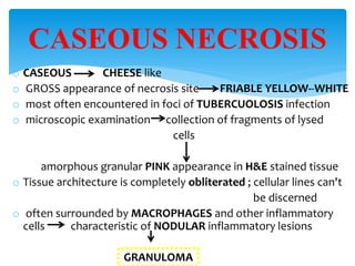
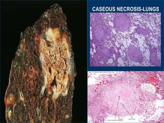
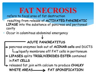
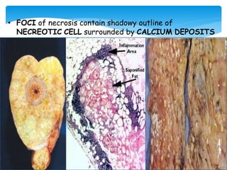
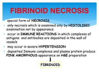
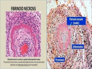
I. Form of cell death
 CELL MEMBRANE fell apart
 CELLULAR ENZYMES leak out
which digest cell
 Culmination of REVERSIBLE
INJURY that cannot be corrected
DEFINATION
Failure of energy generation in the form of ATP
 reduced O2 supply
 mitochondrial damage
Damage to membranes leakage of cellular
lysosomal membrane contents including
plasma membrane ENZYMES
 IRREVERSIBLE DAMAGE to cellular lipid ,proteins
,nucleic acids caused by reactive oxygen species
MECHANISM
 cell show increased EOSINOPHILIA
 loss of BASOPHILIC NUCLEIC ACID (RNA & DNA) in cytoplasm
 cell may have appearance loss of staining
GLASSY GLYCOGEN particle
HOMOGENEOUS
 MYELIN FIGURES more prominent in necrotic cell than cell
with reversible injury
 CYOPLASM becomes VACUOLATED
 Discontinuity in plasma and organelle membranes
 Dilation of MITOCHONDRIA appearance of amorphous intramito-
chondrial densities
 disruption of LYSOSOMES
CYTOPLASMIC CHANGES DURING
NECROSIS
It occurs in 3 patterns
 PYKNOSIS nuclear shrinkage
DNA shrunk into dark mass
increased basophilia
KARYORRHEXIS pyknotic nucleus undergoes destructive
fragmentation
 KARYOLYSIS basophilia fades due to digestion of
DNA by deoxyribonuclease (DNAse)
In 1-2 days nucleus is completely dead and disappears
NUCLEAR CHANGES
FATE OF NECROTIC CELL
may be digested by ENZYMES and disappears
may persist for sometimes
MYELIN FIGURE phagocytized by other cell
further degraded into FATTY ACID
which binds with CALCIUM to form
CALCIUM SALT
dead cell is CALCIFIED
Comes from
LYSOSOMES of DYING CELL
LEUKOCYTES as a part of
INFLAMMAION
REACTION
NECROSIS
&
TYPES
 affected tissue takes firm structure
 tissue architecture is preserved several days after cell death
 denatured STRUCURAL PROTIENS and ENZYMES
block PROTOLYSIS of cell EOSINOPHILIC ,
ANUCLEATE CELL persist
for days and weeks
 LEUKOCYTES digest cell by LYSOSOMAL ENZYMES
 CELL DEBRIS is phagocytized primarily by MACROPHAGES
NEUTROPHILS
 characteristic of INFARCT : occur in all solid organs except BRAIN
COAGULATIVE NECROSIS
 seen in BACTERIAL INFECTION
occasionally in FUNGAL INFECTION
 also evoked by HYPOXIC DEATH of cells in CNS
Dead cells are completely digested transforming tissue into
VISCOUS LIQUID
 if initiated by active inflammation i.e. BACTERIAL INFECTIONS
the material is frequently CREAMY and YELLOW
PUS
LIQUEFACTIVE
NECROSIS
 no distinctive pattern of CELL DEATH
 used to refer condition of limbs usually LOWER LIMBS
that has lost BLOOD SUPPLY undergone COAGULATIVE
necrosis
MULTIPLE TISSUE LAYERS
 when superimposed by BACTERIAL INFECTION
destructive content of BACTERIA and attracted LEUKOCYTES
morphologic appearance changes WET
to LIQUEFACTIVE necrosis GANGRENE
GANGRENOUS NECROSIS
o CASEOUS CHEESE like
o GROSS appearance of necrosis site FRIABLE YELLOW--WHITE
o most often encountered in foci of TUBERCUOLOSIS infection
o microscopic examination collection of fragments of lysed
cells
amorphous granular PINK appearance in H&E stained tissue
o Tissue architecture is completely obliterated ; cellular lines can't
be discerned
o often surrounded by MACROPHAGES and other inflammatory
cells characteristic of NODULAR inflammatory lesions
GRANULOMA
CASEOUS NECROSIS
• refers to focal area of fat destruction
• resulting from release of ACTIVATED PANCREATIC
LIPASE into the substance of pancreas and peritoneal
cavity
• Occur in calamitous abdominal emergency
ACUTE PANCREATITUS
pancreas enzymes leak out of ACINAR cells and DUCTS
liquefy membrane off FAT cells in peritoneum
LIPASES splits TRIGLYCERIDES ESTER contained
in FAT CELLS
released fat join with calcium to produce CHALKY
WHITE AREAS FAT SPONIFICATION
FAT NECROSIS
• FOCI of necrosis contain shadowy outline of
NECREOTIC CELL surrounded by CALCIUM DEPOSITS
 special form of NECROSIS
 only necrosis which is examined only by HISTOLIGIC
examination not by appearance
 occur in IMMUNE REACTIONS in which complexes of
antigens and antibodies are deposited in the wall of
vessels
 may occur in severe HYPERTENSION
 deposited Immune complexes and plasma protein produce
PINK AMORPHOUS appearance on H&E preparation
FIBRINOID
FIBRINOID NECROSIS
Necrosis and types
Necrosis and types
Necrosis and types

Necrosis and types

  • 4.
    I. Form ofcell death  CELL MEMBRANE fell apart  CELLULAR ENZYMES leak out which digest cell  Culmination of REVERSIBLE INJURY that cannot be corrected DEFINATION
  • 5.
    Failure of energygeneration in the form of ATP  reduced O2 supply  mitochondrial damage Damage to membranes leakage of cellular lysosomal membrane contents including plasma membrane ENZYMES  IRREVERSIBLE DAMAGE to cellular lipid ,proteins ,nucleic acids caused by reactive oxygen species MECHANISM
  • 6.
     cell showincreased EOSINOPHILIA  loss of BASOPHILIC NUCLEIC ACID (RNA & DNA) in cytoplasm  cell may have appearance loss of staining GLASSY GLYCOGEN particle HOMOGENEOUS  MYELIN FIGURES more prominent in necrotic cell than cell with reversible injury  CYOPLASM becomes VACUOLATED  Discontinuity in plasma and organelle membranes  Dilation of MITOCHONDRIA appearance of amorphous intramito- chondrial densities  disruption of LYSOSOMES CYTOPLASMIC CHANGES DURING NECROSIS
  • 7.
    It occurs in3 patterns  PYKNOSIS nuclear shrinkage DNA shrunk into dark mass increased basophilia KARYORRHEXIS pyknotic nucleus undergoes destructive fragmentation  KARYOLYSIS basophilia fades due to digestion of DNA by deoxyribonuclease (DNAse) In 1-2 days nucleus is completely dead and disappears NUCLEAR CHANGES
  • 8.
    FATE OF NECROTICCELL may be digested by ENZYMES and disappears may persist for sometimes MYELIN FIGURE phagocytized by other cell further degraded into FATTY ACID which binds with CALCIUM to form CALCIUM SALT dead cell is CALCIFIED
  • 9.
    Comes from LYSOSOMES ofDYING CELL LEUKOCYTES as a part of INFLAMMAION REACTION
  • 10.
  • 11.
     affected tissuetakes firm structure  tissue architecture is preserved several days after cell death  denatured STRUCURAL PROTIENS and ENZYMES block PROTOLYSIS of cell EOSINOPHILIC , ANUCLEATE CELL persist for days and weeks  LEUKOCYTES digest cell by LYSOSOMAL ENZYMES  CELL DEBRIS is phagocytized primarily by MACROPHAGES NEUTROPHILS  characteristic of INFARCT : occur in all solid organs except BRAIN COAGULATIVE NECROSIS
  • 13.
     seen inBACTERIAL INFECTION occasionally in FUNGAL INFECTION  also evoked by HYPOXIC DEATH of cells in CNS Dead cells are completely digested transforming tissue into VISCOUS LIQUID  if initiated by active inflammation i.e. BACTERIAL INFECTIONS the material is frequently CREAMY and YELLOW PUS LIQUEFACTIVE NECROSIS
  • 15.
     no distinctivepattern of CELL DEATH  used to refer condition of limbs usually LOWER LIMBS that has lost BLOOD SUPPLY undergone COAGULATIVE necrosis MULTIPLE TISSUE LAYERS  when superimposed by BACTERIAL INFECTION destructive content of BACTERIA and attracted LEUKOCYTES morphologic appearance changes WET to LIQUEFACTIVE necrosis GANGRENE GANGRENOUS NECROSIS
  • 17.
    o CASEOUS CHEESElike o GROSS appearance of necrosis site FRIABLE YELLOW--WHITE o most often encountered in foci of TUBERCUOLOSIS infection o microscopic examination collection of fragments of lysed cells amorphous granular PINK appearance in H&E stained tissue o Tissue architecture is completely obliterated ; cellular lines can't be discerned o often surrounded by MACROPHAGES and other inflammatory cells characteristic of NODULAR inflammatory lesions GRANULOMA CASEOUS NECROSIS
  • 19.
    • refers tofocal area of fat destruction • resulting from release of ACTIVATED PANCREATIC LIPASE into the substance of pancreas and peritoneal cavity • Occur in calamitous abdominal emergency ACUTE PANCREATITUS pancreas enzymes leak out of ACINAR cells and DUCTS liquefy membrane off FAT cells in peritoneum LIPASES splits TRIGLYCERIDES ESTER contained in FAT CELLS released fat join with calcium to produce CHALKY WHITE AREAS FAT SPONIFICATION FAT NECROSIS
  • 20.
    • FOCI ofnecrosis contain shadowy outline of NECREOTIC CELL surrounded by CALCIUM DEPOSITS
  • 21.
     special formof NECROSIS  only necrosis which is examined only by HISTOLIGIC examination not by appearance  occur in IMMUNE REACTIONS in which complexes of antigens and antibodies are deposited in the wall of vessels  may occur in severe HYPERTENSION  deposited Immune complexes and plasma protein produce PINK AMORPHOUS appearance on H&E preparation FIBRINOID FIBRINOID NECROSIS