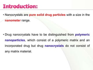
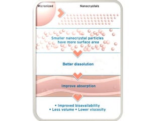
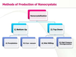
• Nanocrystals are pure solid drug particles with a size in the
nanometer range.
• Drug nanocrystals have to be distinguished from polymeric
nanoparticles, which consist of a polymeric matrix and an
incorporated drug but drug nanocrystals do not consist of
any matrix material.
• Universal method to increase solubility of poorly soluble
drugs
• No use of surfactants
• Produces a drug powder which can be incorporated into a
variety of dosage forms
• Stable
• Cost effective process
• Uses generally regarded as safe (GRAS) materials
Nanocrystals are stabilized against agglomeration by (GRAS)
stabilizing agents such as lecithin, tween 80, poloxamer 188,
sodium glycocholate or low molecular weight
polyvinylpyrrolidone (PVP), also make the nanocrystals less
susceptible to electrolytes in the body.
Nanocrystallization
1) Bottom Up
A) Precipitation B) Cryo- vacuum
2) Top Down
A) Wet Milling
B) High Pressure
Homogenization
The drug is dissolved in a solvent which is subsequently added
to a nonsolvent (water) with efficient stirring to precipitate the
nanocrystals that can be removed from the solution by filtration
and then dried in air.
The use of solvents
Difficulty to avoid growth from nanocrystals to microparticles
• An aqueous, organic, or aqueous–organic cosolvent solution;
aqueous–organic emulsion; or drug suspension is atomized
into a cryogenic liquid such as liquid nitrogen at -196oC to
produce frozen nanocrystals which then lyophilized at -22oC
to obtain free-flowing powder.
• The method yields very pure nanocrystals since there is no
need to use surfactants or harmful reagents.
• Milling chamber, at temp. < 40oC, is charged with milling
media, (dispersion medium ‘normally water’, stabilizer, and
the drug). The most common models are a tumbling ball mill
and a stirred media mill.
• Milling times range from hours to days depending on the
drug's hardness.
One problem of this method is the degradation of mill
surfaces and subsequent product contamination.
• Homogenisation can be
performed in water
(DissoCubes®)
• Or in non-aqueous media as
liquid PEG or oils (Nanopure®)
• The particles are redueced by
cavitation and shear forces.
• High cost of instrument limits
its use
The particle size obtained during the homogenization
process depends on:
1. The nature of the drug
2. The pressure applied
3. The number of homogenization cycles
• Enhanced oral bioavailability
• Rapid onset of action
• Improved dose proportionality
• Reduction of fed/fasted effects
• The use of mucoadhesive polymers in the dispersion
medium can increase residence time
• Carrier-free nanocrystals enable potential higher loading
capacity (Large dose/Small volume).
• Avoidance of undesired side effects associated with the use of
cosolvents and/or high surfactant contents.
 Drug nanocrystals increase the concentration gradient
between the formulation& the skin.
 This effect can be enhanced by the use of positively
charged polymers. The opposite charge leads to an
increased affinity of the drug nanocrystals to the negatively
charged stratum corneum.
They are fluorescent semiconductor nanocrystals with diameters of
2–10 nanometers, when excited by ultraviolet light, they emit a
particular fluorescent color and brightness, depending on the dot's
size.
Reactive functional
groups such as
amines, carboxylic
acids, alcohols, and
thiols
These core-shell quantum dots are not water soluble, which
limits their use in biological studies. They can be rendered
water soluble by providing a shell of functionalized silica, or
linkers, such as mercaptoacetic acid , dihydrolipoic acid,
or amphiphilic polymers such as modified polyacrylic acid
.
Quantum
Dots
Drug Delivery&
Cancer
Treatment
Diagnostic Tools
(Imaging)
Lung cancer
GI malignancies
Cancer Cells
Apoptosis
Fluorescence Resonance Energy Transfer
Light
 QDs are protected by both coordinating ligand, tri-n-
octylphosphine oxide (TOPO), and an amphiphilic triblock
copolymer coating.
These two layers ‘bond’ to each other and form a
hydrophobic protection structure that resists hydrolysis and
enzymatic degradation.
• Polybutylacrylate (hydrophobic)
• Polyethylacrylate (hydrophobic)
• Polymethacrylic acid (hydrophilic)
• Hydrophobic hydrocarbon side chain
• For improved biocompatibility
• Prevents non specific binding
• For in vivo protection
• Prevent particles aggregation
• Prevent fluorescence loss
• Make them water soluble
• peptides, antibodies, oligonucleotides etc.
• For tumor antigen recognition
Much slower and less efficient
Based on tumor permeation, uptake& retention
Polymer-protected QDs are cleared from the body by slow filtration
and excretion through the kidney
Mannose or Galactosamine
 As optimism in exploiting QDs clinical potential is high, but
there is need to assess their cytotoxicity, in vivo
distribution, and excretion.
 A much more work needs to be done to combat QDs
inherent toxicity before they are applied in the clinical
settings.
Nanocrystals

Nanocrystals

  • 2.
    • Nanocrystals arepure solid drug particles with a size in the nanometer range. • Drug nanocrystals have to be distinguished from polymeric nanoparticles, which consist of a polymeric matrix and an incorporated drug but drug nanocrystals do not consist of any matrix material.
  • 4.
    • Universal methodto increase solubility of poorly soluble drugs • No use of surfactants • Produces a drug powder which can be incorporated into a variety of dosage forms • Stable • Cost effective process • Uses generally regarded as safe (GRAS) materials
  • 5.
    Nanocrystals are stabilizedagainst agglomeration by (GRAS) stabilizing agents such as lecithin, tween 80, poloxamer 188, sodium glycocholate or low molecular weight polyvinylpyrrolidone (PVP), also make the nanocrystals less susceptible to electrolytes in the body.
  • 6.
    Nanocrystallization 1) Bottom Up A)Precipitation B) Cryo- vacuum 2) Top Down A) Wet Milling B) High Pressure Homogenization
  • 8.
    The drug isdissolved in a solvent which is subsequently added to a nonsolvent (water) with efficient stirring to precipitate the nanocrystals that can be removed from the solution by filtration and then dried in air. The use of solvents Difficulty to avoid growth from nanocrystals to microparticles
  • 9.
    • An aqueous,organic, or aqueous–organic cosolvent solution; aqueous–organic emulsion; or drug suspension is atomized into a cryogenic liquid such as liquid nitrogen at -196oC to produce frozen nanocrystals which then lyophilized at -22oC to obtain free-flowing powder. • The method yields very pure nanocrystals since there is no need to use surfactants or harmful reagents.
  • 11.
    • Milling chamber,at temp. < 40oC, is charged with milling media, (dispersion medium ‘normally water’, stabilizer, and the drug). The most common models are a tumbling ball mill and a stirred media mill. • Milling times range from hours to days depending on the drug's hardness. One problem of this method is the degradation of mill surfaces and subsequent product contamination.
  • 12.
    • Homogenisation canbe performed in water (DissoCubes®) • Or in non-aqueous media as liquid PEG or oils (Nanopure®) • The particles are redueced by cavitation and shear forces. • High cost of instrument limits its use
  • 13.
    The particle sizeobtained during the homogenization process depends on: 1. The nature of the drug 2. The pressure applied 3. The number of homogenization cycles
  • 14.
    • Enhanced oralbioavailability • Rapid onset of action • Improved dose proportionality • Reduction of fed/fasted effects • The use of mucoadhesive polymers in the dispersion medium can increase residence time
  • 15.
    • Carrier-free nanocrystalsenable potential higher loading capacity (Large dose/Small volume). • Avoidance of undesired side effects associated with the use of cosolvents and/or high surfactant contents.
  • 16.
     Drug nanocrystalsincrease the concentration gradient between the formulation& the skin.  This effect can be enhanced by the use of positively charged polymers. The opposite charge leads to an increased affinity of the drug nanocrystals to the negatively charged stratum corneum.
  • 17.
    They are fluorescentsemiconductor nanocrystals with diameters of 2–10 nanometers, when excited by ultraviolet light, they emit a particular fluorescent color and brightness, depending on the dot's size.
  • 19.
    Reactive functional groups suchas amines, carboxylic acids, alcohols, and thiols
  • 20.
    These core-shell quantumdots are not water soluble, which limits their use in biological studies. They can be rendered water soluble by providing a shell of functionalized silica, or linkers, such as mercaptoacetic acid , dihydrolipoic acid, or amphiphilic polymers such as modified polyacrylic acid .
  • 21.
  • 22.
    Lung cancer GI malignancies CancerCells Apoptosis Fluorescence Resonance Energy Transfer Light
  • 23.
     QDs areprotected by both coordinating ligand, tri-n- octylphosphine oxide (TOPO), and an amphiphilic triblock copolymer coating. These two layers ‘bond’ to each other and form a hydrophobic protection structure that resists hydrolysis and enzymatic degradation.
  • 24.
    • Polybutylacrylate (hydrophobic) •Polyethylacrylate (hydrophobic) • Polymethacrylic acid (hydrophilic) • Hydrophobic hydrocarbon side chain • For improved biocompatibility • Prevents non specific binding • For in vivo protection • Prevent particles aggregation • Prevent fluorescence loss • Make them water soluble • peptides, antibodies, oligonucleotides etc. • For tumor antigen recognition
  • 25.
    Much slower andless efficient Based on tumor permeation, uptake& retention Polymer-protected QDs are cleared from the body by slow filtration and excretion through the kidney
  • 26.
  • 27.
     As optimismin exploiting QDs clinical potential is high, but there is need to assess their cytotoxicity, in vivo distribution, and excretion.  A much more work needs to be done to combat QDs inherent toxicity before they are applied in the clinical settings.