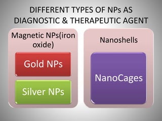
METALLIC NANOPARTICLE
ANUSIKTA GHOSH
18MPH102
DEPT OF P’CEUTICS
NANO & MOLECULAR PHARMACEUTICS
INTRODUCTION
• Nanotechnology refers to the branch of science &
engineering dedicated to materials, having
dimensions in the order of the 100th of nm or
less.
• Nanotechnology has been embraced by industrial
sectors due to its applications in the field of
electronic storage systems, biotechnology,
magnetic separation & pre concentration of
target analytes, targeted drug delivery & vehicles
for gene & drug delivery.
• Nanoparticles which are used in
biotechnology range in particle size of 10 to
500nm.
• Nanosize particles allows various
communications with biomolecules on the cell
surfaces & within the cells in a way which can
be decoded, designated to various
biochemical & physicochemical properties of
cells.
• Potential applications of nanoparticles in drug
delivery system & non invasive imaging
offered various advantages over conventional
pharmaceutical products.
IDEAL PROPERTIES OF
NANOPARTICULATE SYSTEM
Specific sites in
the body after
systemic
administration
Selectively
directed
stable
biocompatible
TARGETED THERAPY
• The targeted cells such as the cancer cells are
recognized by conjugating the nanoparticle
with an appropriate ligand, which has a
specific binding activity w.r.t target cells.
• Nanoparticles allows to attach multiple copies
of therapeutic substance and increase the
concentration of therapeutic & diagnostic
substances at the pathological site.
ADVANCES IN FIELD OF
BIOTECHNOLOGY
• Once targeted (active or passive) nanocarriers
can be designed in a way to act as imaging
probes using variety of techniques such as
•
Ultrasound(US) X-Ray
Computed
Tomography(CT)
Positron Emission
Tomography(PET)
MRI
Optical Imaging
Surface –enhanced Raman Imaging(SERS)
• Hence these molecular imaging probes can be used non-
invasively to provide valuable information about differentiate
abnormalities in body structures & organs to find out the level
of disease and usefulness of treatment.
DIFFERENT TYPES OF NPs AS
DIAGNOSTIC & THERAPEUTIC AGENT
Magnetic NPs(iron
oxide)
Gold NPs
Silver NPs
Nanoshells
NanoCages
IRON OXIDE NPs
• Iron(III) oxide is a reddish brown inorganic
compound
• Paramagnetic in nature
• The Fe3O4 which occurs naturally as the
mineral magnetite is superparamagnetic in
nature.
• Due to their ultrafine size, magnetic
properties & biocompatibility,
superparamagnetic oxide
nanoparticles(SPION) have emerged as
hopeful candidates for various biomedical
applications such as
Enhanced
resolution contrast
agents for MRI
Targeted drug
delivery & imaging
hyperthermia
Gene therapy Stem Cell tracking
Molecular/cellular
tracking
Magnetic separation
technologies(e.g.Rapid
DNA sequencing)
Early detection of
inflammatory
Cancer, Diabetes Atherosclerosis
• All these Biomedical applications require that
the nanoparticles have high magnetization
values so as to provide high resolution MR
images.
• Superparamagnetic NPs resembles excellent
imaging probes to be used as MRI contrast
agents since the MR signal intensity is
significantly modulated w/o any compromise
in its in-vivo stability.
ADVANTAGES OVER NON SPECIFICALLY
TARGETED MOLECULAR IMAGING
PROBES
• Traditional contrast agents distribute
nonspecifically while targeted molecular
imaging probes based on iron oxide
nanoparticles have been developed that
specifically target body tissue or cells.
• In order to improve the cellular uptake, the iron
oxide nanoparticles can be modified with a
peculiar surface coating so that they can be easily
conjugated to drugs, proteins, enzymes,
antibodies or nucleotides & can be directed to an
organ, tissue or tumor.
• These studies provide a new insight to the
bioreactivity of engineered iron nanoparticles
which can provide potential applications in
medical imaging & drug delivery.
• However, the toxicity of these nanoparticles in
certain neuronal cells is still the matter of
concern.
GOLD NANOPARTICLES
Colloidal gold also k/a gold NPs is a suspension
of nanometer sized particles of gold.
Colloidal solution is an intense red colour for
particles less than 100nm or a dirty yellowish
colour for larger particles.
• The different optical properties of these gold
NPs are due to their interaction with light.
• El Sayed et.al have established that gold NPs
can be used for cancer imaging by selectively
transporting AuNPs into the cancer cell
nucleus.
• Qian et.al reported development of tumor
targeted AuNPs as a probe for Raman scatters
in vivo.
SILVER NANOPARTICLES
• Silver NPs are silver particles with size varying
from 1 to 100nm.
• They are frequently described as being “silver”
some of them are composed of a large
percentage of silver oxide due to their large
surface to bulk silver atoms ratio.
• Efforts are being made to incorporate AgNPs in a
wide range of medical devices including bone
cements, surgical instruments, surgical masks
• Ionic silver used in the right quantities are
found to be suitable for treating wounds.
• Silver NPs are now being replaced replaced
by silver sulfadiazines as an effective agent in
treating of wounds.
METALLIC NANOPARTICLES FOR
CANCER THERAPY
• The new drugs which are synthesized for
cancer have therapeutic & toxicological
limitations such as
Barrier effect of the cell membrane
Drug resistance developed by the cell
Drug disposition behavior
• The distribution of anti cancer drugs
essentially depends on physicochemical
factors such as pKa, hydrophilicity &
electrostatic charges; however all of these
criteria does not fit in the domain of a tumuor
cell.
• Large amounts of the drug are distributed
towards a healthy tissue or organ rather than
the target area which is a main limiting factor
of the conventional chemotherapy.
• One more problem associated with the
conventional chemotherapy is the property of
insolubility of the anticancer drugs in water
which makes it necessary for the use of the
pharmaceutical solvents which is again an
another life – threatening problem.
• During the intravenous injection of anticancer
drugs the delivery system is opsonised &
rapidly cleared from the bloodstream
• Consequently, conventional anticancer
therapy by systemic delivery of
chemotherapeutic agents often fails or is
inadequately delivered to the target
cell/tissue and has a tremendous impact on
reducing the quality and expectancy of life.
• Some of the disadvantages of current
conventional anticancer therapy include
Inefficient cell entry
uptake by the immune
system and mononuclear
phagocyte
system
accumulation in non-
targeted organs and
tissues,
non-selective with high
toxicity against normal
tissues
• The effectiveness of cancer therapeutic device
is measured by its ability to reduce tumors
without damaging healthy tissue.
• Metallic nanoparticles for drug delivery are
solid colloidal particles ranging in size from 10
to 1000 nm that contain a therapeutic agent
that is dispersed in a polymer carrier matrix,
encapsulated within a polymer shell,
covalently attached or adsorbed to the
particle surface, or encapsulated within a
structure.
• Metallic nanoparticles increase the therapeutic
index of drugs through site specificity preventing
multi drug resistance & delivering therapeutic
agents efficiently.
• Metallic NPs are emerging as potential
application for diagnosis of cancer, e.g. MRI &
colloidal mediators for cancer magnetic
hyperthermia.
• The metallic NPs used as probes for the
treatment of cancer is mainly derived from their
potential to carry a large dose of drug at the
targeted site, which results in high concentration
of the drug at the desired site.
• This results in high concentration of the
anticancer drugs at the desired site thus
avoiding the toxicity in the whole body &
other painful side effects due to high
concentration of drugs in the body.
ADVANTAGES OF METALLIC NPs
• They are designed to contain tumor targeting
ligands which bind to particular cells within
the tumor to fasten the NP in the solid tumor.
• Metallic NP DDS are capable of delivering
anticancer drugs within the tumor cells which
reduce the accumulation of the drugs in the
healthy organs.
TARGETED DELIVERY & BIOLOGICAL
CONSIDERATION
• Conventionally, anticancer drugs have been
designed to have a relatively low molecular mass
and an agreement between the hydrophilic and
lyphophilic balance (HLB), hence allowing
partitioning across the lipid membrane very
easily.
• Therefore, drug within the systemic circulation is
rapidly distributed throughout the body, reaching
the target and non-target tissue/organ, and is
also rapidly metabolised by the liver and/ or
rapidly excreted by the kidney.
• For effective targeting, it is essential that a drug-
targeting system should not be cleared out
quickly from the body.
• Ideally, a drug carrier should provide a
pharmacokinetic profile that will allow the drug
to interact with its target.
• The performance of nanoparticles inside the
vascular compartment is controlled by complex
factors such as their shape, density, size
distribution and surface characteristics. All these
factors control the flow properties of
nanoparticles, bifurcation in the vascular
compartment, modulation of circulation time,
and mode of entry into the cell.
ACHIEVING TARGETED DELIVERY
• A major barrier that a drug delivery system must
be able to avoid in the systemic circulation is the
removal of nanoparticles by phagocytic cells of
the mononuclear phagocyte system (MPS).
• Nanoparticles will usually be taken up by the
liver, spleen and other parts of the RES depending
on their surface characteristics and undergo
opsonisation in the blood and clearance by the
RES(reticuloendothelial system)
• Therefore, the MPS presents a significant
barrier to effective drug targeting, because it
has the ability to filter out and destroy a drug
delivery system unless appropriate
formulation approaches are used to avoid this.
• Therefore, the nanoparticles should be
designed to avoid these interactions,
particularly opsonisation, and possible
clearance of the drug delivery system from the
vascular compartment.
• Opsonisation is a process in which the surface
of the foreign particles such as bacteria and
particulate drug carrier is coated with blood
proteins, known as opsonins. The
phagocytosis of these tagged particles is
enhanced because surface receptors present
on phagocytes bind to opsonins and the
foreign particle is engulfed and untimely
digested by various lysozymes.
• A more practical approach to avoid RES uptake
and clearance of nanoparticles is the
modification of nanoparticles’ surface. For
example, increasing the surface hydrophilicity,
that is, adding a hydrophilic polymer to the
metallic nanoparticle carriers, made them
invisible to the RES and thus reduced
opsonisation and led to suppression of
macrophage recognition. This coating is
referred to as the stealth moiety. The most
commonly used stealth agent is polyethylene
glycol (PEG) and its block copolymer.
• Another factor affecting the opsonins’ binding
are physicochemical properties of the
nanoparticles (i.e., surface characteristics such
as size, surface charge which is a major factor
in the characteristic biodistribution and
residence times of these particles in vivo.
• Neutral systems tend to remain longer in
blood circulation, whereas their charged
counterparts are cleared out by the RES.
• Similarly, particles of size ~ 1 -- 2 μm undergo
phagocytosis, and higher sizes of ~ 6 μm are
trapped in lung capillaries.
• Therefore, to avoid clearance by the RES,
metallic nanoparticles should be formulated
to be not more than 100 nm in size and should
have a sterically stabilised, preferably neutral,
surface.
PASSIVE TARGETING OF TUMOURS
USING METALLIC NANOPARTICLES
• In passive targeting, the distribution of
nanoparticles is mediated by theMPS’s
physiological condition.
• In passive targeting, advantage is taken of the
pathological condition of the tumour to allow the
accumulation of drug carriers at the target site.
• For example, the pH or specific enzymes present
within the tumour cells can be used to facilitate
the release of drugs from nanoparticles. Enzymes
such as alkaline phosphatase and plasmins are
present at a higher level at the tumour site.
ACTIVE TARGETING OF TUMOUR
USING METALLIC NANOPARTICLES
• As passive targeting does not necessarily
guarantee the internalisation of nanoparticles by
the targeted cell, nanoparticles are modified with
molecular targeting ligands for active targeting of
tumour.
• Active targeting of metallic nanoparticles involves
an interaction between peripherally conjugated
targeting moiety and a corresponding receptor to
facilitate the targeting of a carrier to a specific
malignant cell.
• Drug delivery to the tumour cell can be
achieved by means of molecules that are
specific to antigens or receptors expressed on
the surface of a tumour cell.
• Ligand can be designed to have specificity for
receptors that are expressed on a tumour cell
but are minimally expressed on normal cells.
• The introduction of targeting ligand enhances
the internalisation of metallic nanoparticles
into the tumour cell.
• Care must be taken when selecting ligands for
receptors on the tumour cell, as ligand--
receptor interaction can affect the rate of
internalisation, which in turn affects the
accumulation of metallic nanoparticles in
cancer cells.
• Therefore, ligands used for receptor targeting
in cancer treatment must have the function of
inducing receptor-mediated endocytosis
(RME).
• Various molecules are used to facilitate active
targeting of nanoparticles, such as aptamers,
proteins and antibodies.
• Bioconjugation of ligands, such as monoclonal
antibodies, proteins, or peptides, with the
nanocarrier’s surface has been exploited in
many nanoparticles for the purpose of
concentrating therapeutic action on the
specific tumour cell.
RECENT DEVELOPMENTS AND
APPLICATIONS OF METALLIC
NANOPARTICLES IN CANCER
THERANOSTICS
• Metallic nanoparticles have the ability to treat
as well as to diagnose the disease, the ability
of nanoparticles both to diagnose and to
deliver the targeted drug is an emerging
concept in the nanoplatform. These
nanoparticles are called theranostic
(therapeutic plus diagnostic).
1.Theranostic nanovector represents an
emerging class of imaging and therapeutic
that may provide a personalised therapeutic
response.
• For example, light-activated theranostic
nanoparticles have been reported for imaging
and treatment of brain tumours.
2.The roles of theranostic agent in tumour
diagnosis, monitoring tumour progression and
assessment of therapeutic effect have resulted
in an enhanced role.
• For example, ex vivo imaging of oncology
biomarkers in a preclinical study was reported
by Makino et al. for the visualisation and
monitoring of tumour progression by coupling
the near-infrared fluorescence (NIRF) and
nanoparticles in a targeted drug delivery
system for hepatic tumour.
3. Biodistribution of gold nanoparticles coated
with gadolinium chelate was studied by Alric
et al., they reported that functionalised gold
nanoparticle freely circulate in the blood
vessels without undesirable accumulation in
any major organ.
4. Iron nanoparticles have been used as
theranostic agents with specific application as
contrasting agents for MRI and magnetically
targeted drug deliver to the tumour cell.
5. Christopher et al. reported the synthesis and
use of magnetic nanoparticle hydrogel
(MagNaGel; Alnis Biosciences, Emeryville,
California, USA) as a powerful cancer
treatment regimen. They demonstrated that
these particles had the characteristics of
ability to load chemotherapeutic agent,
tumour associated biomolecular binding and
good magnetic susceptibility.
CONCLUSION
• The development of metallic nanoparticles is
rapid and multidirectional. Metallic
nanotechnology has clearly impacted the
development of new theranostics in oncology
disorders. Recent advances in the field of metallic
nanoparticles indeed offer the promise of better
diagnostic and therapeutic options. Metallic
nanoparticles are attracting attention in cancer
therapeutics owing to their unique prospects for
targeted delivery in imaging and drug delivery to
the desired site.
CONT…
Drug delivery based on metallic
nanotechnology seeks to increase the
therapeutic index of drugs, both by reaching
their in vivo target and by exposing the drugs
to malignant cells. Metallic nanotechnology
combines nanobiotechnology with molecular
imaging techniques, which has led to the
development of multifunctional metallic
nanoparticles for cancer imaging and therapy.
REFERENCES
• Ahmad, M. Z., Akhter, S., Jain, G. K., Rahman, M.,
Pathan, S. A., Ahmad, F. J., & Khar, R. K. (2010).
Metallic nanoparticles: technology overview &
drug delivery applications in oncology. Expert
Opinion on Drug Delivery, 7(8), 927–942.
doi:10.1517/17425247.2010.498473
• Vicky V. Mody, Rodney Siwale, Ajay Singh,1 and
Hardik R. Mody2 Introduction to metallic
nanoparticles, J Pharm Bioallied Sci. 2010 Oct-
Dec; 2(4): 282–289.
doi: 10.4103/0975-7406.72127
THANK YOU

Metallic nanoparticle

  • 1.
    METALLIC NANOPARTICLE ANUSIKTA GHOSH 18MPH102 DEPTOF P’CEUTICS NANO & MOLECULAR PHARMACEUTICS
  • 2.
    INTRODUCTION • Nanotechnology refersto the branch of science & engineering dedicated to materials, having dimensions in the order of the 100th of nm or less. • Nanotechnology has been embraced by industrial sectors due to its applications in the field of electronic storage systems, biotechnology, magnetic separation & pre concentration of target analytes, targeted drug delivery & vehicles for gene & drug delivery.
  • 3.
    • Nanoparticles whichare used in biotechnology range in particle size of 10 to 500nm. • Nanosize particles allows various communications with biomolecules on the cell surfaces & within the cells in a way which can be decoded, designated to various biochemical & physicochemical properties of cells.
  • 4.
    • Potential applicationsof nanoparticles in drug delivery system & non invasive imaging offered various advantages over conventional pharmaceutical products.
  • 5.
    IDEAL PROPERTIES OF NANOPARTICULATESYSTEM Specific sites in the body after systemic administration Selectively directed stable biocompatible
  • 6.
    TARGETED THERAPY • Thetargeted cells such as the cancer cells are recognized by conjugating the nanoparticle with an appropriate ligand, which has a specific binding activity w.r.t target cells. • Nanoparticles allows to attach multiple copies of therapeutic substance and increase the concentration of therapeutic & diagnostic substances at the pathological site.
  • 7.
    ADVANCES IN FIELDOF BIOTECHNOLOGY • Once targeted (active or passive) nanocarriers can be designed in a way to act as imaging probes using variety of techniques such as • Ultrasound(US) X-Ray Computed Tomography(CT) Positron Emission Tomography(PET) MRI
  • 8.
    Optical Imaging Surface –enhancedRaman Imaging(SERS) • Hence these molecular imaging probes can be used non- invasively to provide valuable information about differentiate abnormalities in body structures & organs to find out the level of disease and usefulness of treatment.
  • 9.
    DIFFERENT TYPES OFNPs AS DIAGNOSTIC & THERAPEUTIC AGENT Magnetic NPs(iron oxide) Gold NPs Silver NPs Nanoshells NanoCages
  • 10.
    IRON OXIDE NPs •Iron(III) oxide is a reddish brown inorganic compound • Paramagnetic in nature • The Fe3O4 which occurs naturally as the mineral magnetite is superparamagnetic in nature.
  • 11.
    • Due totheir ultrafine size, magnetic properties & biocompatibility, superparamagnetic oxide nanoparticles(SPION) have emerged as hopeful candidates for various biomedical applications such as Enhanced resolution contrast agents for MRI Targeted drug delivery & imaging hyperthermia Gene therapy Stem Cell tracking
  • 12.
  • 13.
    • All theseBiomedical applications require that the nanoparticles have high magnetization values so as to provide high resolution MR images. • Superparamagnetic NPs resembles excellent imaging probes to be used as MRI contrast agents since the MR signal intensity is significantly modulated w/o any compromise in its in-vivo stability.
  • 14.
    ADVANTAGES OVER NONSPECIFICALLY TARGETED MOLECULAR IMAGING PROBES • Traditional contrast agents distribute nonspecifically while targeted molecular imaging probes based on iron oxide nanoparticles have been developed that specifically target body tissue or cells.
  • 15.
    • In orderto improve the cellular uptake, the iron oxide nanoparticles can be modified with a peculiar surface coating so that they can be easily conjugated to drugs, proteins, enzymes, antibodies or nucleotides & can be directed to an organ, tissue or tumor. • These studies provide a new insight to the bioreactivity of engineered iron nanoparticles which can provide potential applications in medical imaging & drug delivery. • However, the toxicity of these nanoparticles in certain neuronal cells is still the matter of concern.
  • 16.
    GOLD NANOPARTICLES Colloidal goldalso k/a gold NPs is a suspension of nanometer sized particles of gold. Colloidal solution is an intense red colour for particles less than 100nm or a dirty yellowish colour for larger particles.
  • 17.
    • The differentoptical properties of these gold NPs are due to their interaction with light. • El Sayed et.al have established that gold NPs can be used for cancer imaging by selectively transporting AuNPs into the cancer cell nucleus. • Qian et.al reported development of tumor targeted AuNPs as a probe for Raman scatters in vivo.
  • 18.
    SILVER NANOPARTICLES • SilverNPs are silver particles with size varying from 1 to 100nm. • They are frequently described as being “silver” some of them are composed of a large percentage of silver oxide due to their large surface to bulk silver atoms ratio. • Efforts are being made to incorporate AgNPs in a wide range of medical devices including bone cements, surgical instruments, surgical masks
  • 19.
    • Ionic silverused in the right quantities are found to be suitable for treating wounds. • Silver NPs are now being replaced replaced by silver sulfadiazines as an effective agent in treating of wounds.
  • 20.
    METALLIC NANOPARTICLES FOR CANCERTHERAPY • The new drugs which are synthesized for cancer have therapeutic & toxicological limitations such as Barrier effect of the cell membrane Drug resistance developed by the cell Drug disposition behavior
  • 21.
    • The distributionof anti cancer drugs essentially depends on physicochemical factors such as pKa, hydrophilicity & electrostatic charges; however all of these criteria does not fit in the domain of a tumuor cell. • Large amounts of the drug are distributed towards a healthy tissue or organ rather than the target area which is a main limiting factor of the conventional chemotherapy.
  • 22.
    • One moreproblem associated with the conventional chemotherapy is the property of insolubility of the anticancer drugs in water which makes it necessary for the use of the pharmaceutical solvents which is again an another life – threatening problem. • During the intravenous injection of anticancer drugs the delivery system is opsonised & rapidly cleared from the bloodstream
  • 23.
    • Consequently, conventionalanticancer therapy by systemic delivery of chemotherapeutic agents often fails or is inadequately delivered to the target cell/tissue and has a tremendous impact on reducing the quality and expectancy of life.
  • 24.
    • Some ofthe disadvantages of current conventional anticancer therapy include Inefficient cell entry uptake by the immune system and mononuclear phagocyte system accumulation in non- targeted organs and tissues, non-selective with high toxicity against normal tissues
  • 25.
    • The effectivenessof cancer therapeutic device is measured by its ability to reduce tumors without damaging healthy tissue. • Metallic nanoparticles for drug delivery are solid colloidal particles ranging in size from 10 to 1000 nm that contain a therapeutic agent that is dispersed in a polymer carrier matrix, encapsulated within a polymer shell, covalently attached or adsorbed to the particle surface, or encapsulated within a structure.
  • 26.
    • Metallic nanoparticlesincrease the therapeutic index of drugs through site specificity preventing multi drug resistance & delivering therapeutic agents efficiently. • Metallic NPs are emerging as potential application for diagnosis of cancer, e.g. MRI & colloidal mediators for cancer magnetic hyperthermia. • The metallic NPs used as probes for the treatment of cancer is mainly derived from their potential to carry a large dose of drug at the targeted site, which results in high concentration of the drug at the desired site.
  • 27.
    • This resultsin high concentration of the anticancer drugs at the desired site thus avoiding the toxicity in the whole body & other painful side effects due to high concentration of drugs in the body.
  • 28.
    ADVANTAGES OF METALLICNPs • They are designed to contain tumor targeting ligands which bind to particular cells within the tumor to fasten the NP in the solid tumor. • Metallic NP DDS are capable of delivering anticancer drugs within the tumor cells which reduce the accumulation of the drugs in the healthy organs.
  • 29.
    TARGETED DELIVERY &BIOLOGICAL CONSIDERATION • Conventionally, anticancer drugs have been designed to have a relatively low molecular mass and an agreement between the hydrophilic and lyphophilic balance (HLB), hence allowing partitioning across the lipid membrane very easily. • Therefore, drug within the systemic circulation is rapidly distributed throughout the body, reaching the target and non-target tissue/organ, and is also rapidly metabolised by the liver and/ or rapidly excreted by the kidney.
  • 30.
    • For effectivetargeting, it is essential that a drug- targeting system should not be cleared out quickly from the body. • Ideally, a drug carrier should provide a pharmacokinetic profile that will allow the drug to interact with its target. • The performance of nanoparticles inside the vascular compartment is controlled by complex factors such as their shape, density, size distribution and surface characteristics. All these factors control the flow properties of nanoparticles, bifurcation in the vascular compartment, modulation of circulation time, and mode of entry into the cell.
  • 31.
    ACHIEVING TARGETED DELIVERY •A major barrier that a drug delivery system must be able to avoid in the systemic circulation is the removal of nanoparticles by phagocytic cells of the mononuclear phagocyte system (MPS). • Nanoparticles will usually be taken up by the liver, spleen and other parts of the RES depending on their surface characteristics and undergo opsonisation in the blood and clearance by the RES(reticuloendothelial system)
  • 32.
    • Therefore, theMPS presents a significant barrier to effective drug targeting, because it has the ability to filter out and destroy a drug delivery system unless appropriate formulation approaches are used to avoid this. • Therefore, the nanoparticles should be designed to avoid these interactions, particularly opsonisation, and possible clearance of the drug delivery system from the vascular compartment.
  • 33.
    • Opsonisation isa process in which the surface of the foreign particles such as bacteria and particulate drug carrier is coated with blood proteins, known as opsonins. The phagocytosis of these tagged particles is enhanced because surface receptors present on phagocytes bind to opsonins and the foreign particle is engulfed and untimely digested by various lysozymes.
  • 34.
    • A morepractical approach to avoid RES uptake and clearance of nanoparticles is the modification of nanoparticles’ surface. For example, increasing the surface hydrophilicity, that is, adding a hydrophilic polymer to the metallic nanoparticle carriers, made them invisible to the RES and thus reduced opsonisation and led to suppression of macrophage recognition. This coating is referred to as the stealth moiety. The most commonly used stealth agent is polyethylene glycol (PEG) and its block copolymer.
  • 35.
    • Another factoraffecting the opsonins’ binding are physicochemical properties of the nanoparticles (i.e., surface characteristics such as size, surface charge which is a major factor in the characteristic biodistribution and residence times of these particles in vivo. • Neutral systems tend to remain longer in blood circulation, whereas their charged counterparts are cleared out by the RES.
  • 36.
    • Similarly, particlesof size ~ 1 -- 2 μm undergo phagocytosis, and higher sizes of ~ 6 μm are trapped in lung capillaries. • Therefore, to avoid clearance by the RES, metallic nanoparticles should be formulated to be not more than 100 nm in size and should have a sterically stabilised, preferably neutral, surface.
  • 37.
    PASSIVE TARGETING OFTUMOURS USING METALLIC NANOPARTICLES • In passive targeting, the distribution of nanoparticles is mediated by theMPS’s physiological condition. • In passive targeting, advantage is taken of the pathological condition of the tumour to allow the accumulation of drug carriers at the target site. • For example, the pH or specific enzymes present within the tumour cells can be used to facilitate the release of drugs from nanoparticles. Enzymes such as alkaline phosphatase and plasmins are present at a higher level at the tumour site.
  • 38.
    ACTIVE TARGETING OFTUMOUR USING METALLIC NANOPARTICLES • As passive targeting does not necessarily guarantee the internalisation of nanoparticles by the targeted cell, nanoparticles are modified with molecular targeting ligands for active targeting of tumour. • Active targeting of metallic nanoparticles involves an interaction between peripherally conjugated targeting moiety and a corresponding receptor to facilitate the targeting of a carrier to a specific malignant cell.
  • 39.
    • Drug deliveryto the tumour cell can be achieved by means of molecules that are specific to antigens or receptors expressed on the surface of a tumour cell. • Ligand can be designed to have specificity for receptors that are expressed on a tumour cell but are minimally expressed on normal cells. • The introduction of targeting ligand enhances the internalisation of metallic nanoparticles into the tumour cell.
  • 40.
    • Care mustbe taken when selecting ligands for receptors on the tumour cell, as ligand-- receptor interaction can affect the rate of internalisation, which in turn affects the accumulation of metallic nanoparticles in cancer cells. • Therefore, ligands used for receptor targeting in cancer treatment must have the function of inducing receptor-mediated endocytosis (RME).
  • 41.
    • Various moleculesare used to facilitate active targeting of nanoparticles, such as aptamers, proteins and antibodies. • Bioconjugation of ligands, such as monoclonal antibodies, proteins, or peptides, with the nanocarrier’s surface has been exploited in many nanoparticles for the purpose of concentrating therapeutic action on the specific tumour cell.
  • 42.
    RECENT DEVELOPMENTS AND APPLICATIONSOF METALLIC NANOPARTICLES IN CANCER THERANOSTICS • Metallic nanoparticles have the ability to treat as well as to diagnose the disease, the ability of nanoparticles both to diagnose and to deliver the targeted drug is an emerging concept in the nanoplatform. These nanoparticles are called theranostic (therapeutic plus diagnostic).
  • 43.
    1.Theranostic nanovector representsan emerging class of imaging and therapeutic that may provide a personalised therapeutic response. • For example, light-activated theranostic nanoparticles have been reported for imaging and treatment of brain tumours.
  • 44.
    2.The roles oftheranostic agent in tumour diagnosis, monitoring tumour progression and assessment of therapeutic effect have resulted in an enhanced role. • For example, ex vivo imaging of oncology biomarkers in a preclinical study was reported by Makino et al. for the visualisation and monitoring of tumour progression by coupling the near-infrared fluorescence (NIRF) and nanoparticles in a targeted drug delivery system for hepatic tumour.
  • 45.
    3. Biodistribution ofgold nanoparticles coated with gadolinium chelate was studied by Alric et al., they reported that functionalised gold nanoparticle freely circulate in the blood vessels without undesirable accumulation in any major organ. 4. Iron nanoparticles have been used as theranostic agents with specific application as contrasting agents for MRI and magnetically targeted drug deliver to the tumour cell.
  • 46.
    5. Christopher etal. reported the synthesis and use of magnetic nanoparticle hydrogel (MagNaGel; Alnis Biosciences, Emeryville, California, USA) as a powerful cancer treatment regimen. They demonstrated that these particles had the characteristics of ability to load chemotherapeutic agent, tumour associated biomolecular binding and good magnetic susceptibility.
  • 47.
    CONCLUSION • The developmentof metallic nanoparticles is rapid and multidirectional. Metallic nanotechnology has clearly impacted the development of new theranostics in oncology disorders. Recent advances in the field of metallic nanoparticles indeed offer the promise of better diagnostic and therapeutic options. Metallic nanoparticles are attracting attention in cancer therapeutics owing to their unique prospects for targeted delivery in imaging and drug delivery to the desired site.
  • 48.
    CONT… Drug delivery basedon metallic nanotechnology seeks to increase the therapeutic index of drugs, both by reaching their in vivo target and by exposing the drugs to malignant cells. Metallic nanotechnology combines nanobiotechnology with molecular imaging techniques, which has led to the development of multifunctional metallic nanoparticles for cancer imaging and therapy.
  • 49.
    REFERENCES • Ahmad, M.Z., Akhter, S., Jain, G. K., Rahman, M., Pathan, S. A., Ahmad, F. J., & Khar, R. K. (2010). Metallic nanoparticles: technology overview & drug delivery applications in oncology. Expert Opinion on Drug Delivery, 7(8), 927–942. doi:10.1517/17425247.2010.498473 • Vicky V. Mody, Rodney Siwale, Ajay Singh,1 and Hardik R. Mody2 Introduction to metallic nanoparticles, J Pharm Bioallied Sci. 2010 Oct- Dec; 2(4): 282–289. doi: 10.4103/0975-7406.72127
  • 50.