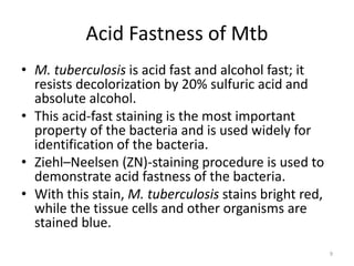
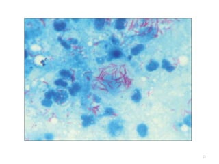
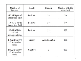
Tuberculosis
M.Sc. Biotechnology Part-II (Sem III)
Paper II - Unit II
Mumbai University
By: Mayur D. Chauhan
1
Introduction
• Tuberculosis, MTB, or TB in the past also
called phthisis, phthisis pulmonalis, or
consumption, is a widespread, and in many
cases fatal, infectious disease caused by
various strains of mycobacteria, usually
Mycobacterium tuberculosis.
• Tuberculosis typically attacks the lungs, but
can also affect other parts of the body.
2
Types of Tuberculosis Infection
• Infection mainly affects
the lungs
Pulmonary
• Pleura, CNS,
Genitourinary, Lymphatic
system, Bones and Joints
etc
Extra-
pulmonary
3
Extra-pulmonary Sites
• Pleura (Tuberculous pleurisy)
• Central nervous system (Tuberculous meningitis)
• Lymphatic system (Scrofula of the neck)
• Genitourinary system (Urogenital tuberculosis)
• Bones and joints (Pott disease of the spine).
When it spreads to the bones, it is also known as
"osseous tuberculosis“, a form of osteomyelitis.
• A potentially more serious, widespread form of
TB is called "disseminated" TB, commonly known
as miliary tuberculosis.
4
Symptoms of TB
• General signs and symptoms include fever,
chills, night sweats, loss of appetite, weight
loss, and fatigue.
• Significant nail clubbing may also occur.
5
6
Causative Agent and it’s Properties
• Mycobacterium tuberculosis is the causative agent for
TB and it belongs to the group of Mycobacteriaceae.
• M. tuberculosis organisms are straight or slightly
curved rods
• occurring singly, in pairs, or in small clumps.
• They measure about 3 0.3 m in size; sometimes long
filamentous forms are also seen. M. bovis is usually
shorter and stouter than M. tuberculosis.
• They are Gram positive but are difficult to stain. They
stain poorly with Gram staining.
• They are nonmotile, nonsporing, and noncapsulated
7
Mycobacterium tuberculosis Complex
• M. bovis• M. cannettii
• M.
tuberculosis
• M. microti
Bacillus-
Calmette
Guerin
M. caprae
M.
africannum
M.
pinnipedii
8
Acid Fastness of Mtb
• M. tuberculosis is acid fast and alcohol fast; it
resists decolorization by 20% sulfuric acid and
absolute alcohol.
• This acid-fast staining is the most important
property of the bacteria and is used widely for
identification of the bacteria.
• Ziehl–Neelsen (ZN)-staining procedure is used to
demonstrate acid fastness of the bacteria.
• With this stain, M. tuberculosis stains bright red,
while the tissue cells and other organisms are
stained blue.
9
C] Ziehl- Neelsen Staining and Grading
10
11
12
• These bacilli can also be stained by Auramine
• O stain and examined under a fluorescent
microscope. They
• appear as bright fluorescent rods against a
dark background
13
Culturing Mtb
• M. tuberculosis is an obligate aerobe and
characteristically grows very slowly in media.
• Optimum temperature is 37°C, and the
bacteria do not grow below 25°C or above
40°C.
• Optimum pH is 6.4–7.0. M. bovis is
microaerophilic on primary isolation, which
subsequently becomes aerobic on subculture
14
• M. tuberculosis is a slow-growing bacillus with
an average generation time of 14–15 hours
• Prolonged incubation is therefore necessary
for demonstrating growth of the bacteria.
• The colonies usually appear in almost 2 weeks,
but sometimes require incubation up to 8
weeks to appear.
• M. tuberculosis can grow on a wide range of
enriched solid and liquid media.
15
Culture on solid media
• The examples of solid media are
(i) egg-containing media (Lowenstein–Jensen [LJ]
medium,Petragnani and Dorset egg medium)
(ii) blood-containing media (Tarshis medium),
serum-combining media (Loeffler’s serum slope),
(iii) potato-based media (Pawlowsky medium).
• On these media, M. tuberculosis produces dry,
rough, raised, and irregular colonies with a
wrinkled surface.
16
Lowenstein-Jensen Medium
• LJ medium without starch is most widely used and is also
recommended by the International Union against
Tuberculosis (IUAT).
• The LJ medium consists of coagulated whole egg,
asparagines, malachite green, mineral salt, and glycerol or
sodium pyruvate.
• Malachite green inhibits growth of bacteria other than
mycobacterium.
• Addition of 0.75% glycerol enhances growth of M.
tuberculosis, but is inhibitory to growth of M. bovis.
• On the LJ medium after 6–8 weeks of incubation, M.
tuberculosis produces yellowish or buff-colored colonies.
They are tenacious not easily emulsified
17
18
Culture in liquid media
• The commonly used liquid media include
Soloac’s solutions, Dubos medium, and
Middlebrook’s and Beck’s medium.
• In liquid media, M. tuberculosis produces
growth that appears first at the bottom and
then grips up t the sides and produces a
surface pellicle that may extend along the
sides above the medium.
19
• The liquid media are not used for routine
culture of the bacilli.
• They are usually used for (i) preparation of
mycobacterial antigens for vaccines and (ii) to
assess antibiotic sensitivity of M. tuberculosis
20
Biochemical reactions
• Niacin test is an important test to identify niacin-positive
human strains of M. tuberculosis and differentiate them
from niacin-negative M. bovis strains. M. tuberculosis
human strains are niacin test positive. M. simiae and a few
strains of M. chelonae are also niacin positive. They
produce niacin as a metabolic by-product when grown on
an egg-containing solid medium.
• M. tuberculosis is usually catalase positive. They lose their
catalase activity when they become resistant to isoniazid
(INH). Catalase-negative strains of M. tuberculosis are not
virulent for guinea pigs.
• M. tuberculosis is amidase positive. It produces amidase
enzymes, such as nicotinamidase and pyrazinamidase,
which split various amide substrates.
21
• M. tuberculosis is positive for nitrate reduction
test. It produces the enzyme nitrate
reductase, thereby reducing nitrate to nitrite.
M. bovis and M. avium lack the enzyme
nitroreductase and therefore are negative for
nitrate reduction test.
• M. tuberculosis as well as M. bovis are neutral
red test positive. They have the property to
bind neutral red in alkaline buffer solution.
22
• M. tuberculosis is arylsulfate test negative. If
arylsulfatase is produced, it splits the
phenolphthalein substrate, releasing free
phenolphthalein, which turns pink to red
when alkali is added to the medium.
23
Other properties
• Mycobacteria are killed by heating at 60°C for 15–20
minutes.
• Killing of the bacteria is dependent on the nature of
the clinical specimen in which the bacteria are present.
• Mycobacteria in the sputum may survive for 20–30
hours, but in dried sputum protected from the sunlight
they may survive longer, up to 6 months.
• In droplet nuclei, they may remain alive for 8–10 days.
• Bacteria are killed when exposed to direct sunlight for
2 hours, but they remain viable at room temperature
for 6–8 months.
24
• Tubercle bacilli are sensitive to formaldehyde and
glutaraldehyde.
• They are destroyed by tincture of iodine in 5
minutes and by 80% ethanol in 2–10 minutes.
• They are generally more resistant to chemical
disinfectants than other nonsporeforming bacilli.
• They can survive exposure to 5% phenol, 15%
sulfuric acid, 3% nitric acid, 5% oxalic acid, and
4% sodium hydroxide.
• M. tuberculosis is susceptible to pyrazinamide,
while M. bovis and other mycobacteria are
resistant. M. tuberculosis is resistant to
thiophene-2-carboxylic acid hydrazide (TCH),
while M. bovis is susceptible.
25
Cell Wall of Mtb
• The cell wall of M. tuberculosis consists of four
layers—(i) peptidoglycan layer,
(ii) arabinogalactan layer,
(iii) mycolic acid layer,
(iv) mycosides
26
27
• The peptidoglycan layer is the innermost layer
covalently linked with arabinogalactan
(polysaccharide) and its terminal ends are
linked to mycolic acid.
• This layer is overlaid with polypeptides and a
layer of mycolic acid consisting of free lipids,
glycolipids, and peptidoglycolipids. These
lipids constitute nearly 60% of the dry weight
of the cell wall.
28
• The peptide chains present in the outer layer are
important antigens, which stimulate cell-
mediated immunity (CMI) in infected humans.
• PPDs are extracted and partially purified
preparation of these proteins, which are used as
antigens in tuberculosis skin test.
• The mycolic acid fraction of the lipids of the cell
wall is responsible for many of the characteristic
properties of the bacilli. These include (a) acid
fastness, (b) slow growth by delaying permeation
of nutrients, (c) resistance to commonly used
antibiotics, (d) resistance to detergents, (e)
unusual resistance to killing by acids and alkalies,
and (f) clumping or cord formation.
29
Antigenic Structure
• Mycobacteria possess two types of antigens:
(a) cell wall insoluble polysaccharide antigens
and (b) cytoplasmic soluble protein antigens.
• Cell Wall Antigens include arabinogalactans,
lipoarabinomannan, and also lipids, glycolipids
and peptidoglycolipids.
• Cytoplasmic antigens include antigen 5,
antigen 6, antigen 14, antigen 19, antigen 33,
antigen 38, an antigen 60
30
Transmission of
Tuberculosis
• Person to Person
• Present in Air-borne
particles termed as
Droplet Nuclei.
• 1-5 µm in size
• Generated when
infected patients cough
or sneeze
• Bacteria may remain
viable in the
environment uptill 8
months.
31
Pathogenesis of Tuberculosis
32
33
Virulence factors
• The factors determining the virulence of M.
tuberculosis are poorly understood. M. tuberculosis
does not produce any toxin.
• Although cord factor and sulfolipids are toxic
substances produced by mycobacterium, their
existence as virulence factors is doubtful.
• Cord factor: Cord factor was so called because of the
false belief that it is responsible for producing
serpentine cords typically found on the surface of
liquid or on solid media by M. tuberculosis. The cord
factor was originally thought to be a virulent factor,
which no longer holds true.
34
• Sulfolipids: Sulfolipids are of doubtful virulence
factor. Their exact role in pathogenesis of the
disease is not known.
• They are found to be associated with virulence of
tubercle bacilli by preventing fusion of
phagosome and lysosome inside the
macrophages, thereby allowing the bacteria to
multiply within the macrophages.
• The main pathology in the infected tissue caused
by mycobacterial infection is primarily due to
responses of the host to M. tuberculosis infection
rather than any virulence factor produced by it.
35
Host Immunity
• M. tuberculosis infection in an infected host
induces CMI.
• The CMI is manifested either as delayed
tuberculin hypersensitivity or as resistance to
infection.
• The course of infection is determined by the
interaction of hypersensitivity or immunity
36
Tuberculin hypersensitivity reaction
• This was first described by Robert Koch in
experimentally infected animals,such as
guinea pigs.
• Demonstration of this tuberculin reactivity in
guinea pigs is known as Koch’s phenomenon.
• Koch phenomenon is demonstrated by
subcutaneous injection of pure culture of
virulent tubercle bacilli in a normal guinea pig.
37
• Initially, no immediate visible reaction is
observed at the site of inoculation in the
guinea pigs.
• But after 10–14 days, a hard nodule appears
at the site of inoculation, which soon breaks
down to from an ulcer that persists till the
animal dies of progressive tuberculosis.
• The regional lymph nodes draining the region
become enlarged and caseous.
38
• In contrast, when a guinea pig already
inoculated 4–6 weeks earlier by tubercle
bacilli is injected with tubercle bacilli, an
individual lesion develops at the site of second
inoculation within 24–48 hours.
• The lesion undergoes necrosis in another day
to produce a shallow ulcer that heals rapidly,
involving the regional lymph nodes and other
tissues.
39
• The DTH can be induced by live attenuated
and killed bacilli, bacillary products, and
tubercular protein.
• Usually live o killed bacilli or tubercular
protein (tuberculin) are employed for
demonstration of hypersensitivity reaction
• This hypersensitivity reaction can be
transferred passively by cells, but not by
serum.
40
Tuberculin test
• Tuberculin test is performed to demonstrate
delayed type IV (DTH) or cell-mediated (CMI)
hypersensitivity reaction to tubercle bacilli.
• Originally, the tuberculin test was performed
by using a protein known as old tuberculin
(OT) prepared by Robert Koch.
41
• The OT is a protein component of tubercle
bacilli prepared from a 6–8 week culture
filtrate of M. tuberculosis cultured in 5%
glycerol, which is concentrated 10-fold by
evaporation on a steam bath.
• The OT is a crude protein, which consists of
protein as a constituent but varies from batch
to batch in its purity and potency.
• This has now been replaced by the use of PPD
of tubercle bacilli.
42
Humoral Immunity
• Humoral immunity is characterized by the
development of antibodies in serum, but they do
not play any role in conferring immunity against
the bacteria.
• Antibodies against polysaccharide, proteins, and
phosphate antigens of tubercle bacilli have been
demonstrated in serum of patients with
tuberculosis.
• These serum antibodies, however, are not
protective.
43
Habitat
• M. tuberculosis inhabits primarily the
respiratory tract of the infected human host
• The droplet nuclei consisting of M.
tuberculosis have been found in the terminal
air spaces of the lung.
44
Reservoir, source, and transmission
of infection
• Human beings are the only source and reservoir for M.
tuberculosis infection.
• The infectiousness of the source is of primary
importance, which determines the possibility of
transmission of the disease.
• This depends on bacillary load of lesions and also on
the morphology of the lesion. Lesions with cavities
have 100–10,000 bacilli; therefore, cases with cavitary
lesions are potentially highly infectious.
• Cases treated with antitubercular therapy are less
infectious than those who are not treated with any
antitubercular drugs.
45
• The decrease in infectiousness is primarily due to
reduction in the bacillary load in the lungs.
• Humans acquire M. tuberculosis infection most
frequently by inhalation of infectious aerosolized
droplets.
• These infective droplets are usually coughed or
sneezed into environment by a patient suffering
from pulmonary tuberculosis.
• The acts of coughing, sneezing, and speaking
release a large number of droplets containing as
many as 3000 infectious airborne droplet nuclei
per cough.
46
• The droplet nucleus is small, measures 5 μm or less, and
may contain approximately 1–10 tubercle bacilli.
• Theoretically, although a single tubercle bacillus may cause
disease, in practice 5–200 inhaled bacilli are essential for
infection.
• These droplets by virtue of their small size remain
suspended in the air for a very long period of time.
• The infection is acquired rarely by inoculation.
• M. bovis infection is transmitted to humans by ingestion of
raw milk of the cows infected with M. bovis.
• The infection among animals is spread by aerosolized bacilli
in moist cough sprays.
• The infected animals usually excrete the bacilli in their milk.
Person-to-person transmission of M. bovis usually does not
occur.
47
References
• Textbook of Microbiology and Immunology by
Subhash Parija
48
49

Tuberculosis - Diagnosis and Treatment

  • 1.
    Tuberculosis M.Sc. Biotechnology Part-II(Sem III) Paper II - Unit II Mumbai University By: Mayur D. Chauhan 1
  • 2.
    Introduction • Tuberculosis, MTB,or TB in the past also called phthisis, phthisis pulmonalis, or consumption, is a widespread, and in many cases fatal, infectious disease caused by various strains of mycobacteria, usually Mycobacterium tuberculosis. • Tuberculosis typically attacks the lungs, but can also affect other parts of the body. 2
  • 3.
    Types of TuberculosisInfection • Infection mainly affects the lungs Pulmonary • Pleura, CNS, Genitourinary, Lymphatic system, Bones and Joints etc Extra- pulmonary 3
  • 4.
    Extra-pulmonary Sites • Pleura(Tuberculous pleurisy) • Central nervous system (Tuberculous meningitis) • Lymphatic system (Scrofula of the neck) • Genitourinary system (Urogenital tuberculosis) • Bones and joints (Pott disease of the spine). When it spreads to the bones, it is also known as "osseous tuberculosis“, a form of osteomyelitis. • A potentially more serious, widespread form of TB is called "disseminated" TB, commonly known as miliary tuberculosis. 4
  • 5.
    Symptoms of TB •General signs and symptoms include fever, chills, night sweats, loss of appetite, weight loss, and fatigue. • Significant nail clubbing may also occur. 5
  • 6.
  • 7.
    Causative Agent andit’s Properties • Mycobacterium tuberculosis is the causative agent for TB and it belongs to the group of Mycobacteriaceae. • M. tuberculosis organisms are straight or slightly curved rods • occurring singly, in pairs, or in small clumps. • They measure about 3 0.3 m in size; sometimes long filamentous forms are also seen. M. bovis is usually shorter and stouter than M. tuberculosis. • They are Gram positive but are difficult to stain. They stain poorly with Gram staining. • They are nonmotile, nonsporing, and noncapsulated 7
  • 8.
    Mycobacterium tuberculosis Complex •M. bovis• M. cannettii • M. tuberculosis • M. microti Bacillus- Calmette Guerin M. caprae M. africannum M. pinnipedii 8
  • 9.
    Acid Fastness ofMtb • M. tuberculosis is acid fast and alcohol fast; it resists decolorization by 20% sulfuric acid and absolute alcohol. • This acid-fast staining is the most important property of the bacteria and is used widely for identification of the bacteria. • Ziehl–Neelsen (ZN)-staining procedure is used to demonstrate acid fastness of the bacteria. • With this stain, M. tuberculosis stains bright red, while the tissue cells and other organisms are stained blue. 9
  • 10.
    C] Ziehl- NeelsenStaining and Grading 10
  • 11.
  • 12.
  • 13.
    • These bacillican also be stained by Auramine • O stain and examined under a fluorescent microscope. They • appear as bright fluorescent rods against a dark background 13
  • 14.
    Culturing Mtb • M.tuberculosis is an obligate aerobe and characteristically grows very slowly in media. • Optimum temperature is 37°C, and the bacteria do not grow below 25°C or above 40°C. • Optimum pH is 6.4–7.0. M. bovis is microaerophilic on primary isolation, which subsequently becomes aerobic on subculture 14
  • 15.
    • M. tuberculosisis a slow-growing bacillus with an average generation time of 14–15 hours • Prolonged incubation is therefore necessary for demonstrating growth of the bacteria. • The colonies usually appear in almost 2 weeks, but sometimes require incubation up to 8 weeks to appear. • M. tuberculosis can grow on a wide range of enriched solid and liquid media. 15
  • 16.
    Culture on solidmedia • The examples of solid media are (i) egg-containing media (Lowenstein–Jensen [LJ] medium,Petragnani and Dorset egg medium) (ii) blood-containing media (Tarshis medium), serum-combining media (Loeffler’s serum slope), (iii) potato-based media (Pawlowsky medium). • On these media, M. tuberculosis produces dry, rough, raised, and irregular colonies with a wrinkled surface. 16
  • 17.
    Lowenstein-Jensen Medium • LJmedium without starch is most widely used and is also recommended by the International Union against Tuberculosis (IUAT). • The LJ medium consists of coagulated whole egg, asparagines, malachite green, mineral salt, and glycerol or sodium pyruvate. • Malachite green inhibits growth of bacteria other than mycobacterium. • Addition of 0.75% glycerol enhances growth of M. tuberculosis, but is inhibitory to growth of M. bovis. • On the LJ medium after 6–8 weeks of incubation, M. tuberculosis produces yellowish or buff-colored colonies. They are tenacious not easily emulsified 17
  • 18.
  • 19.
    Culture in liquidmedia • The commonly used liquid media include Soloac’s solutions, Dubos medium, and Middlebrook’s and Beck’s medium. • In liquid media, M. tuberculosis produces growth that appears first at the bottom and then grips up t the sides and produces a surface pellicle that may extend along the sides above the medium. 19
  • 20.
    • The liquidmedia are not used for routine culture of the bacilli. • They are usually used for (i) preparation of mycobacterial antigens for vaccines and (ii) to assess antibiotic sensitivity of M. tuberculosis 20
  • 21.
    Biochemical reactions • Niacintest is an important test to identify niacin-positive human strains of M. tuberculosis and differentiate them from niacin-negative M. bovis strains. M. tuberculosis human strains are niacin test positive. M. simiae and a few strains of M. chelonae are also niacin positive. They produce niacin as a metabolic by-product when grown on an egg-containing solid medium. • M. tuberculosis is usually catalase positive. They lose their catalase activity when they become resistant to isoniazid (INH). Catalase-negative strains of M. tuberculosis are not virulent for guinea pigs. • M. tuberculosis is amidase positive. It produces amidase enzymes, such as nicotinamidase and pyrazinamidase, which split various amide substrates. 21
  • 22.
    • M. tuberculosisis positive for nitrate reduction test. It produces the enzyme nitrate reductase, thereby reducing nitrate to nitrite. M. bovis and M. avium lack the enzyme nitroreductase and therefore are negative for nitrate reduction test. • M. tuberculosis as well as M. bovis are neutral red test positive. They have the property to bind neutral red in alkaline buffer solution. 22
  • 23.
    • M. tuberculosisis arylsulfate test negative. If arylsulfatase is produced, it splits the phenolphthalein substrate, releasing free phenolphthalein, which turns pink to red when alkali is added to the medium. 23
  • 24.
    Other properties • Mycobacteriaare killed by heating at 60°C for 15–20 minutes. • Killing of the bacteria is dependent on the nature of the clinical specimen in which the bacteria are present. • Mycobacteria in the sputum may survive for 20–30 hours, but in dried sputum protected from the sunlight they may survive longer, up to 6 months. • In droplet nuclei, they may remain alive for 8–10 days. • Bacteria are killed when exposed to direct sunlight for 2 hours, but they remain viable at room temperature for 6–8 months. 24
  • 25.
    • Tubercle bacilliare sensitive to formaldehyde and glutaraldehyde. • They are destroyed by tincture of iodine in 5 minutes and by 80% ethanol in 2–10 minutes. • They are generally more resistant to chemical disinfectants than other nonsporeforming bacilli. • They can survive exposure to 5% phenol, 15% sulfuric acid, 3% nitric acid, 5% oxalic acid, and 4% sodium hydroxide. • M. tuberculosis is susceptible to pyrazinamide, while M. bovis and other mycobacteria are resistant. M. tuberculosis is resistant to thiophene-2-carboxylic acid hydrazide (TCH), while M. bovis is susceptible. 25
  • 26.
    Cell Wall ofMtb • The cell wall of M. tuberculosis consists of four layers—(i) peptidoglycan layer, (ii) arabinogalactan layer, (iii) mycolic acid layer, (iv) mycosides 26
  • 27.
  • 28.
    • The peptidoglycanlayer is the innermost layer covalently linked with arabinogalactan (polysaccharide) and its terminal ends are linked to mycolic acid. • This layer is overlaid with polypeptides and a layer of mycolic acid consisting of free lipids, glycolipids, and peptidoglycolipids. These lipids constitute nearly 60% of the dry weight of the cell wall. 28
  • 29.
    • The peptidechains present in the outer layer are important antigens, which stimulate cell- mediated immunity (CMI) in infected humans. • PPDs are extracted and partially purified preparation of these proteins, which are used as antigens in tuberculosis skin test. • The mycolic acid fraction of the lipids of the cell wall is responsible for many of the characteristic properties of the bacilli. These include (a) acid fastness, (b) slow growth by delaying permeation of nutrients, (c) resistance to commonly used antibiotics, (d) resistance to detergents, (e) unusual resistance to killing by acids and alkalies, and (f) clumping or cord formation. 29
  • 30.
    Antigenic Structure • Mycobacteriapossess two types of antigens: (a) cell wall insoluble polysaccharide antigens and (b) cytoplasmic soluble protein antigens. • Cell Wall Antigens include arabinogalactans, lipoarabinomannan, and also lipids, glycolipids and peptidoglycolipids. • Cytoplasmic antigens include antigen 5, antigen 6, antigen 14, antigen 19, antigen 33, antigen 38, an antigen 60 30
  • 31.
    Transmission of Tuberculosis • Personto Person • Present in Air-borne particles termed as Droplet Nuclei. • 1-5 µm in size • Generated when infected patients cough or sneeze • Bacteria may remain viable in the environment uptill 8 months. 31
  • 32.
  • 33.
  • 34.
    Virulence factors • Thefactors determining the virulence of M. tuberculosis are poorly understood. M. tuberculosis does not produce any toxin. • Although cord factor and sulfolipids are toxic substances produced by mycobacterium, their existence as virulence factors is doubtful. • Cord factor: Cord factor was so called because of the false belief that it is responsible for producing serpentine cords typically found on the surface of liquid or on solid media by M. tuberculosis. The cord factor was originally thought to be a virulent factor, which no longer holds true. 34
  • 35.
    • Sulfolipids: Sulfolipidsare of doubtful virulence factor. Their exact role in pathogenesis of the disease is not known. • They are found to be associated with virulence of tubercle bacilli by preventing fusion of phagosome and lysosome inside the macrophages, thereby allowing the bacteria to multiply within the macrophages. • The main pathology in the infected tissue caused by mycobacterial infection is primarily due to responses of the host to M. tuberculosis infection rather than any virulence factor produced by it. 35
  • 36.
    Host Immunity • M.tuberculosis infection in an infected host induces CMI. • The CMI is manifested either as delayed tuberculin hypersensitivity or as resistance to infection. • The course of infection is determined by the interaction of hypersensitivity or immunity 36
  • 37.
    Tuberculin hypersensitivity reaction •This was first described by Robert Koch in experimentally infected animals,such as guinea pigs. • Demonstration of this tuberculin reactivity in guinea pigs is known as Koch’s phenomenon. • Koch phenomenon is demonstrated by subcutaneous injection of pure culture of virulent tubercle bacilli in a normal guinea pig. 37
  • 38.
    • Initially, noimmediate visible reaction is observed at the site of inoculation in the guinea pigs. • But after 10–14 days, a hard nodule appears at the site of inoculation, which soon breaks down to from an ulcer that persists till the animal dies of progressive tuberculosis. • The regional lymph nodes draining the region become enlarged and caseous. 38
  • 39.
    • In contrast,when a guinea pig already inoculated 4–6 weeks earlier by tubercle bacilli is injected with tubercle bacilli, an individual lesion develops at the site of second inoculation within 24–48 hours. • The lesion undergoes necrosis in another day to produce a shallow ulcer that heals rapidly, involving the regional lymph nodes and other tissues. 39
  • 40.
    • The DTHcan be induced by live attenuated and killed bacilli, bacillary products, and tubercular protein. • Usually live o killed bacilli or tubercular protein (tuberculin) are employed for demonstration of hypersensitivity reaction • This hypersensitivity reaction can be transferred passively by cells, but not by serum. 40
  • 41.
    Tuberculin test • Tuberculintest is performed to demonstrate delayed type IV (DTH) or cell-mediated (CMI) hypersensitivity reaction to tubercle bacilli. • Originally, the tuberculin test was performed by using a protein known as old tuberculin (OT) prepared by Robert Koch. 41
  • 42.
    • The OTis a protein component of tubercle bacilli prepared from a 6–8 week culture filtrate of M. tuberculosis cultured in 5% glycerol, which is concentrated 10-fold by evaporation on a steam bath. • The OT is a crude protein, which consists of protein as a constituent but varies from batch to batch in its purity and potency. • This has now been replaced by the use of PPD of tubercle bacilli. 42
  • 43.
    Humoral Immunity • Humoralimmunity is characterized by the development of antibodies in serum, but they do not play any role in conferring immunity against the bacteria. • Antibodies against polysaccharide, proteins, and phosphate antigens of tubercle bacilli have been demonstrated in serum of patients with tuberculosis. • These serum antibodies, however, are not protective. 43
  • 44.
    Habitat • M. tuberculosisinhabits primarily the respiratory tract of the infected human host • The droplet nuclei consisting of M. tuberculosis have been found in the terminal air spaces of the lung. 44
  • 45.
    Reservoir, source, andtransmission of infection • Human beings are the only source and reservoir for M. tuberculosis infection. • The infectiousness of the source is of primary importance, which determines the possibility of transmission of the disease. • This depends on bacillary load of lesions and also on the morphology of the lesion. Lesions with cavities have 100–10,000 bacilli; therefore, cases with cavitary lesions are potentially highly infectious. • Cases treated with antitubercular therapy are less infectious than those who are not treated with any antitubercular drugs. 45
  • 46.
    • The decreasein infectiousness is primarily due to reduction in the bacillary load in the lungs. • Humans acquire M. tuberculosis infection most frequently by inhalation of infectious aerosolized droplets. • These infective droplets are usually coughed or sneezed into environment by a patient suffering from pulmonary tuberculosis. • The acts of coughing, sneezing, and speaking release a large number of droplets containing as many as 3000 infectious airborne droplet nuclei per cough. 46
  • 47.
    • The dropletnucleus is small, measures 5 μm or less, and may contain approximately 1–10 tubercle bacilli. • Theoretically, although a single tubercle bacillus may cause disease, in practice 5–200 inhaled bacilli are essential for infection. • These droplets by virtue of their small size remain suspended in the air for a very long period of time. • The infection is acquired rarely by inoculation. • M. bovis infection is transmitted to humans by ingestion of raw milk of the cows infected with M. bovis. • The infection among animals is spread by aerosolized bacilli in moist cough sprays. • The infected animals usually excrete the bacilli in their milk. Person-to-person transmission of M. bovis usually does not occur. 47
  • 48.
    References • Textbook ofMicrobiology and Immunology by Subhash Parija 48
  • 49.