Mycobacterial diseases
•Download as PPTX, PDF•
4 likes•2,710 views

This document describes Mycobacterium tuberculosis, the bacteria that causes tuberculosis. It discusses how M. tuberculosis infects and replicates within macrophages, evading the immune system. This can lead to the formation of granulomas and caseation in the lungs. If not controlled, the infection can spread throughout the body and cause serious illness. Effective treatment requires a strong T-helper 1 immune response and cytokines like IFN-γ to activate macrophages and control the infection. The pathology of primary and secondary tuberculosis is also summarized.

Recommended
More Related Content
What's hot
What's hot (20)
Similar to Mycobacterial diseases
Similar to Mycobacterial diseases (20)
More from Guvera Vasireddy
More from Guvera Vasireddy (8)
Recently uploaded
Recently uploaded (20)
Mycobacterial diseases
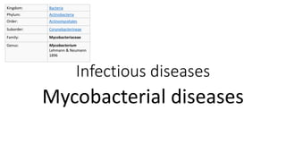
- 1. Kingdom: Bacteria Phylum: Actinobacteria Order: Actinomycetales Suborder: Corynebacterineae Family: Mycobacteriaceae Genus: Mycobacterium Lehmann & Neumann 1896 Infectious diseases Mycobacterial diseases
- 3. Tuberculosis • Mycobacterium tuberculosis is responsible for most cases of tuberculosis; • Oropharyngeal and intestinal tuberculosis contracted by drinking milk contaminated with M. bovis. • Tuberculosis flourishes wherever there is poverty, crowding, and chronic debilitating illness.
- 4. Pathogenesis • Macrophages are the primary cells infected by M. tuberculosis. • M. tuberculosis enters macrophages by endocytosis mediated by mannose receptors bind lipoarabinomannan, a glycolipid in the bacterial cell wall, and complement receptors bind opsonized mycobacteria. • Once inside the macrophage, M. tuberculosis organisms replicate within the phagosome by blocking fusion of the phagosome and lysosome • In some people with polymorphisms in the NRAMP1 gene, the disease may progress due to the absence of an effective immune response.
- 5. Primary immune response • About 3 weeks after infection, a T-helper 1 (TH1) response is mounted that activates macrophages to become bactericidal • Mature TH1 cells, both in lymph nodes and in the lung, produce IFN-γ. • In addition to stimulating macrophages to kill mycobacteria, the TH1 response orchestrates the formation of granulomas and caseous necrosis. • Macrophages activated by IFN-γ differentiate into the “epithelioid histiocytes” that characterize the granulomatous response, and may fuse to form giant cells. • In addition to the TH1 response, NK-T cells that recognize mycobacterial lipid antigens bound to CD1 on antigen-presenting cells, or T cells that express a γδ T-cell receptor, also make IFN-γ.
- 6. The sequence of events in primary pulmonary tuberculosis, commencing with inhalation of virulent Mycobacterium tuberculosis organisms and culminating with the development of cell-mediated immunity to the organism. A, Events occurring in the first 3 weeks after exposure. B, Events thereafter. The development of resistance to the organism is accompanied by the appearance of a positive tuberculin test
- 7. Primary tuberculosis • Develops in a previously unexposed, and therefore unsensitized, person. • About 5% of newly infected people develop clinically significant disease. • The elderly and immunosuppressed persons may develop primary tuberculosis more than once. • With primary tuberculosis the source of the organism is exogenous. • Primary tuberculosis in immunocompromised and elderly often resembles an acute bacterial pneumonia. • Lymphohematogenous dissemination may result in the development of tuberculous meningitis and miliary tuberculosis.
- 8. Morphology – Primary tuberculosis • Inhaled bacilli implant in the distal airspaces of the lower part of the upper lobe or the upper part of the lower lobe, usually close to the pleura. • As sensitization develops, a 1- to 1.5-cm area of gray-white inflammation with consolidation emerges, known as the Ghon focus. • Tubercle bacilli, either free or within phagocytes, drain to the regional nodes, which also often caseate. • This combination of parenchymal lung lesion and nodal involvement is referred to as the Ghon complex . • Ghon complex undergoes progressive fibrosis, often followed by radiologically detectable calcification (Ranke complex), and despite seeding of other organs, no lesions develop.
- 9. Primary pulmonary tuberculosis, Ghon complex. The gray-white parenchymal focus is under the pleura in the lower part of the upper lobe. Hilar lymph nodes with caseation are seen on the left.
- 10. Primary Tuberculosis - Histopathology • Marked by a characteristic granulomatous inflammatory reaction that forms both caseating and noncaseating tubercles. • Individual tubercles are microscopic; it is only when multiple granulomas coalesce that they become macroscopically visible. • The granulomas are usually enclosed within a fibroblastic rim punctuated by lymphocytes. • Multinucleate giant cells are present in the granulomas. • Immunocompromised people do not form the characteristic granulomas.
- 11. Progressive pulmonary tuberculosis • may ensue in the elderly and immunosuppressed. • The apical lesion expands into adjacent lung and eventually erodes into bronchi and vessels. • Erosion of blood vessels results in hemoptysis. • Miliary pulmonary disease occurs when organisms draining through lymphatics enter the venous blood and circulate back to the lung. • Individual lesions are either microscopic or small, visible (2-mm) foci of yellow-white consolidation scattered through the lung parenchyma. • With progressive pulmonary tuberculosis, the pleural cavity is invariably involved, and serous pleural effusions, tuberculous empyema, or obliterative fibrous pleuritis may develop.
- 12. Secondary tuberculosis • Pattern of disease that arises in a previously sensitized host. • Secondary pulmonary tuberculosis classically involves the apex of the upper lobes of one or both lungs. • Regional lymph nodes are less prominently involved early in secondary disease than they are in primary tuberculosis. • Cavitation occurs readily in the secondary form. • Erosion of the cavities into an airway is an important source of infection because the person now coughs sputum that contains bacteria.
- 13. Secondary Tuberculosis – disease course • The initial lesion is usually a small focus of consolidation, less than 2 cm in diameter, within 1 to 2 cm of the apical pleura. • Such foci are sharply circumscribed, firm, gray-white to yellow areas that have a variable amount of central caseation and peripheral fibrosis. • In immunocomptetent individuals, the initial parenchymal focus undergoes progressive fibrous encapsulation, leaving only fibrocalcific scars. • Histologically, the active lesions show characteristic coalescent tubercles with central caseation. • Tubercle bacilli can often be identified with acid-fast stains in early exudative and caseous phases of granuloma formation but are usually too few to be found in the late, fibrocalcific stages. • Localized, apical, secondary pulmonary tuberculosis may heal with fibrosis either spontaneously or after therapy, or the disease may progress and extend along several different pathways.
- 14. The morphologic spectrum of tuberculosis. (A)tubercle at low magnification (B)central caseation surrounded by epithelioid and multinucleated giant cells. (C)central caseation (D) sheets of foamy macrophages are seen that are packed with mycobacteria (demonstrable with acid-fast stains).
- 15. Miliary tuberculosis of the spleen. The cut surface shows numerous gray-white tubercles. Secondary pulmonary tuberculosis. The upper parts of both lungs are riddled with gray-white areas of caseation and multiple areas of softening and cavitation
- 16. Mycobacterium avium and intracellulare • Infections are similar that they are simply referred to as M. avium-intracellulare complex, or MAC. • MAC is common in soil, water, dust, and domestic animals. • Clinically significant infection with MAC is uncommon except among people with AIDS and low numbers of CD4+ lymphocytes (<60 cells/mm3). • In AIDS patients MAC causes widely disseminated infections, and organisms proliferate abundantly in many organs, including the lungs and gastrointestinal system. • Unchecked by the immune response, the organisms reach very high levels: up to 104 organisms/mL of blood and 106 organisms/gm in tissue. • Patients are feverish, with drenching night sweats and weight loss.
- 17. The hallmark of MAC infections in patients with HIV is abundant acid-fast bacilli within macrophages
- 19. Mycobacterium leprae - leprosy • Slowly progressive infection caused by Mycobacterium leprae that mainly affects the skin and peripheral nerves and results in disabling deformities. • M. leprae is likely to be transmitted from person to person through aerosols from asymptomatic lesions in the upper respiratory tract. • Inhaled M. leprae, is taken up by alveolar macrophages and disseminates through the blood, but replicates only in relatively cool tissues of the skin and extremities. • Leprosy pursues an extremely slow course, spanning decades, most patients die with leprosy rather than of it
- 20. General characteristics • M. leprae is an acid-fast obligate intracellular organism that grows very poorly in culture but can be propagated in the armadillo. • It proliferates best at 32° to 34°C, the temperature of the human skin and the core temperature of armadillos. • Like M. tuberculosis, M. leprae secretes no toxins, and its virulence is based on properties of its cell wall. • The cell wall is similar enough to that of M. tuberculosis that immunization with BCG confers some protection against M. leprae infection. • Cell-mediated immunity is reflected by delayed-type hypersensitivity reactions to dermal injections of a bacterial extract called lepromin.
- 21. Pathogenesis • The T-helper lymphocyte response to M. leprae determines whether an individual has tuberculoid or lepromatous leprosy. • People with tuberculoid leprosy have a TH1 response associated with production of IL-2 and IFN-γ. • Lepromatous leprosy is associated with a weak TH1 response and, in some cases, a relative increase in the TH2 response. • In the lepromatous form, antibodies are produced against M. leprae antigens and these antibodies form immune complexes with free antigens that can lead to erythema nodosum, vasculitis, and glomerulonephritis.
- 22. Morphology - Tuberculoid leprosy • Tuberculoid leprosy begins with localized flat, red skin lesions that enlarge and develop irregular shapes with indurated, elevated, hyperpigmented margins and depressed pale centers (central healing). • Neuronal involvement dominates tuberculoid leprosy. Nerves become enclosed within granulomatous inflammatory reactions and, if small (e.g., the peripheral twigs), are destroyed. • Nerve degeneration causes skin anesthesias and skin and muscle atrophy that render the person liable to trauma of the affected parts, leading to the development of chronic skin ulcers. • On microscopic examination, all sites of involvement have granulomatous lesions closely resembling those found in tuberculosis, and bacilli are almost never found, hence the name “paucibacillary” leprosy. • The presence of granulomas and absence of bacteria reflect strong T-cell immunity.
- 23. Lepromatous leprosy (LL) • Lepromatous leprosy involves the skin, peripheral nerves, anterior chamber of the eye, upper airways (down to the larynx), testes, hands, and feet. • Lepromatous lesions contain large aggregates of lipid-laden macrophages (lepra cells), often filled with masses (“globi”) of acid-fast bacilli. • Because of the abundant bacteria, lepromatous leprosy is referred to as “multibacillary”. • Macular, papular, or nodular lesions form on the face, ears, wrists, elbows, and knees. • With progression, the nodular lesions coalesce to yield a distinctive leonine facies. • Most skin lesions are hypoesthetic or anesthetic. • Lesions in the nose may cause persistent inflammation and bacilli-laden discharge.
- 24. Morphology of lesions in LL • The peripheral nerves, particularly the ulnar and peroneal nerves where they approach the skin surface, are symmetrically invaded with mycobacteria, with minimal inflammation. • Loss of sensation and trophic changes in the hands and feet follow the nerve lesions. • Lymph nodes contain aggregates of bacteria-filled foamy macrophages in the paracortical (T-cell) areas and reactive germinal centers. • In advanced disease, aggregates of macrophages are also present in the splenic red pulp and the liver. • The testes are usually extensively involved, leading to destruction of the seminiferous tubules and consequent sterility.
- 25. Acid-fast bacilli (“red snappers”) within macrophages A, Peripheral nerve. Note the inflammatory cell infiltrates in the endoneural and epineural compartments. B, Cells within the endoneurium contain acid-fast positive lepra bacilli.
- 26. Kingdom: Bacteria Phylum: Actinobacteria Order: Actinomycetales Suborder: Corynebacterineae Family: Mycobacteriaceae Genus: Mycobacterium Lehmann & Neumann 1896