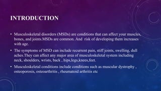
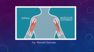
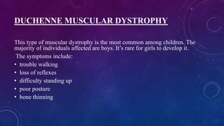
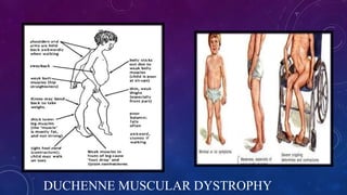
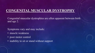
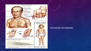
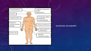
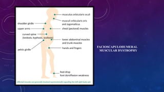
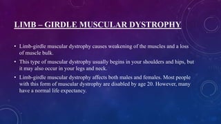
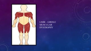
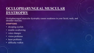
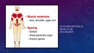
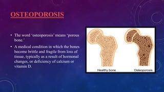
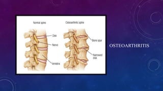
MUSCULO SKELETAL
SYSTEM DISORDERS
INTRODUCTION
• Musculoskeletal disorders (MSDs) are conditions that can affect your muscles,
bones, and joints.MSDs are common. And risk of developing them increases
with age.
• The symptoms of MSD can include recurrent pain, stiff joints, swelling, dull
aches.They can affect any major area of musculoskeletal system including
neck, shoulders, wrists, back , hips,legs,knees,feet.
• Musculoskeletal conditions include conditions such as muscular dystrophy ,
osteoporosis, osteoarthritis , rheumatoid arthritis etc
MUSCULO SKELETAL SYSTEM DISORDER
• Musculoskeletal Disorders or MSDs are injuries and disorders that affect the
human body’s movement or musculoskeletal system
• Other common names for MSDs are “repetitive motion injury”, “repetitive
stress injury”, “overuse injury” and many more.
SYMPTOMS OF MSD
Some of the most common symptoms of MSDs are:
• Stiffness, weakness and pain – all of them often persistent.
• Decreased range of motion – limiting mobility, dexterity and functional abilities.
• Noises in the joints – where early diagnosis and treatment are not available, joint
deformity may be visualised.
• Inflammation – along with pain and impaired function, there is redness, swelling
and warmth in the overlying skin area.
CAUSES OF MSD
• age
• occupation
• activity level
• lifestyle
• family history
• Certain activities can cause wear and tear on your musculoskeletal system,
leading to MSDs. These include:
•
• sitting in the same position at a computer every day
• engaging in repetitive motions
• lifting heavy weights
• maintaining poor posture at work
PREVENTION AND MANAGEMENT
• Most musculoskeletal disorders share the same risk factors
such as lack of physical activity, obesity, poor nutrition, and
smoking. Management generally requires specialist care and,
in many cases, surgical intervention. Some MSDs can be
resolved with primary care including psychological therapies,
weight management, exercise, and other Pharmacological
therapies.
TYPES OF MUSCULO
SKELETAL SYSTEM
DISORDERS
MUSCULAR DYSTROPHY
• Muscular dystrophy is a group of inherited diseases in which the
muscles that control movement progressively weaken.
• The prefix, dys-, means abnormal.
• The root, -trophy, refers to maintaining normal nourishment,
structure and function.
• They are genetically and clinically heterogeneous group of rare
muscle disorders.
• That cause progressive weakness and breakdown of skeletal
muscles over time.
• The damage and weakness is due to the lack of a protein called
dystrophin,
• Muscular dystrophy can occur at any age, but most diagnoses
occur in childhood. The most common form in children is called
Duchenne muscular dystrophy and affects only males.
• Young boys are more likely to have this disease than girls.
• Most individuals with muscular dystrophy do lose the ability to
walk and eventually require a wheelchair.
Fig : Muscular Dystrophy
TYPES OF MUSCULAR DYSTROPHY
There are more than 30 kinds of muscular dystrophy, and each is different
based on:
• The genes that cause it
• The muscles it affects
• The age when symptoms first appear
• How quickly the disease gets worse
People usually get one of nine major forms of the disease:
DUCHENNE MUSCULAR DYSTROPHY
This type of muscular dystrophy is the most common among children. The
majority of individuals affected are boys. It’s rare for girls to develop it.
The symptoms include:
• trouble walking
• loss of reflexes
• difficulty standing up
• poor posture
• bone thinning
• scoliosis, which is an abnormal curvature of your spine
• mild intellectual impairment
• breathing difficulties
• swallowing problems
• lung and heart weakness
• People with Duchenne muscular dystrophy typically require a
wheelchair before their teenage years. The life expectancy for those
with this disease is late teens or 20s.
DUCHENNE MUSCULAR DYSTROPHY
BECKER MUSCULAR DYSTROPHY
Muscular dystrophy also more commonly affects boys. Muscle weakness
occurs mostly in your arms and legs, with symptoms appearing between
age 11 and 25.
Other symptoms of Becker muscular dystrophy include:
• walking on your toes
• frequent falls
• muscle cramps
• trouble getting up from the floor
BECKER
MUSCULAR
DYSTROPHY
CONGENITAL MUSCULAR DYSTROPHY
Congenital muscular dystrophies are often apparent between birth
and age 2.
Symptoms vary and may include:
• muscle weakness
• poor motor control
• inability to sit or stand without support
• trouble swallowing
• respiratory problems
• vision problems
• speech problems
• intellectual impairment
• scoliosis
• foot deformities
MYOTONIC DYSTROPHY
• Myotonic dystrophy is also called Steinert’s disease or dystrophia
myotonica. This form of muscular dystrophy causes myotonia, which
is an inability to relax your muscles after they contract.
• Myotonic dystrophy can affect facial muscles , central nervous
system, adrenal glands , heart , thyroid , eyes , gastrointestinal tract.
• This dystrophy type may also cause impotence and testicular atrophy
in males.
• In women, it may cause irregular periods and infertility.
SYMPTOMS
• drooping muscles in your face, producing a thin, haggard look
• difficulty lifting your neck due to weak neck muscles
• difficulty swallowing
• droopy eyelids, or ptosis
• early baldness in the front area of your scalp
• poor vision, including cataracts
• weight loss
• increased sweating
MYOTONIC DYSTROPHY
MYOTONIC DYSTROPHY
FACIOSCAPULOHUMERAL MUSCULAR
DYSTROPHY
Facioscapulohumeral muscular dystrophy (FSHD) is also known as Landouzy-
Dejerine disease. This type of muscular dystrophy affects the muscles in your
face, shoulders, and upper arms.
FSHD may cause:
• difficulty chewing or swallowing
• slanted shoulders
• a crooked appearance of the mouth
• a wing-like appearance of the shoulder blades
FACIOSCAPULOHUMERAL
MUSCULAR DYSTROPHY
LIMB – GIRDLE MUSCULAR DYSTROPHY
• Limb-girdle muscular dystrophy causes weakening of the muscles and a loss
of muscle bulk.
• This type of muscular dystrophy usually begins in your shoulders and hips, but
it may also occur in your legs and neck.
• Limb-girdle muscular dystrophy affects both males and females. Most people
with this form of muscular dystrophy are disabled by age 20. However, many
have a normal life expectancy.
LIMB – GIRDLE
MUSCULAR
DYSTROPHY
OCULOPHARYNGEAL MUSCULAR
DYSTROPHY
Oculopharyngeal muscular dystrophy causes weakness in your facial, neck, and
shoulder muscles.
SYMPTOMS
• drooping eyelids
• trouble swallowing
• voice changes
• vision problems
• heart problems
• difficulty walkin
OCULOPHARYNGEAL
MUSCULAR
DYSTROPHY
DISTAL MUSCULAR DYSTROPHY
Distal muscular dystrophy is also called distal myopathy. It affects the muscles in
your:
• forearms
• hands
• calves
• Feet
Most people, both male and female, are diagnosed with distal muscular
dystrophy between the ages of 40 and 60.
DISTAL MUSCULAR
DYSTROPHY
EMERY – DREIFUSS MUSCULAR
DYSTROPHY
Emery-Dreifuss muscular dystrophy tends to affect more boys than girls. This
type of muscular dystrophy usually begins in childhood.
SYMPTOMS
• weakness in your upper arm and lower leg muscles
• breathing problems
• heart problems
• shortening of the muscles in your spine, neck, ankles, knees, and elbows
EMERY – DREIFUSS
MUSCULAR DYSTROPHY
DIAGNOSIS OF MUSCULAR DYSTROPHY
A number of different tests can help your doctor diagnose a muscular dystrophy.
• Blood tests
• Electromyography or EMG
• Muscle biopsy
• Electrocardiogram
• MRI, or magnetic resonance imaging
• Ultrasound
TREATMENT FOR MUSCULAR DYSTROPHY
Treatment options include:
• corticosteroid drugs, which help strengthen your muscles and slow muscle
deterioration
• assisted ventilation if respiratory muscles are affected
• medication for heart problems
• surgery to help correct the shortening of your muscles
• surgery to repair cataracts
• surgery to treat scoliosis
• surgery to treat cardiac problems
OSTEOPOROSIS
• The word ‘osteoporosis’ means ‘porous
bone.’
• A medical condition in which the bones
become brittle and fragile from loss of
tissue, typically as a result of hormonal
changes, or deficiency of calcium or
vitamin D.
SYMPTOMS
• Back pain, caused by a fractured or collapsed vertebra
• Loss of height over time
• A stooped posture
• A bone that breaks much more easily than expected
• Shortness of breath
RISK FACTORS
Unchangeable risks
Some risk factors for osteoporosis are out of your control, including:
• Your sex. Women are much more likely to develop osteoporosis than are men.
• Age. The older you get, the greater your risk of osteoporosis.
• Race. You're at greatest risk of osteoporosis if you're white or of Asian descent.
• Family history. Having a parent or sibling with osteoporosis puts you at greater risk, especially
if your mother or father fractured a hip.
• Body frame size. Men and women who have small body frames tend to have a higher risk
because they might have less bone mass to draw from as they age
Hormone levels
Osteoporosis is more common in people who have too much or too little of certain hormones in their
bodies. Examples include:
• Sex hormones. Lowered sex hormone levels tend to weaken bone. The reduction of estrogen levels
in women at menopause is one of the strongest risk factors for developing osteoporosis.
• Men have a gradual reduction in testosterone levels as they age. Treatments for prostate cancer that
reduce testosterone levels in men and treatments for breast cancer that reduce estrogen levels in
women are likely to accelerate bone loss.
• Thyroid problems. Too much thyroid hormone can cause bone loss. This can occur if your thyroid
is overactive or if you take too much thyroid hormone medication to treat an underactive thyroid.
• Other glands. Osteoporosis has also been associated with overactive parathyroid and adrenal
glands.
Dietary factors
Osteoporosis is more likely to occur in people who have:
• Low calcium intake. A lifelong lack of calcium plays a role in the development
of osteoporosis. Low calcium intake contributes to diminished bone density, early
bone loss and an increased risk of fractures.
• Eating disorders. Severely restricting food intake and being underweight
weakens bone in both men and women.
• Gastrointestinal surgery. Surgery to reduce the size of your stomach or to
remove part of the intestine limits the amount of surface area available to absorb
nutrients, including calcium. These surgeries include those to help you lose weight
and for other gastrointestinal disorders.
Steroids and other medications
Long-term use of oral or injected corticosteroid medications, such as
prednisone and cortisone, interferes with the bone-rebuilding process.
Osteoporosis has also been associated with medications used to
combat or prevent:
• Seizures
• Gastric reflux
• Cancer
• Transplant rejection
Medical conditions
The risk of osteoporosis is higher in people who have certain medical problems,
including:
• Celiac disease
• Inflammatory bowel disease
• Kidney or liver disease
• Cancer
• Lupus
• Multiple myeloma
• Rheumatoid arthritis
COMPLICATIONS
• Bone fractures, particularly in the spine or
hip, are the most serious complications of
osteoporosis.
• Hip fractures often are caused by a fall and
can result in disability and even an
increased risk of death within the first year
after the injury.
• In some cases, spinal fractures can occur
even if you haven't fallen.
TREATMENT
Treatment aims to:
• slow or prevent the development of osteoporosis
• maintain healthy bone mineral density and bone mass
• prevent fractures
• reduce pain
• maximize the person’s ability to continue with their daily life
People at risk of osteoporosis and fractures can use preventive lifestyle
measures, supplements, and certain medications to achieve these goals.
Drugs that can to prevent osteoporosis include:
• Bisphosphonates: These are antiresorptive drugs that slow bone loss
and reduce a person’s fracture risk.
OSTEOARTHRITIS
• Arthritis is a general term that means inflammation of the
joints.
• Osteoarthritis, commonly known as wear and tear arthritis,
is the most common type of arthritis.
• It is associated with a breakdown of cartilage in joints and
can occur in almost any joint in the body.
OSTEOARTHRITIS
SYMPTOMS
• Pain
• Stiffness
• Tenderness
• Loss of flexibility
• Grating sensation
• Bone spurs
• Swelling
RISK FACTORS
• Older age
• Sex
• Obesity
• Joint injuries
• Repeated stress on the joints
• Genetics
• Bone deformities
• Certain metabolic diseases
DIAGNOSIS
Imaging tests
• X-rays. Cartilage doesn't show up on X-ray images, but cartilage loss is
revealed by a narrowing of the space between the bones in your joint. An X-
ray can also show bone spurs around a joint.
• Magnetic resonance imaging (MRI). An MRI uses radio waves and a strong
magnetic field to produce detailed images of bone and soft tissues, including
cartilage. An MRI isn't commonly needed to diagnose osteoarthritis but can
help provide more information in complex cases.
Lab tests
• Blood tests. Although there's no blood test for osteoarthritis,
certain tests can help rule out other causes of joint pain, such as
rheumatoid arthritis.
• Joint fluid analysis. Your doctor might use a needle to draw fluid
from an affected joint. The fluid is then tested for inflammation and
to determine whether your pain is caused by gout or an infection
rather than osteoarthritis.
TREATMENT
Medications
• Medications that can help relieve osteoarthritis symptoms, primarily pain, include:
• Acetaminophen. Acetaminophen (Tylenol, others) has been shown to help some people with
osteoarthritis who have mild to moderate pain. Taking more than the recommended dose of
acetaminophen can cause liver damage.
• Nonsteroidal anti-inflammatory drugs (NSAIDs). Over-the-counter NSAIDs, such as
ibuprofen (Advil, Motrin IB, others) and naproxen sodium (Aleve, others), taken at the
recommended doses, typically relieve osteoarthritis pain.
• Duloxetine (Cymbalta). Normally used as an antidepressant, this medication is also approved
to treat chronic pain, including osteoarthritis pain.
Therapy
• Physical therapy. A physical therapist can show you exercises to strengthen the
muscles around your joint, increase your flexibility and reduce pain. Regular
gentle exercise that you do on your own, such as swimming or walking, can be
equally effective.
• Occupational therapy. An occupational therapist can help you discover ways to
do everyday tasks without putting extra stress on your already painful joint. For
instance, a toothbrush with a large grip could make brushing your teeth easier if
you have osteoarthritis in your hands. A bench in your shower could help relieve
the pain of standing if you have knee osteoarthritis.
OSTEOARTHRITIS
RHEUMATIOD ARTHRITIS
• Rheumatoid arthritis (RA) is an autoimmune disease that can cause
joint pain and damage throughout your body.
• An autoimmune disorder, rheumatoid arthritis occurs when your
immune system mistakenly attacks your own body's tissues.
• Rheumatoid arthritis affects the lining of your joints, causing a
painful swelling that can eventually result in bone erosion and joint
deformity.
RHEUMATIOD ARTHRITIS
SYMPTOMS
• Tender, warm, swollen joints
• Joint stiffness that is usually worse in the mornings and after
inactivity
• Fatigue, fever and loss of appetite
As the disease progresses, symptoms often spread to the wrists,
knees, ankles, elbows, hips and shoulders.
CAUSES
• Rheumatoid arthritis occurs when your immune system attacks the
synovium — the lining of the membranes that surround your joints.
• The resulting inflammation thickens the synovium, which can
eventually destroy the cartilage and bone within the joint.
• The tendons and ligaments that hold the joint together weaken and
stretch. Gradually, the joint loses its shape and alignment.
RISK FACTORS
• Sex
• Age
• Family history
• Smoking
• Environmental exposures
• Obesity
COMPLICATIONS
Rheumatoid arthritis increases your risk of developing:
• Osteoporosis
• Rheumatoid nodules
• Dry eyes and mouth
• Infections
• Abnormal body composition
• Heart problems
• Lung diseases
• Lymphoma
DIAGNOSIS
Blood tests
• Rheumatoid factor test
• Anticitrullinated protein antibody test (anti-CCP).
• Antinuclear antibody test
• Erythrocyte sedimentation rate.
• C-reactive protein test.
TREATMENT
Medications
• Nonsteroidal anti-inflammatory drugs (NSAIDs)
• Disease-modifying antirheumatic drugs (DMARDs
• Steroids
Surgery
• Synovectomy
• Tendon repair
• Joint fusion
• Total joint replacement
THANK YOU

Musculo skeletal disorders.pptx

  • 1.
  • 2.
    INTRODUCTION • Musculoskeletal disorders(MSDs) are conditions that can affect your muscles, bones, and joints.MSDs are common. And risk of developing them increases with age. • The symptoms of MSD can include recurrent pain, stiff joints, swelling, dull aches.They can affect any major area of musculoskeletal system including neck, shoulders, wrists, back , hips,legs,knees,feet. • Musculoskeletal conditions include conditions such as muscular dystrophy , osteoporosis, osteoarthritis , rheumatoid arthritis etc
  • 3.
    MUSCULO SKELETAL SYSTEMDISORDER • Musculoskeletal Disorders or MSDs are injuries and disorders that affect the human body’s movement or musculoskeletal system • Other common names for MSDs are “repetitive motion injury”, “repetitive stress injury”, “overuse injury” and many more.
  • 4.
    SYMPTOMS OF MSD Someof the most common symptoms of MSDs are: • Stiffness, weakness and pain – all of them often persistent. • Decreased range of motion – limiting mobility, dexterity and functional abilities. • Noises in the joints – where early diagnosis and treatment are not available, joint deformity may be visualised. • Inflammation – along with pain and impaired function, there is redness, swelling and warmth in the overlying skin area.
  • 5.
    CAUSES OF MSD •age • occupation • activity level • lifestyle • family history • Certain activities can cause wear and tear on your musculoskeletal system, leading to MSDs. These include: • • sitting in the same position at a computer every day • engaging in repetitive motions • lifting heavy weights • maintaining poor posture at work
  • 6.
    PREVENTION AND MANAGEMENT •Most musculoskeletal disorders share the same risk factors such as lack of physical activity, obesity, poor nutrition, and smoking. Management generally requires specialist care and, in many cases, surgical intervention. Some MSDs can be resolved with primary care including psychological therapies, weight management, exercise, and other Pharmacological therapies.
  • 7.
    TYPES OF MUSCULO SKELETALSYSTEM DISORDERS
  • 8.
    MUSCULAR DYSTROPHY • Musculardystrophy is a group of inherited diseases in which the muscles that control movement progressively weaken. • The prefix, dys-, means abnormal. • The root, -trophy, refers to maintaining normal nourishment, structure and function. • They are genetically and clinically heterogeneous group of rare muscle disorders. • That cause progressive weakness and breakdown of skeletal muscles over time.
  • 9.
    • The damageand weakness is due to the lack of a protein called dystrophin, • Muscular dystrophy can occur at any age, but most diagnoses occur in childhood. The most common form in children is called Duchenne muscular dystrophy and affects only males. • Young boys are more likely to have this disease than girls. • Most individuals with muscular dystrophy do lose the ability to walk and eventually require a wheelchair.
  • 10.
    Fig : MuscularDystrophy
  • 11.
    TYPES OF MUSCULARDYSTROPHY There are more than 30 kinds of muscular dystrophy, and each is different based on: • The genes that cause it • The muscles it affects • The age when symptoms first appear • How quickly the disease gets worse People usually get one of nine major forms of the disease:
  • 12.
    DUCHENNE MUSCULAR DYSTROPHY Thistype of muscular dystrophy is the most common among children. The majority of individuals affected are boys. It’s rare for girls to develop it. The symptoms include: • trouble walking • loss of reflexes • difficulty standing up • poor posture • bone thinning
  • 13.
    • scoliosis, whichis an abnormal curvature of your spine • mild intellectual impairment • breathing difficulties • swallowing problems • lung and heart weakness • People with Duchenne muscular dystrophy typically require a wheelchair before their teenage years. The life expectancy for those with this disease is late teens or 20s.
  • 14.
  • 15.
    BECKER MUSCULAR DYSTROPHY Musculardystrophy also more commonly affects boys. Muscle weakness occurs mostly in your arms and legs, with symptoms appearing between age 11 and 25. Other symptoms of Becker muscular dystrophy include: • walking on your toes • frequent falls • muscle cramps • trouble getting up from the floor
  • 16.
  • 17.
    CONGENITAL MUSCULAR DYSTROPHY Congenitalmuscular dystrophies are often apparent between birth and age 2. Symptoms vary and may include: • muscle weakness • poor motor control • inability to sit or stand without support
  • 18.
    • trouble swallowing •respiratory problems • vision problems • speech problems • intellectual impairment • scoliosis • foot deformities
  • 19.
    MYOTONIC DYSTROPHY • Myotonicdystrophy is also called Steinert’s disease or dystrophia myotonica. This form of muscular dystrophy causes myotonia, which is an inability to relax your muscles after they contract. • Myotonic dystrophy can affect facial muscles , central nervous system, adrenal glands , heart , thyroid , eyes , gastrointestinal tract. • This dystrophy type may also cause impotence and testicular atrophy in males. • In women, it may cause irregular periods and infertility.
  • 20.
    SYMPTOMS • drooping musclesin your face, producing a thin, haggard look • difficulty lifting your neck due to weak neck muscles • difficulty swallowing • droopy eyelids, or ptosis • early baldness in the front area of your scalp • poor vision, including cataracts • weight loss • increased sweating
  • 21.
  • 22.
  • 23.
    FACIOSCAPULOHUMERAL MUSCULAR DYSTROPHY Facioscapulohumeral musculardystrophy (FSHD) is also known as Landouzy- Dejerine disease. This type of muscular dystrophy affects the muscles in your face, shoulders, and upper arms. FSHD may cause: • difficulty chewing or swallowing • slanted shoulders • a crooked appearance of the mouth • a wing-like appearance of the shoulder blades
  • 24.
  • 25.
    LIMB – GIRDLEMUSCULAR DYSTROPHY • Limb-girdle muscular dystrophy causes weakening of the muscles and a loss of muscle bulk. • This type of muscular dystrophy usually begins in your shoulders and hips, but it may also occur in your legs and neck. • Limb-girdle muscular dystrophy affects both males and females. Most people with this form of muscular dystrophy are disabled by age 20. However, many have a normal life expectancy.
  • 26.
  • 27.
    OCULOPHARYNGEAL MUSCULAR DYSTROPHY Oculopharyngeal musculardystrophy causes weakness in your facial, neck, and shoulder muscles. SYMPTOMS • drooping eyelids • trouble swallowing • voice changes • vision problems • heart problems • difficulty walkin
  • 28.
  • 29.
    DISTAL MUSCULAR DYSTROPHY Distalmuscular dystrophy is also called distal myopathy. It affects the muscles in your: • forearms • hands • calves • Feet Most people, both male and female, are diagnosed with distal muscular dystrophy between the ages of 40 and 60.
  • 30.
  • 31.
    EMERY – DREIFUSSMUSCULAR DYSTROPHY Emery-Dreifuss muscular dystrophy tends to affect more boys than girls. This type of muscular dystrophy usually begins in childhood. SYMPTOMS • weakness in your upper arm and lower leg muscles • breathing problems • heart problems • shortening of the muscles in your spine, neck, ankles, knees, and elbows
  • 32.
  • 33.
    DIAGNOSIS OF MUSCULARDYSTROPHY A number of different tests can help your doctor diagnose a muscular dystrophy. • Blood tests • Electromyography or EMG • Muscle biopsy • Electrocardiogram • MRI, or magnetic resonance imaging • Ultrasound
  • 34.
    TREATMENT FOR MUSCULARDYSTROPHY Treatment options include: • corticosteroid drugs, which help strengthen your muscles and slow muscle deterioration • assisted ventilation if respiratory muscles are affected • medication for heart problems • surgery to help correct the shortening of your muscles • surgery to repair cataracts • surgery to treat scoliosis • surgery to treat cardiac problems
  • 35.
    OSTEOPOROSIS • The word‘osteoporosis’ means ‘porous bone.’ • A medical condition in which the bones become brittle and fragile from loss of tissue, typically as a result of hormonal changes, or deficiency of calcium or vitamin D.
  • 36.
    SYMPTOMS • Back pain,caused by a fractured or collapsed vertebra • Loss of height over time • A stooped posture • A bone that breaks much more easily than expected • Shortness of breath
  • 37.
    RISK FACTORS Unchangeable risks Somerisk factors for osteoporosis are out of your control, including: • Your sex. Women are much more likely to develop osteoporosis than are men. • Age. The older you get, the greater your risk of osteoporosis. • Race. You're at greatest risk of osteoporosis if you're white or of Asian descent. • Family history. Having a parent or sibling with osteoporosis puts you at greater risk, especially if your mother or father fractured a hip. • Body frame size. Men and women who have small body frames tend to have a higher risk because they might have less bone mass to draw from as they age
  • 38.
    Hormone levels Osteoporosis ismore common in people who have too much or too little of certain hormones in their bodies. Examples include: • Sex hormones. Lowered sex hormone levels tend to weaken bone. The reduction of estrogen levels in women at menopause is one of the strongest risk factors for developing osteoporosis. • Men have a gradual reduction in testosterone levels as they age. Treatments for prostate cancer that reduce testosterone levels in men and treatments for breast cancer that reduce estrogen levels in women are likely to accelerate bone loss. • Thyroid problems. Too much thyroid hormone can cause bone loss. This can occur if your thyroid is overactive or if you take too much thyroid hormone medication to treat an underactive thyroid. • Other glands. Osteoporosis has also been associated with overactive parathyroid and adrenal glands.
  • 39.
    Dietary factors Osteoporosis ismore likely to occur in people who have: • Low calcium intake. A lifelong lack of calcium plays a role in the development of osteoporosis. Low calcium intake contributes to diminished bone density, early bone loss and an increased risk of fractures. • Eating disorders. Severely restricting food intake and being underweight weakens bone in both men and women. • Gastrointestinal surgery. Surgery to reduce the size of your stomach or to remove part of the intestine limits the amount of surface area available to absorb nutrients, including calcium. These surgeries include those to help you lose weight and for other gastrointestinal disorders.
  • 40.
    Steroids and othermedications Long-term use of oral or injected corticosteroid medications, such as prednisone and cortisone, interferes with the bone-rebuilding process. Osteoporosis has also been associated with medications used to combat or prevent: • Seizures • Gastric reflux • Cancer • Transplant rejection
  • 41.
    Medical conditions The riskof osteoporosis is higher in people who have certain medical problems, including: • Celiac disease • Inflammatory bowel disease • Kidney or liver disease • Cancer • Lupus • Multiple myeloma • Rheumatoid arthritis
  • 42.
    COMPLICATIONS • Bone fractures,particularly in the spine or hip, are the most serious complications of osteoporosis. • Hip fractures often are caused by a fall and can result in disability and even an increased risk of death within the first year after the injury. • In some cases, spinal fractures can occur even if you haven't fallen.
  • 43.
    TREATMENT Treatment aims to: •slow or prevent the development of osteoporosis • maintain healthy bone mineral density and bone mass • prevent fractures • reduce pain • maximize the person’s ability to continue with their daily life People at risk of osteoporosis and fractures can use preventive lifestyle measures, supplements, and certain medications to achieve these goals. Drugs that can to prevent osteoporosis include: • Bisphosphonates: These are antiresorptive drugs that slow bone loss and reduce a person’s fracture risk.
  • 44.
    OSTEOARTHRITIS • Arthritis isa general term that means inflammation of the joints. • Osteoarthritis, commonly known as wear and tear arthritis, is the most common type of arthritis. • It is associated with a breakdown of cartilage in joints and can occur in almost any joint in the body.
  • 45.
  • 46.
    SYMPTOMS • Pain • Stiffness •Tenderness • Loss of flexibility • Grating sensation • Bone spurs • Swelling
  • 47.
    RISK FACTORS • Olderage • Sex • Obesity • Joint injuries • Repeated stress on the joints • Genetics • Bone deformities • Certain metabolic diseases
  • 48.
    DIAGNOSIS Imaging tests • X-rays.Cartilage doesn't show up on X-ray images, but cartilage loss is revealed by a narrowing of the space between the bones in your joint. An X- ray can also show bone spurs around a joint. • Magnetic resonance imaging (MRI). An MRI uses radio waves and a strong magnetic field to produce detailed images of bone and soft tissues, including cartilage. An MRI isn't commonly needed to diagnose osteoarthritis but can help provide more information in complex cases.
  • 49.
    Lab tests • Bloodtests. Although there's no blood test for osteoarthritis, certain tests can help rule out other causes of joint pain, such as rheumatoid arthritis. • Joint fluid analysis. Your doctor might use a needle to draw fluid from an affected joint. The fluid is then tested for inflammation and to determine whether your pain is caused by gout or an infection rather than osteoarthritis.
  • 50.
    TREATMENT Medications • Medications thatcan help relieve osteoarthritis symptoms, primarily pain, include: • Acetaminophen. Acetaminophen (Tylenol, others) has been shown to help some people with osteoarthritis who have mild to moderate pain. Taking more than the recommended dose of acetaminophen can cause liver damage. • Nonsteroidal anti-inflammatory drugs (NSAIDs). Over-the-counter NSAIDs, such as ibuprofen (Advil, Motrin IB, others) and naproxen sodium (Aleve, others), taken at the recommended doses, typically relieve osteoarthritis pain. • Duloxetine (Cymbalta). Normally used as an antidepressant, this medication is also approved to treat chronic pain, including osteoarthritis pain.
  • 51.
    Therapy • Physical therapy.A physical therapist can show you exercises to strengthen the muscles around your joint, increase your flexibility and reduce pain. Regular gentle exercise that you do on your own, such as swimming or walking, can be equally effective. • Occupational therapy. An occupational therapist can help you discover ways to do everyday tasks without putting extra stress on your already painful joint. For instance, a toothbrush with a large grip could make brushing your teeth easier if you have osteoarthritis in your hands. A bench in your shower could help relieve the pain of standing if you have knee osteoarthritis.
  • 52.
  • 53.
    RHEUMATIOD ARTHRITIS • Rheumatoidarthritis (RA) is an autoimmune disease that can cause joint pain and damage throughout your body. • An autoimmune disorder, rheumatoid arthritis occurs when your immune system mistakenly attacks your own body's tissues. • Rheumatoid arthritis affects the lining of your joints, causing a painful swelling that can eventually result in bone erosion and joint deformity.
  • 54.
  • 55.
    SYMPTOMS • Tender, warm,swollen joints • Joint stiffness that is usually worse in the mornings and after inactivity • Fatigue, fever and loss of appetite As the disease progresses, symptoms often spread to the wrists, knees, ankles, elbows, hips and shoulders.
  • 56.
    CAUSES • Rheumatoid arthritisoccurs when your immune system attacks the synovium — the lining of the membranes that surround your joints. • The resulting inflammation thickens the synovium, which can eventually destroy the cartilage and bone within the joint. • The tendons and ligaments that hold the joint together weaken and stretch. Gradually, the joint loses its shape and alignment.
  • 57.
    RISK FACTORS • Sex •Age • Family history • Smoking • Environmental exposures • Obesity
  • 58.
    COMPLICATIONS Rheumatoid arthritis increasesyour risk of developing: • Osteoporosis • Rheumatoid nodules • Dry eyes and mouth • Infections • Abnormal body composition • Heart problems • Lung diseases • Lymphoma
  • 59.
    DIAGNOSIS Blood tests • Rheumatoidfactor test • Anticitrullinated protein antibody test (anti-CCP). • Antinuclear antibody test • Erythrocyte sedimentation rate. • C-reactive protein test.
  • 60.
    TREATMENT Medications • Nonsteroidal anti-inflammatorydrugs (NSAIDs) • Disease-modifying antirheumatic drugs (DMARDs • Steroids Surgery • Synovectomy • Tendon repair • Joint fusion • Total joint replacement
  • 61.