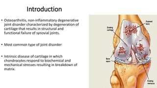
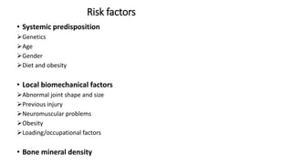
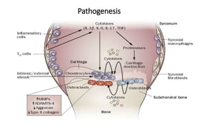
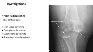
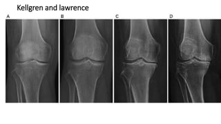
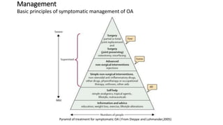
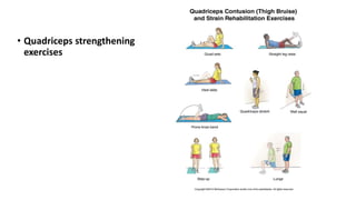
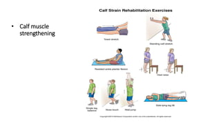
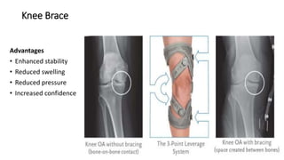
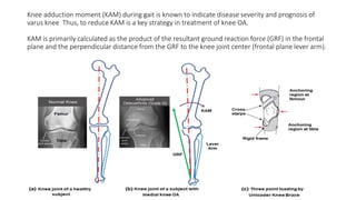
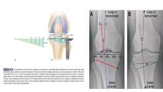
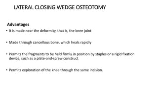
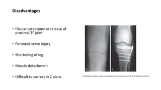
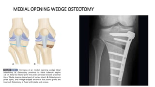
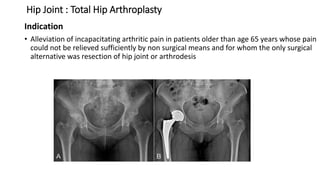
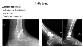
Osteoarthritis
Moderator: Assoc Prof Dr Suresh Uprety
Presenter: Dr Ajay shah
MS orthopedics
1st year Resident
Content
• Anatomy
• Introduction
• Aetiology and risk factors
• Pathogenesis
• Pathology
• Symptoms and Signs
• Investigations
• Management
ANATOMY
 Usually a condition of synovial joints
 Components
 Articular cartilage
 Synovial membrane
• Synoviocytes
• Produce lubricants, hyaluronic acid,
cytokines and growth factors
Fibrous capsule
• Lined by synovial membrane
• Continuous with the periosteum
• Provide stability throughout the ROM of joints
Synovial Fluid
• Viscous slippery fluid rich in albumin and hyaluronic acid
Intra articular discs of fibrocartilage
• Hip joint- labrum-deepens the articulation
• Knee joint-mensci- improve congruency
• Protect from both compressive and shear forces
Introduction
• Osteoarthritis, non-inflammatory degenerative
joint disorder characterized by degeneration of
cartilage that results in structural and
functional failure of synovial joints.
• Most common type of joint disorder
• Intrinsic disease of cartilage in which
chondrocytes respond to biochemical and
mechanical stresses resulting in breakdown of
matrix.
Joints involvement
Knee joint
• MC joint to involve
• MC sites: anteromedial compartment- Tibiofemoral joint
lateral facet- Patellofemoral joint
 Hip joint
• Superolateral aspect of joint
Hand
• Distal interphalangeal joints, PIP joints
 Feet -1ST MTP
Spine
• Facet joints of cervical and lumbosacral spine
Prevalence
• Internationally, OA is the most common articular
disease
• On the basis of the radiographic criteria for
osteoarthritis more than 50% of adults older
than 65 years are affected by the disease
• In individual older than 55 years, the prevalence
of OA is higher among women than among men
• Leading cause of chronic disability in those older
than 70 years, costing US greater than $100
billion annually
Types of OA
1 Primary/Idiopathic
• When there is no obvious predisposing factors
• Common form of OA
• Its mainly due to wear and tear changes occurring in old ages mainly in weight
bearing joints
2 Secondary OA
• When degenerative joint changes occur in response to recognizable local or
systemic factors
Causes of secondary OA
• Developmental
Developmental dysplasia of hip
Legg-calves perthe’s disease
Epiphyseal dysplasia
• Mechanical
Hypermobility syndromes
Leg length discrepancy
Mal-alignment
• Trauma (acute or chronic)
Sports injury
Iatrogenic
Occupational
Accidental
• Metabolic
Hemochromatosis
Gout
Pseudogout
Calcium crystal deposition
• Endocrine
Acromegaly
Hyperparathyroidism
Hypothyroidism
• Inflammatory
Any systemic rheumatic disease
Septic arthritis
• Miscellaneous
Hemophilia
Osteonecrosis
Paget’s disease
Neuropathic arthropathy
OA Variants
Atrophic destructive OA including
rapidly progressive hip OA and
Milwaukee shoulder
 Rapid period with joint damage progresses
to extensive bone loss
Whole head of femur and humerus may
disappear
• Hypertrophic OA
Particularly common at hip and knee joints
Characterized by massive osteophyte formation
It may be associated with
 calcium pyrophosphate dehydrate crystal deposition
 diffuse idiopathic skeletal hyperstosis (DISH)
• Erosive inflammatory OA of
interphalangeal joints
Involves terminal interphalangeal joints of hands
Associated with signs of inflammation and
development of joint erosions.
Radiograph shows erosion of joint
( sea gull appearance)
• Kashin Beck disease
Rare polyarticular form of OA
Joint pain, polyarticular swelling and deformity from childhood
Adults have short stature
Radiographs reveal distorted epiphyses and tubular long bones
Risk factors
• Systemic predisposition
Genetics
Age
Gender
Diet and obesity
• Local biomechanical factors
Abnormal joint shape and size
Previous injury
Neuromuscular problems
Obesity
Loading/occupational factors
• Bone mineral density
Pathogenesis
Pathological Changes
• Focal areas of loss of articular cartilage
• Bone growth at the joint margins
• Sclerosis of underlying bone
• Cyst formation in underlying bone
• Loss of bone
• Varying degrees of synovial inflammation
• Fibrosis and thickening of joint capsule
Gross Appearance
Microscopic Features
Symptoms and signs at different joint sites
Knee Joint
• MC site medial patellofemoral joint
• Pain: globally over knee and proximal tibia
• In isolated patellofemoral OA, pain is anteriorly over knee and is often worst
during ascending and descending stairs
• Symptoms of instability
• Swelling and stiffness
• Antalgic gait
• Weakness/Wasting of quadriceps muscles
• Varus deformity
• Restricted extension and flexion
• Audible crepitus
Hip joint
• Pain usually felt in groin, radiates down anterolateral aspect of thigh
• Pain is worse on exercise and walking distance is reduced
• Morning and rest stiffness
• Antalgic gait
• Weakness of hip abductors
• Restricted internal rotation with flexion
Hand joint
• MC affected joints are DIPs and thumb base
( radiocarpal and radiotrapezoid joints)
• Strongly associated with knee OA and genetic
predisposition, suggesting as feature of
generalized OA
• More common in women and often start
around the time of menopause( sometimes
called as menopausal OA)
• Bouchard's nodes are a classic sign of OA of the
hand.
• Named after the French pathologist Charles-
Joseph Bouchard, who studied arthritis patients
in the 19th century.
• Bouchard nodes are bony enlargements of the
proximal interphalangeal (PIP) joints.
• Heberden’s nodes are similar bony swellings
that develop at the distal interphalangeal (DIP)
joint. Bouchard's nodes are less common than
Heberden's nodes.
FOOT
• Hallux rigidus: 1st MTP joint OA
• Pain and tenderness over
dorsum of joint
• Limited dorsiflexon secondary to
large osteophytes
• Stiffness of 1st MTP joint and
compensatory hyperextension of
IP joint
• Grind test
Investigations
• Plain Radiographhs
Four cardinal signs
Joint space narrowing
Osteophytes formation
Subchondral bone cysts
Sclerosis of underlying bone
Kellgren and lawrence
• Grade 0 : Normal
• Grade 1: Minimal osteophyte, doubtful significance
• Grade 2: Definite osteophyte, no loss of joint space
• Grade 3: Some diminution of joint space
• Grade 4: Advanced joint space loss and sclerosis of bone
X-rays
Blood Tests
• Raised CRP ( in some cases)
• Normal ESR
• Rheumatoid factor and Antinuclear antibodies are negative
MRI
• Early cartilage and subchondral bone changes, although it is not routinely used due to
cost
Arthroscopy
• Reveal early fissuring and surface erosions
Synovial Fluid Analysis
• Viscous with low turbidity
• Can be used to exclude gout, CPPD or septic arthritis if diagnosis is in doubt
Outerbridge classification
Grade 0 : Normal cartilage
Grade I : Softening and swelling of cartilage
Grade II: Fragmentation and fissuring of the cartilage in an area less
than ½ inch in diameter
Grade III: Fragmentation and fissuring of the cartilage in an area more than ½ inch in
diameter
Grade IV: Exposure of underlying bone
Modified Outerbridge Classification
Grade 0 : Intact cartilage
Grade I : Chondral softening or blistering with intact surface
Grade II : Superficial ulceration,fibrillation, or fissuring less than 50% of depth of
cartilage
Grade III : Deep ulceration, fibrillation, fissuring, or chondral flap more than 50% of
cartilage without exposed bone
Grade IV : Full thickness wear with exposed subchondral bone
Secondary OA: Special Tests
• Ochronosis: Presence of homogenistic acid in urine which turns black in
exposure to air
• Wilson’s disease: Reduced serum ceruloplasmin, increased urinary excretion of
copper
• Hemochromatosis: Raised serum iron and ferritin
• Gout: Raised serum urate
• Hypothyroidism: Low T3 ,T4 and raised TSH
• Hyperparathyroidism: Raised PTH,calcium and low phosphate
• Tabes dorsalis: Positive VDRL
• DM: Abnormal glucose tolerance test
Management
Basic principles of symptomatic management of OA
Pyramid of treatment for symptomatic OA ( From Dieppe and Lohmander,2005)
Based on the UK National Institute for Health and Care Excellence
(NICE) 2014 guideline.
Step 1: Take a holistic approach and encourage self-management
• Assessing the impact of OA on the individual’s quality of life, function, mood ,
relationships and activities
• Self-management strategies include
Alterations of diet (lose weight)
Alteration in activities
Changing footwear
Step 2: Introduce the “core treatments” appropriate for most people
with OA
• These includes
Provision of information about the condition and its management
To increase their exercise level
Footwear advice
To do specific exercises to strengthen muscles around affected joints
Step 3: Introduce specific non-surgical interventions
These may be pharmacological or non-pharmacological
Non-pharmacological
• Supervised courses of physical therapy
• Use of aids and devices
Braces and splints
Walking aids such as sticks or crutches
Electrotherapy such as TENS
PHYSIOTHERAPY
• Hamstring stretching
exercises
• Quadriceps strengthening
exercises
• Calf muscle
strengthening
Knee Brace
Advantages
• Enhanced stability
• Reduced swelling
• Reduced pressure
• Increased confidence
Knee adduction moment (KAM) during gait is known to indicate disease severity and prognosis of
varus knee Thus, to reduce KAM is a key strategy in treatment of knee OA.
KAM is primarily calculated as the product of the resultant ground reaction force (GRF) in the frontal
plane and the perpendicular distance from the GRF to the knee joint center (frontal plane lever arm).
Pharmacological
 NSAIDs
• Non-selective NSAID
• COX-2 selective
 Tramadol, opioids
Intra-articular injection
• Local anaesthetic
• Corticosteroids
Supplements
• Glucosamine
• Chondroitin sulfate etc
Step 4: Consider surgical options
Arthroscopic procedures
• Joint debridement
• Removal of mechanical obstructions
• Joint lavage
• Drilling of sclerotic lesions
• Abrasion chondroplasty
• Autologous chondrocyte transplantation
Proximal Tibial Osteotomy
• Indications for proximal tibial osteotomy are
Pain and disability resulting from osteoarthritis that significantly interfere with
high-demand employment or recreation
Evidence on weight-bearing radiographs of degenerative arthritis that is confined
to one compartment with a corresponding varus or valgus deformity
Contraindications to a proximal tibial osteotomy
• Narrowing of lateral compartment cartilage space
• Lateral tibial subluxation of more than 1 cm
• Medial compartment tibial bone loss of more than 2 or 3 mm
• Flexion contracture of more than 15 degrees
• Knee flexion of less than 90 degrees
• More than 20 degrees of correction needed
• Inflammatory arthritis
• Significant peripheral vascular disease.
LATERAL CLOSING WEDGE OSTEOTOMY
Advantages
• It is made near the deformity, that is, the knee joint
• Made through cancellous bone, which heals rapidly
• Permits the fragments to be held firmly in position by staples or a rigid fixation
device, such as a plate-and-screw construct
• Permits exploration of the knee through the same incision.
Disadvantages
• Fibular osteotomy or release of
proximal TF joint
• Peroneal nerve injury
• Shortening of leg
• Muscle detachment
• Difficult to correct in 2 plans
MEDIAL OPENING WEDGE OSTEOTOMY
Proximal Fibular Osteotomy
Proximal fibular osteotomy is an alternative treatment to high tibial osteotomy.
It is a surgical procedure for medial compartment knee osteoarthritis
Total Knee Arthroplasty After PTO
• At 10 to 15 years after proximal tibial osteotomy, 40% of patients require
conversion to total knee arthroplasty
• Studies have shown that the outcome of total knee arthroplasty in patients with
previous high tibial osteotomies was not significantly different from outcomes
after primary total knee arthroplasty
• Although total knee arthroplasty after high tibial osteotomy is technically
demanding and is a longer operative procedure.
• Unicompartmental arthroplasty has poor results after high tibial osteotomy (28%
failure at 5 years).
• Indications of TKA
Relieve pain caused by severe
arthritis, with or without deformity
Failed high tibial osteotomy
Kellgren and Lawrence grade IV
Arthrodesis Of Knee
• Most frequent indication for knee arthrodesis is failed total knee arthrodesis,
secondary to infection
• Salvage procedure after failed TKA, expected to have some inferior results
compared with primary knee arthrodesis including
Lower fusion rates
Higher infection rates
Shortening of leg
Techniques
• Compression arthrodesis with external fixation
• Arthrodesis with intramedullary rod fixation
• Arthrodesis with plate fixation
Hip Joint : Total Hip Arthroplasty
Indication
• Alleviation of incapacitating arthritic pain in patients older than age 65 years whose pain
could not be relieved sufficiently by non surgical means and for whom the only surgical
alternative was resection of hip joint or arthrodesis
Ankle joint
Surgical Treatment
• Arthroscopic debridement
• Arthrodesis
• Total ankle replacement
Foot
Arthrodesis
• Subtalar arthrodesis
• Calcaneocuboid arthrodesis
• Talonavicular arthrodesis
• Double arthrodesis
• Triple arthrodesis
First MTP Joint OA
Surgical Treatment
• Cheilectomy
• Osteotomy -Dorsiflexion phalangeal
osteotomy
-Metatarsal osteotomy
• Arthrodesis
• Arthroplasty -Hemiarthroplasty
-Total joint arthroplasty
References
• Apley’s system of orthopedics and fractures, 9th edition
• Robbins and cotran pathologic basis of disease,9th edition
• Campbell’s operative orthopedics, 13th edition

Osteoarthritis slideshare

  • 1.
    Osteoarthritis Moderator: Assoc ProfDr Suresh Uprety Presenter: Dr Ajay shah MS orthopedics 1st year Resident
  • 2.
    Content • Anatomy • Introduction •Aetiology and risk factors • Pathogenesis • Pathology • Symptoms and Signs • Investigations • Management
  • 3.
    ANATOMY  Usually acondition of synovial joints  Components  Articular cartilage  Synovial membrane • Synoviocytes • Produce lubricants, hyaluronic acid, cytokines and growth factors
  • 4.
    Fibrous capsule • Linedby synovial membrane • Continuous with the periosteum • Provide stability throughout the ROM of joints Synovial Fluid • Viscous slippery fluid rich in albumin and hyaluronic acid Intra articular discs of fibrocartilage • Hip joint- labrum-deepens the articulation • Knee joint-mensci- improve congruency • Protect from both compressive and shear forces
  • 5.
    Introduction • Osteoarthritis, non-inflammatorydegenerative joint disorder characterized by degeneration of cartilage that results in structural and functional failure of synovial joints. • Most common type of joint disorder • Intrinsic disease of cartilage in which chondrocytes respond to biochemical and mechanical stresses resulting in breakdown of matrix.
  • 6.
    Joints involvement Knee joint •MC joint to involve • MC sites: anteromedial compartment- Tibiofemoral joint lateral facet- Patellofemoral joint  Hip joint • Superolateral aspect of joint Hand • Distal interphalangeal joints, PIP joints  Feet -1ST MTP Spine • Facet joints of cervical and lumbosacral spine
  • 7.
    Prevalence • Internationally, OAis the most common articular disease • On the basis of the radiographic criteria for osteoarthritis more than 50% of adults older than 65 years are affected by the disease • In individual older than 55 years, the prevalence of OA is higher among women than among men • Leading cause of chronic disability in those older than 70 years, costing US greater than $100 billion annually
  • 8.
    Types of OA 1Primary/Idiopathic • When there is no obvious predisposing factors • Common form of OA • Its mainly due to wear and tear changes occurring in old ages mainly in weight bearing joints 2 Secondary OA • When degenerative joint changes occur in response to recognizable local or systemic factors
  • 9.
    Causes of secondaryOA • Developmental Developmental dysplasia of hip Legg-calves perthe’s disease Epiphyseal dysplasia • Mechanical Hypermobility syndromes Leg length discrepancy Mal-alignment • Trauma (acute or chronic) Sports injury Iatrogenic Occupational Accidental • Metabolic Hemochromatosis Gout Pseudogout Calcium crystal deposition
  • 10.
    • Endocrine Acromegaly Hyperparathyroidism Hypothyroidism • Inflammatory Anysystemic rheumatic disease Septic arthritis • Miscellaneous Hemophilia Osteonecrosis Paget’s disease Neuropathic arthropathy
  • 11.
    OA Variants Atrophic destructiveOA including rapidly progressive hip OA and Milwaukee shoulder  Rapid period with joint damage progresses to extensive bone loss Whole head of femur and humerus may disappear
  • 12.
    • Hypertrophic OA Particularlycommon at hip and knee joints Characterized by massive osteophyte formation It may be associated with  calcium pyrophosphate dehydrate crystal deposition  diffuse idiopathic skeletal hyperstosis (DISH)
  • 13.
    • Erosive inflammatoryOA of interphalangeal joints Involves terminal interphalangeal joints of hands Associated with signs of inflammation and development of joint erosions. Radiograph shows erosion of joint ( sea gull appearance)
  • 14.
    • Kashin Beckdisease Rare polyarticular form of OA Joint pain, polyarticular swelling and deformity from childhood Adults have short stature Radiographs reveal distorted epiphyses and tubular long bones
  • 15.
    Risk factors • Systemicpredisposition Genetics Age Gender Diet and obesity • Local biomechanical factors Abnormal joint shape and size Previous injury Neuromuscular problems Obesity Loading/occupational factors • Bone mineral density
  • 16.
  • 18.
    Pathological Changes • Focalareas of loss of articular cartilage • Bone growth at the joint margins • Sclerosis of underlying bone • Cyst formation in underlying bone • Loss of bone • Varying degrees of synovial inflammation • Fibrosis and thickening of joint capsule
  • 19.
  • 20.
  • 21.
    Symptoms and signsat different joint sites Knee Joint • MC site medial patellofemoral joint • Pain: globally over knee and proximal tibia • In isolated patellofemoral OA, pain is anteriorly over knee and is often worst during ascending and descending stairs • Symptoms of instability • Swelling and stiffness
  • 22.
    • Antalgic gait •Weakness/Wasting of quadriceps muscles • Varus deformity • Restricted extension and flexion • Audible crepitus
  • 23.
    Hip joint • Painusually felt in groin, radiates down anterolateral aspect of thigh • Pain is worse on exercise and walking distance is reduced • Morning and rest stiffness • Antalgic gait • Weakness of hip abductors • Restricted internal rotation with flexion
  • 24.
    Hand joint • MCaffected joints are DIPs and thumb base ( radiocarpal and radiotrapezoid joints) • Strongly associated with knee OA and genetic predisposition, suggesting as feature of generalized OA • More common in women and often start around the time of menopause( sometimes called as menopausal OA)
  • 25.
    • Bouchard's nodesare a classic sign of OA of the hand. • Named after the French pathologist Charles- Joseph Bouchard, who studied arthritis patients in the 19th century. • Bouchard nodes are bony enlargements of the proximal interphalangeal (PIP) joints. • Heberden’s nodes are similar bony swellings that develop at the distal interphalangeal (DIP) joint. Bouchard's nodes are less common than Heberden's nodes.
  • 26.
    FOOT • Hallux rigidus:1st MTP joint OA • Pain and tenderness over dorsum of joint • Limited dorsiflexon secondary to large osteophytes • Stiffness of 1st MTP joint and compensatory hyperextension of IP joint • Grind test
  • 27.
    Investigations • Plain Radiographhs Fourcardinal signs Joint space narrowing Osteophytes formation Subchondral bone cysts Sclerosis of underlying bone
  • 28.
  • 29.
    • Grade 0: Normal • Grade 1: Minimal osteophyte, doubtful significance • Grade 2: Definite osteophyte, no loss of joint space • Grade 3: Some diminution of joint space • Grade 4: Advanced joint space loss and sclerosis of bone
  • 30.
  • 33.
    Blood Tests • RaisedCRP ( in some cases) • Normal ESR • Rheumatoid factor and Antinuclear antibodies are negative MRI • Early cartilage and subchondral bone changes, although it is not routinely used due to cost Arthroscopy • Reveal early fissuring and surface erosions Synovial Fluid Analysis • Viscous with low turbidity • Can be used to exclude gout, CPPD or septic arthritis if diagnosis is in doubt
  • 34.
    Outerbridge classification Grade 0: Normal cartilage Grade I : Softening and swelling of cartilage Grade II: Fragmentation and fissuring of the cartilage in an area less than ½ inch in diameter Grade III: Fragmentation and fissuring of the cartilage in an area more than ½ inch in diameter Grade IV: Exposure of underlying bone
  • 35.
    Modified Outerbridge Classification Grade0 : Intact cartilage Grade I : Chondral softening or blistering with intact surface Grade II : Superficial ulceration,fibrillation, or fissuring less than 50% of depth of cartilage Grade III : Deep ulceration, fibrillation, fissuring, or chondral flap more than 50% of cartilage without exposed bone Grade IV : Full thickness wear with exposed subchondral bone
  • 36.
    Secondary OA: SpecialTests • Ochronosis: Presence of homogenistic acid in urine which turns black in exposure to air • Wilson’s disease: Reduced serum ceruloplasmin, increased urinary excretion of copper • Hemochromatosis: Raised serum iron and ferritin • Gout: Raised serum urate • Hypothyroidism: Low T3 ,T4 and raised TSH • Hyperparathyroidism: Raised PTH,calcium and low phosphate • Tabes dorsalis: Positive VDRL • DM: Abnormal glucose tolerance test
  • 37.
    Management Basic principles ofsymptomatic management of OA Pyramid of treatment for symptomatic OA ( From Dieppe and Lohmander,2005)
  • 38.
    Based on theUK National Institute for Health and Care Excellence (NICE) 2014 guideline. Step 1: Take a holistic approach and encourage self-management • Assessing the impact of OA on the individual’s quality of life, function, mood , relationships and activities • Self-management strategies include Alterations of diet (lose weight) Alteration in activities Changing footwear
  • 39.
    Step 2: Introducethe “core treatments” appropriate for most people with OA • These includes Provision of information about the condition and its management To increase their exercise level Footwear advice To do specific exercises to strengthen muscles around affected joints
  • 40.
    Step 3: Introducespecific non-surgical interventions These may be pharmacological or non-pharmacological Non-pharmacological • Supervised courses of physical therapy • Use of aids and devices Braces and splints Walking aids such as sticks or crutches Electrotherapy such as TENS
  • 41.
  • 42.
  • 43.
  • 44.
    Knee Brace Advantages • Enhancedstability • Reduced swelling • Reduced pressure • Increased confidence
  • 45.
    Knee adduction moment(KAM) during gait is known to indicate disease severity and prognosis of varus knee Thus, to reduce KAM is a key strategy in treatment of knee OA. KAM is primarily calculated as the product of the resultant ground reaction force (GRF) in the frontal plane and the perpendicular distance from the GRF to the knee joint center (frontal plane lever arm).
  • 46.
    Pharmacological  NSAIDs • Non-selectiveNSAID • COX-2 selective  Tramadol, opioids Intra-articular injection • Local anaesthetic • Corticosteroids Supplements • Glucosamine • Chondroitin sulfate etc
  • 47.
    Step 4: Considersurgical options Arthroscopic procedures • Joint debridement • Removal of mechanical obstructions • Joint lavage • Drilling of sclerotic lesions • Abrasion chondroplasty • Autologous chondrocyte transplantation
  • 48.
    Proximal Tibial Osteotomy •Indications for proximal tibial osteotomy are Pain and disability resulting from osteoarthritis that significantly interfere with high-demand employment or recreation Evidence on weight-bearing radiographs of degenerative arthritis that is confined to one compartment with a corresponding varus or valgus deformity
  • 49.
    Contraindications to aproximal tibial osteotomy • Narrowing of lateral compartment cartilage space • Lateral tibial subluxation of more than 1 cm • Medial compartment tibial bone loss of more than 2 or 3 mm • Flexion contracture of more than 15 degrees • Knee flexion of less than 90 degrees • More than 20 degrees of correction needed • Inflammatory arthritis • Significant peripheral vascular disease.
  • 51.
    LATERAL CLOSING WEDGEOSTEOTOMY Advantages • It is made near the deformity, that is, the knee joint • Made through cancellous bone, which heals rapidly • Permits the fragments to be held firmly in position by staples or a rigid fixation device, such as a plate-and-screw construct • Permits exploration of the knee through the same incision.
  • 52.
    Disadvantages • Fibular osteotomyor release of proximal TF joint • Peroneal nerve injury • Shortening of leg • Muscle detachment • Difficult to correct in 2 plans
  • 53.
  • 55.
    Proximal Fibular Osteotomy Proximalfibular osteotomy is an alternative treatment to high tibial osteotomy. It is a surgical procedure for medial compartment knee osteoarthritis
  • 57.
    Total Knee ArthroplastyAfter PTO • At 10 to 15 years after proximal tibial osteotomy, 40% of patients require conversion to total knee arthroplasty • Studies have shown that the outcome of total knee arthroplasty in patients with previous high tibial osteotomies was not significantly different from outcomes after primary total knee arthroplasty • Although total knee arthroplasty after high tibial osteotomy is technically demanding and is a longer operative procedure. • Unicompartmental arthroplasty has poor results after high tibial osteotomy (28% failure at 5 years).
  • 58.
    • Indications ofTKA Relieve pain caused by severe arthritis, with or without deformity Failed high tibial osteotomy Kellgren and Lawrence grade IV
  • 60.
    Arthrodesis Of Knee •Most frequent indication for knee arthrodesis is failed total knee arthrodesis, secondary to infection • Salvage procedure after failed TKA, expected to have some inferior results compared with primary knee arthrodesis including Lower fusion rates Higher infection rates Shortening of leg
  • 61.
    Techniques • Compression arthrodesiswith external fixation • Arthrodesis with intramedullary rod fixation • Arthrodesis with plate fixation
  • 65.
    Hip Joint :Total Hip Arthroplasty Indication • Alleviation of incapacitating arthritic pain in patients older than age 65 years whose pain could not be relieved sufficiently by non surgical means and for whom the only surgical alternative was resection of hip joint or arthrodesis
  • 66.
    Ankle joint Surgical Treatment •Arthroscopic debridement • Arthrodesis • Total ankle replacement
  • 67.
    Foot Arthrodesis • Subtalar arthrodesis •Calcaneocuboid arthrodesis • Talonavicular arthrodesis • Double arthrodesis • Triple arthrodesis
  • 68.
    First MTP JointOA Surgical Treatment • Cheilectomy • Osteotomy -Dorsiflexion phalangeal osteotomy -Metatarsal osteotomy • Arthrodesis • Arthroplasty -Hemiarthroplasty -Total joint arthroplasty
  • 69.
    References • Apley’s systemof orthopedics and fractures, 9th edition • Robbins and cotran pathologic basis of disease,9th edition • Campbell’s operative orthopedics, 13th edition