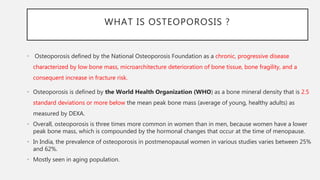
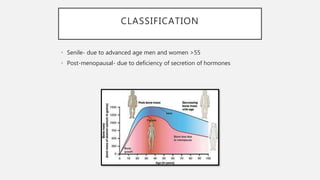
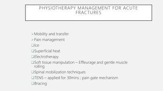
OSTEOPOROSIS
Dr. Ramandeep Kaur Saini (PT)
Assistant Professor - Community Physiotherapy
DPO’s NETT College of Physiotherapy
OBJECTIVES
• At the end of the session you should know:
• What is osteoporosis?
• Classification and risk factors
• Clinical features
• Diagnosis and investigation
• Physiotherapy management
WHAT IS OSTEOPOROSIS ?
• Osteoporosis defined by the National Osteoporosis Foundation as a chronic, progressive disease
characterized by low bone mass, microarchitecture deterioration of bone tissue, bone fragility, and a
consequent increase in fracture risk.
• Osteoporosis is defined by the World Health Organization (WHO) as a bone mineral density that is 2.5
standard deviations or more below the mean peak bone mass (average of young, healthy adults) as
measured by DEXA.
• Overall, osteoporosis is three times more common in women than in men, because women have a lower
peak bone mass, which is compounded by the hormonal changes that occur at the time of menopause.
• In India, the prevalence of osteoporosis in postmenopausal women in various studies varies between 25%
and 62%.
• Mostly seen in aging population.
CLASSIFICATION
• Senile- due to advanced age men and women >55
• Post-menopausal- due to deficiency of secretion of hormones
PATHOGENESIS
• Skeletal fragility can result from:
1. Failure to produce a skeleton of optimal mass and strength
during growth
2. Excessive bone resorption resulting in decreased bone mass and
micro-architectural deterioration of the skeleton
3. An inadequate bone formation response to increased resorption
during bone remodeling.
• Estrogen deficiency was initially proposed to be the sole
mechanism by which bone mass decreases in postmenopausal
women and elderly men.
• Accelerated boss loss in post-menopause is associated with
increased osteoclast activity: bone resorption occurs at a greater
rate than bone formation and balance between both is disturbed
, the net result being bone loss
RISK FACTORS
SIGN AND SYMPTOMS
• Signs and symptoms
Loss of height – A loss of 4cm or more over ten years ; also
with decreased bone mineral density is a clinical marker
Pain -- Rarely painful prior to fracture or compression or
Rib pain seen in established osteoporosis may be due to
costal impingement on pelvic bones
 Shortness of breath and fatigue –Due to posture and shape of the thorax
 Hiatus hernia – Decrease in abdominal volume and associated indigestion, heartburn or
regurgitation
 Protuberant stomach
 Stress incontinence – Increased abdominal pressure –> also due to straining as a result of
constipation provoked by medications especially calcium based
 Transparent skin – Skin is thin in those over age of 60 which is suggestive of
possible or existing osteopenia
 Dowager’s hump – Severe kyphotic deformity .
DIAGNOSIS
• BMD assessment by dual-energy X-ray absorptiometry (DEXA) scan: gold
standard
• skeletal sites such as lumbar spine, hip, and wrist.
• However, by convention, the score then is converted to a T-score and a Z-score.
• T-score: Number of standard deviations above or below BMD of age-matched
controls
• Z-score: Number of standard deviations above or below BMD of young normal
mean
• Radiography :
• Genant grading has been used to categorize the severity of vertebral fractures
(mild, moderate, and severe) based on the reduction in height or reduction in
projected area of the vertebrae
• Quantitative computed tomography (QCT)
• MRI
• Fracture risk assessment tool (FRAX)
• FRAX is the most widely used fracture risk assessment tool that takes into
account nine factors:
1. Age
2. BMD
3. Body mass index (BMI)
4. Prior fragility fracture,
5. Use of oral
6. Glucocorticoids
7. Parental history of HF
8. Current smoking
9. Alcohol intake, and rheumatoid arthritis
WHOM TO SCREEN FOR OSTEOPOROSIS?
• The following patients need to be screened for osteoporosis by DXA based on the
recommendation by the Indian Menopause Society (IMS):
• All postmenopausal women more than 5 years of menopause
• Postmenopausal women <5 years of menopause with risk factors (low BMI, glucocorticoid use,
alcohol, smoking, rheumatoid arthritis, prior history of fragility fracture, and parental history of
HF)
• Women in menopause transition with secondary causes
• Radiological evidence of osteopenia and presence of vertebral compression fracture
• Women with fragility fractures
• Ideally before initiating pharmacotherapy for osteoporosis
MANAGEMENT
• Primary prevention:
- Education: include
- risk factors
- pain management self help techniques
- Medical treatment
- The recommended daily allowance of calcium is 1000–1500 mg
- Vit. D: Cholecalciferol 60,000 U once in every 1–2 months is recommended in all
postmenopausal women with or without osteoporosis
- Exercise
- Nutrition
- Prevention of falls
- Smoking cessation
SECONDARY PREVENTION
• Exercise prescription: ACSM
• FITT RECOMMENDATIONS FOR INDIVIDUALS WITH OSTEOPOROSIS
• Weight bearing exercise
• Resistance training
• Aerobic exercise
OSTEOPOROTIC-RELATED FRACTURES
Pathogenesis of osteoporotic-related fractures. The risk
for fracture is dependent on both skeletal and non-
skeletal risk factors, but fractures result from a structural
failure of bone, wherein the loads applied to bone (most
often from a fall) exceed its strength.
PHYSIOTHERAPY MANAGEMENT FOR ACUTE
FRACTURES
Mobility and transfer
Pain management
Ice
Superficial heat
Electrotherapy
Soft tissue manipulation – Effleurage and gentle muscle
rolling
Spinal mobilization techniques
TENS – applied for 30mins ; pain gate mechanism
Bracing
• Exercises :-
 Aims - (1) Strengthening the supportive axial musculature like spinal extensors
• (2) Proprioceptive training -> improve posture and ambulation and decrease the likelihood of
future falls.
 Exercises should focus on strengthening back extension and may include weighted or unweighted
prone position extension exercises, isometric contraction of the paraspinal muscles, and careful
loading of the upper extremities
REHABILITATION IN OSTEOPOROTIC
VERTEBRAL FRACTURE
• Goals :
Pain control
Prevention of complications
Use of orthopaedic corsets
Specific physiotherapeutic training
Bed mobility
Neuromuscular stabilization exercises for thoraco lumbar spine
Active / passive UL movements along with cervical spine
TLSO for 8-12 wks
Relaxing exercises
Breathing exercises
Rehabilitation phase can start during the corset wearing and has to proceed after the
orthopaedic corset is removed, generally in 8-12 weeks from the acute event back-
extensor muscle strengthening exercises, postural retraining exercises, ergonomic and
balance increasing exercises
• Physical therapy management for hip fractures
 Goals –
Improving ROM of affected hip joint
Early mobilization
Pain free , independent ambulation
 Post op management
Positioning
Bed mobility and transfer
Strengthening of unaffected LL and both UL
AAROM for hip , ATM
Static quads , glutes
Ambulation -> Toe touch to PWB and then FWB over a period of 6 weeks to 4 months
Hydrotherapy
• Physical therapy management for DER fractures
 ROM of shoulder
Pain-free wrist and finger movements
Improve grip strength , hand function
Fall prevention
Lifestyle management
• Physiotherapy in long tem for osteoporosis:
 Goals
Maintain and slow the loss of or increase bone density
Reduce pain
Prevent spinal deformity and vertebral fractures
Prevention of falls
Maintenance of mobility and independence
• Aquatic therapy for osteoporosis
Benefits ->
Decreased stress on weight-bearing joints due to the buoyancy of the water
Increased mobility due to diminished gravitational pull
The ability to use varying levels of resistance for strengthening
Increased sensory stimulation in brain
REFERENCES
• Jeremiah MP, Unwin BK, Greenawald MH, Casiano VE. Diagnosis and management of
osteoporosis. American family physician. 2015 Aug 15;92(4):261-8.
• Rajan R, Paul J, Kapoor N, Cherian KE, Paul TV. Postmenopausal osteoporosis–An
Indian perspective. Current Medical Issues. 2020 Apr 1;18(2):98.
• Shaki O, Rai SK, Kashid M, Chakrabarty BK. Prevalence of osteoporosis in peri- and
post-menopausal women in slum area of Mumbai, India. J Mid-life Health 2018;9:117-
22.
• American College of Sports Medicine. ACSM's guidelines for exercise testing and
prescription. Lippincott Williams & Wilkins; 2013 Mar 4.
• Daly RM, Dalla Via J, Duckham RL, Fraser SF, Helge EW. Exercise for the prevention of
osteoporosis in postmenopausal women: an evidence-based guide to the optimal
prescription. Brazilian journal of physical therapy. 2019 Mar 1;23(2):170-80.
Osteoporosis

Osteoporosis

  • 1.
    OSTEOPOROSIS Dr. Ramandeep KaurSaini (PT) Assistant Professor - Community Physiotherapy DPO’s NETT College of Physiotherapy
  • 2.
    OBJECTIVES • At theend of the session you should know: • What is osteoporosis? • Classification and risk factors • Clinical features • Diagnosis and investigation • Physiotherapy management
  • 3.
    WHAT IS OSTEOPOROSIS? • Osteoporosis defined by the National Osteoporosis Foundation as a chronic, progressive disease characterized by low bone mass, microarchitecture deterioration of bone tissue, bone fragility, and a consequent increase in fracture risk. • Osteoporosis is defined by the World Health Organization (WHO) as a bone mineral density that is 2.5 standard deviations or more below the mean peak bone mass (average of young, healthy adults) as measured by DEXA. • Overall, osteoporosis is three times more common in women than in men, because women have a lower peak bone mass, which is compounded by the hormonal changes that occur at the time of menopause. • In India, the prevalence of osteoporosis in postmenopausal women in various studies varies between 25% and 62%. • Mostly seen in aging population.
  • 4.
    CLASSIFICATION • Senile- dueto advanced age men and women >55 • Post-menopausal- due to deficiency of secretion of hormones
  • 5.
    PATHOGENESIS • Skeletal fragilitycan result from: 1. Failure to produce a skeleton of optimal mass and strength during growth 2. Excessive bone resorption resulting in decreased bone mass and micro-architectural deterioration of the skeleton 3. An inadequate bone formation response to increased resorption during bone remodeling. • Estrogen deficiency was initially proposed to be the sole mechanism by which bone mass decreases in postmenopausal women and elderly men. • Accelerated boss loss in post-menopause is associated with increased osteoclast activity: bone resorption occurs at a greater rate than bone formation and balance between both is disturbed , the net result being bone loss
  • 6.
  • 7.
    SIGN AND SYMPTOMS •Signs and symptoms Loss of height – A loss of 4cm or more over ten years ; also with decreased bone mineral density is a clinical marker Pain -- Rarely painful prior to fracture or compression or Rib pain seen in established osteoporosis may be due to costal impingement on pelvic bones
  • 8.
     Shortness ofbreath and fatigue –Due to posture and shape of the thorax  Hiatus hernia – Decrease in abdominal volume and associated indigestion, heartburn or regurgitation  Protuberant stomach  Stress incontinence – Increased abdominal pressure –> also due to straining as a result of constipation provoked by medications especially calcium based  Transparent skin – Skin is thin in those over age of 60 which is suggestive of possible or existing osteopenia  Dowager’s hump – Severe kyphotic deformity .
  • 9.
    DIAGNOSIS • BMD assessmentby dual-energy X-ray absorptiometry (DEXA) scan: gold standard • skeletal sites such as lumbar spine, hip, and wrist. • However, by convention, the score then is converted to a T-score and a Z-score. • T-score: Number of standard deviations above or below BMD of age-matched controls • Z-score: Number of standard deviations above or below BMD of young normal mean
  • 10.
    • Radiography : •Genant grading has been used to categorize the severity of vertebral fractures (mild, moderate, and severe) based on the reduction in height or reduction in projected area of the vertebrae • Quantitative computed tomography (QCT) • MRI
  • 11.
    • Fracture riskassessment tool (FRAX) • FRAX is the most widely used fracture risk assessment tool that takes into account nine factors: 1. Age 2. BMD 3. Body mass index (BMI) 4. Prior fragility fracture, 5. Use of oral 6. Glucocorticoids 7. Parental history of HF 8. Current smoking 9. Alcohol intake, and rheumatoid arthritis
  • 12.
    WHOM TO SCREENFOR OSTEOPOROSIS? • The following patients need to be screened for osteoporosis by DXA based on the recommendation by the Indian Menopause Society (IMS): • All postmenopausal women more than 5 years of menopause • Postmenopausal women <5 years of menopause with risk factors (low BMI, glucocorticoid use, alcohol, smoking, rheumatoid arthritis, prior history of fragility fracture, and parental history of HF) • Women in menopause transition with secondary causes • Radiological evidence of osteopenia and presence of vertebral compression fracture • Women with fragility fractures • Ideally before initiating pharmacotherapy for osteoporosis
  • 13.
    MANAGEMENT • Primary prevention: -Education: include - risk factors - pain management self help techniques - Medical treatment - The recommended daily allowance of calcium is 1000–1500 mg - Vit. D: Cholecalciferol 60,000 U once in every 1–2 months is recommended in all postmenopausal women with or without osteoporosis - Exercise - Nutrition - Prevention of falls - Smoking cessation
  • 16.
    SECONDARY PREVENTION • Exerciseprescription: ACSM • FITT RECOMMENDATIONS FOR INDIVIDUALS WITH OSTEOPOROSIS
  • 17.
    • Weight bearingexercise • Resistance training • Aerobic exercise
  • 18.
    OSTEOPOROTIC-RELATED FRACTURES Pathogenesis ofosteoporotic-related fractures. The risk for fracture is dependent on both skeletal and non- skeletal risk factors, but fractures result from a structural failure of bone, wherein the loads applied to bone (most often from a fall) exceed its strength.
  • 20.
    PHYSIOTHERAPY MANAGEMENT FORACUTE FRACTURES Mobility and transfer Pain management Ice Superficial heat Electrotherapy Soft tissue manipulation – Effleurage and gentle muscle rolling Spinal mobilization techniques TENS – applied for 30mins ; pain gate mechanism Bracing
  • 21.
    • Exercises :- Aims - (1) Strengthening the supportive axial musculature like spinal extensors • (2) Proprioceptive training -> improve posture and ambulation and decrease the likelihood of future falls.  Exercises should focus on strengthening back extension and may include weighted or unweighted prone position extension exercises, isometric contraction of the paraspinal muscles, and careful loading of the upper extremities
  • 22.
    REHABILITATION IN OSTEOPOROTIC VERTEBRALFRACTURE • Goals : Pain control Prevention of complications Use of orthopaedic corsets Specific physiotherapeutic training
  • 23.
    Bed mobility Neuromuscular stabilizationexercises for thoraco lumbar spine Active / passive UL movements along with cervical spine TLSO for 8-12 wks Relaxing exercises Breathing exercises Rehabilitation phase can start during the corset wearing and has to proceed after the orthopaedic corset is removed, generally in 8-12 weeks from the acute event back- extensor muscle strengthening exercises, postural retraining exercises, ergonomic and balance increasing exercises
  • 24.
    • Physical therapymanagement for hip fractures  Goals – Improving ROM of affected hip joint Early mobilization Pain free , independent ambulation  Post op management Positioning Bed mobility and transfer Strengthening of unaffected LL and both UL AAROM for hip , ATM Static quads , glutes Ambulation -> Toe touch to PWB and then FWB over a period of 6 weeks to 4 months Hydrotherapy
  • 25.
    • Physical therapymanagement for DER fractures  ROM of shoulder Pain-free wrist and finger movements Improve grip strength , hand function Fall prevention Lifestyle management
  • 26.
    • Physiotherapy inlong tem for osteoporosis:  Goals Maintain and slow the loss of or increase bone density Reduce pain Prevent spinal deformity and vertebral fractures Prevention of falls Maintenance of mobility and independence
  • 27.
    • Aquatic therapyfor osteoporosis Benefits -> Decreased stress on weight-bearing joints due to the buoyancy of the water Increased mobility due to diminished gravitational pull The ability to use varying levels of resistance for strengthening Increased sensory stimulation in brain
  • 28.
    REFERENCES • Jeremiah MP,Unwin BK, Greenawald MH, Casiano VE. Diagnosis and management of osteoporosis. American family physician. 2015 Aug 15;92(4):261-8. • Rajan R, Paul J, Kapoor N, Cherian KE, Paul TV. Postmenopausal osteoporosis–An Indian perspective. Current Medical Issues. 2020 Apr 1;18(2):98. • Shaki O, Rai SK, Kashid M, Chakrabarty BK. Prevalence of osteoporosis in peri- and post-menopausal women in slum area of Mumbai, India. J Mid-life Health 2018;9:117- 22. • American College of Sports Medicine. ACSM's guidelines for exercise testing and prescription. Lippincott Williams & Wilkins; 2013 Mar 4. • Daly RM, Dalla Via J, Duckham RL, Fraser SF, Helge EW. Exercise for the prevention of osteoporosis in postmenopausal women: an evidence-based guide to the optimal prescription. Brazilian journal of physical therapy. 2019 Mar 1;23(2):170-80.

Editor's Notes

  • #28 Stand with your feet hip width apart and rest your arms on the surface of the water. Then gently turn your body to the right, swinging your left arm in front of you and your right arm behind you. Keep your elbows straight and in the water throughout. Repeat in the other direction. Stand holding on to the side of the pool. Bend one hip and knee up in front of you, standing tall and keeping your back straight. Then stretch your leg out behind you, keeping your knee straight. Repeat with the other leg. Try taking long steps in the water (forwards and then sideways).