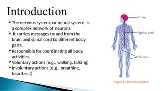
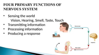
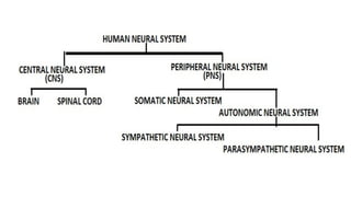
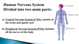
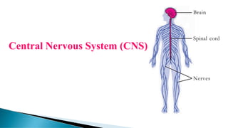
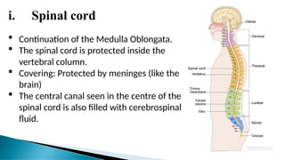
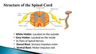
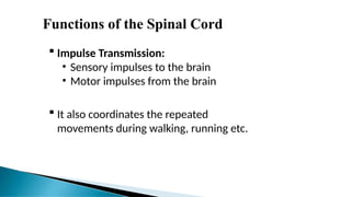
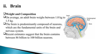
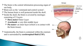
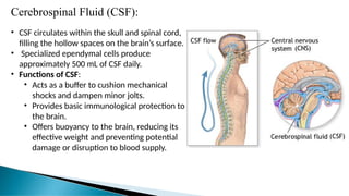
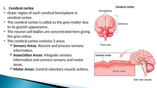
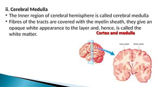
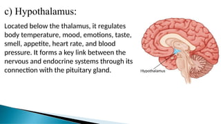
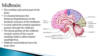
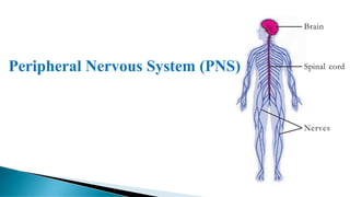
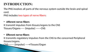
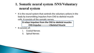
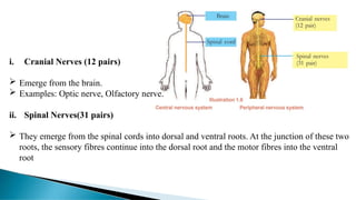
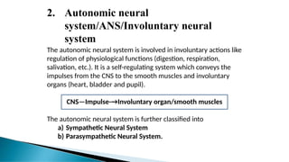
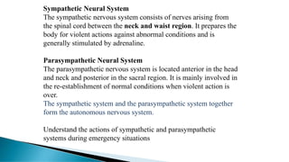
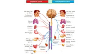
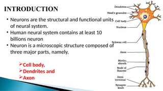
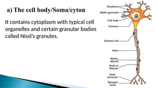
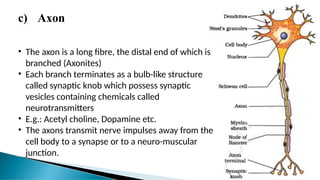
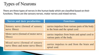
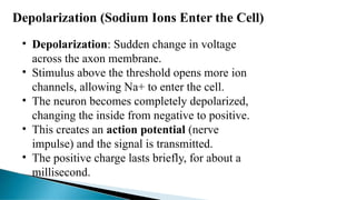
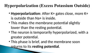
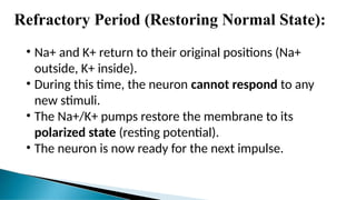
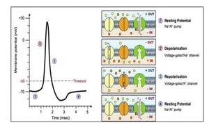
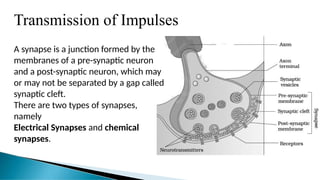
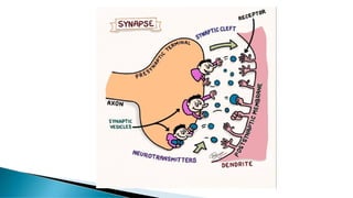
HUMAN NERVOUS
SYSTEM
Minnu Sunny
M.Sc. Dietetics and food service management
I PG Student
Introduction
The nervous system, or neural system, is
a complex network of neurons.
 It carries messages to and from the
brain and spinal cord to different body
parts.
Responsible for coordinating all body
activities.
Voluntary actions (e.g., walking, talking)
Involuntary actions (e.g., breathing,
heartbeat)
FOUR PRIMARY FUNCTIONS OF
NERVOUS SYSTEM
• Sensing the world
Vision, Hearing, Smell, Taste, Touch
• Transmitting information
• Processing information
• Producing a response
Human Nervous System
Divided into two main parts:
a) Central Nervous System (CNS): consists of
the brain and spinal cord
b) Peripheral Nervous System (PNS): includes
all the nerves of the body
Central Nervous System (CNS)
INTODUCTION:
• The Central Nervous System (CNS) is the
central hub of the body.
• It initiates information, commands,
coordinates, and influences all body
activities.
• Often called the central processing unit of
the body.
• Comprises two parts:
i. Brain
ii. Spinal cord
i. Spinal cord
 Continuation of the Medulla Oblongata.
 The spinal cord is protected inside the
vertebral column.
 Covering: Protected by meninges (like the
brain)
 The central canal seen in the centre of the
spinal cord is also filled with cerebrospinal
fluid.
Structure of the Spinal Cord
• White Matter: Located on the outside
• Grey Matter: Located on the inside
• 31 Pairs of Spinal Nerves
• Dorsal Root: Sensory impulses enter
• Ventral Root: Motor impulses exit
Functions of the Spinal Cord
 Impulse Transmission:
• Sensory impulses to the brain
• Motor impulses from the brain
 It also coordinates the repeated
movements during walking, running etc.
ii. Brain
Weight and Composition
On average, an adult brain weighs between 1.0 kg to
1.5 kg.
The brain is predominantly composed of neurons,
which are the fundamental units of the brain and
nervous system.
Recent estimates suggest that the brain contains
between 86 billion to 100 billion neurons.
The brain is the central information processing organ of
our body,
Brain acts as the ‘command and control system’.
The human brain is well protected inside the skull.
Inside the skull, the brain is covered by meninges
consisting of 3 Layers
 Dura mater-Outer Layer
 Arachnoid- a very thin middle layer
 Pia mater- an inner layer (which is in contact with
the brain tissue).
Anatomically, the brain is contained within the cranium
and is surrounded by cerebrospinal fluid (CSF).
Cerebrospinal Fluid (CSF):
• CSF circulates within the skull and spinal cord,
filling the hollow spaces on the brain’s surface.
• Specialized ependymal cells produce
approximately 500 mL of CSF daily.
• Functions of CSF:
• Acts as a buffer to cushion mechanical
shocks and dampen minor jolts.
• Provides basic immunological protection to
the brain.
• Offers buoyancy to the brain, reducing its
effective weight and preventing potential
damage or disruption to blood supply.
The brain can be divided into three major parts:
a) Forebrain
b)Midbrain
c) hindbrain
a) Forebrain:
• The largest and most anterior part of the brain.
Includes:
i. Cerebrum
ii. Thalamus
iii. Hypothalamus
a) Cerebrum:
• Cerebrum forms the human brain.
• A deep cleft divides the cerebrum longitudinally into two
halves, which are termed as the left and right cerebral
hemispheres. The hemispheres are connected by a tract of
nerve fibres called corpus callosum.
• Cerebrum is the seat of mind and intelligence
• Each cerebral hemisphere has two Parts
i. Cerebral cortex-Outer region
ii. Cerebral medulla-Inner region
i. Cerebral cortex
• Outer region of each cerebral hemisphere is
cerebral cortex.
• The cerebral cortex is called as the grey matter due
to its greyish appearance.
• The neuron cell bodies are concentrated here giving
the grey colour.
• The cerebral cortex contains 3 areas
Sensory Areas: Receive and process sensory
information.
Association Areas: Integrate sensory
information and connect sensory and motor
areas.
Motor Areas: Control voluntary muscle actions.
ii. Cerebral Medulla
• The Inner region of cerebral hemisphere is called cerebral medulla
• Fibres of the tracts are covered with the myelin sheath, they give an
opaque white appearance to the layer and, hence, is called the
white matter.
b) Thalamus:
Located above the brainstem, responsible for relaying sensory information
from the sense organs to the cerebrum, and transmitting motor
information for movement and coordination.
c) Hypothalamus:
Located below the thalamus, it regulates
body temperature, mood, emotions, taste,
smell, appetite, heart rate, and blood
pressure. It forms a key link between the
nervous and endocrine systems through its
connection with the pituitary gland.
Midbrain:
• The smallest and central part of the
brain.
• It is located between the
thalamus/hypothalamus of the
forebrain and pons of the hindbrain.
• A canal called the cerebral aqueduct
passess through the midbrain.
• The dorsal portion of the midbrain
consists mainly of four round
swellings (lobes) called corpora
quadrigemina.
• Midbrain and hindbrain form the
brain stem
Hindbrain:
• The lower part of the brain.
• It comprises :
a) Pons (Latin: Bridge),
b)cerebellum
c) and medulla (also called the
medulla oblongata)
a) Pons:
Located between the midbrain and medulla oblongata. It relays signals
between the lower cerebellum, spinal cord, midbrain, cerebrum, and other
brain regions. It also regulates sleep cycles, respiration, and sensations like
taste, hearing, and balance.
b) Cerebellum:
The second largest part of the brain,
located posteriorly to the medulla
and pons. It has an outer grey cortex
with parallel ridges (folia) and an
inner white medulla. The cerebellum
coordinates balance and precision of
voluntary movements, predicts
future positions of the body during
movement, and makes fine
adjustments to motor actions.
c) Medulla Oblongata:
Positioned at the brain’s
lowest region, it controls
autonomic functions such
as heartbeat, breathing, and
digestion. It connects the
spinal cord, pons, and
cerebral cortex and helps
maintain posture and
reflexes.
Peripheral Nervous System (PNS)
INTRODUCTION:
The PNS involves all parts of the nervous system outside the brain and spinal
cord.
PNS includes two types of nerve fibers:
• Afferent nerve fibers
It transmit impulses from tissues/organs to the CNS
Tissues/Organs----- (impulse) ------CNS
• Efferent nerve-fibers
It transmits regulatory impulses from the CNS to the concerned Peripheral
tissues/organs.
CNS------- (impulse) ---→Tissues/Organ
The PNS is divided into two divisions called
1. Somatic neural system and
2. Autonomic neural system.
1. Somatic neural system /SNS/Voluntary
neural system
• It is the neural system that controls the voluntary actions in the
body by transmitting impulses from CNS to skeletal muscle
cells. It consists of the somatic nerves.
It relays impulses from the CNS to skeletal muscles
CNS-Impulse----------→Skeletal Muscle
• It consists of:
i. Cranial Nerves
ii. Spinal Nerves
i. Cranial Nerves (12 pairs)
 Emerge from the brain.
 Examples: Optic nerve, Olfactory nerve.
ii. Spinal Nerves(31 pairs)
 They emerge from the spinal cords into dorsal and ventral roots. At the junction of these two
roots, the sensory fibres continue into the dorsal root and the motor fibres into the ventral
root
2. Autonomic neural
system/ANS/Involuntary neural
system
The autonomic neural system is involved in involuntary actions like
regulation of physiological functions (digestion, respiration,
salivation, etc.). It is a self-regulating system which conveys the
impulses from the CNS to the smooth muscles and involuntary
organs (heart, bladder and pupil).
CNS—Impulse-→Involuntary organ/smooth muscles
The autonomic neural system is further classified into
a) Sympathetic Neural System
b) Parasympathetic Neural System.
Sympathetic Neural System
The sympathetic nervous system consists of nerves arising from
the spinal cord between the neck and waist region. It prepares the
body for violent actions against abnormal conditions and is
generally stimulated by adrenaline.
Parasympathetic Neural System
The parasympathetic nervous system is located anterior in the head
and neck and posterior in the sacral region. It is mainly involved in
the re-establishment of normal conditions when violent action is
over.
The sympathetic system and the parasympathetic system together
form the autonomous nervous system.
Understand the actions of sympathetic and parasympathetic
systems during emergency situations
STRUCTURE OF NEURON
INTRODUCTION
• Neurons are the structural and functional units
of neural system.
• Human neural system contains at least 10
billions neuron
• Neuron is a microscopic structure composed of
three major parts, namely,
Cell body,
Dendrites and
Axon
a) The cell body/Soma/cyton
It contains cytoplasm with typical cell
organelles and certain granular bodies
called Nissl’s granules.
b) Dendrites
Short fibres which branch repeatedly
and project out of the cell body also
contain Nissl’s granules and are called
dendrites.
These fibres transmit impulses
towards the cell body.
c) Axon
• The axon is a long fibre, the distal end of which is
branched (Axonites)
• Each branch terminates as a bulb-like structure
called synaptic knob which possess synaptic
vesicles containing chemicals called
neurotransmitters
• E.g.: Acetyl choline, Dopamine etc.
• The axons transmit nerve impulses away from the
cell body to a synapse or to a neuro-muscular
junction.
Types of Neurons
There are three types of nerves in the human body which are classified based on their
functions. These are the sensory nerves, motor nerves and mixed nerves.
Generation and Conduction of
Nerve Impulse
What is a Nerve Impulse?
• A nerve impulse is an electrical signal that travels along the
neuron.
• It moves from one end of the neuron to the other in about 7
milliseconds, faster than a lightning strike.
• The inside of the axon is electrically different from its
surroundings, creating an electrical difference.
Nerve Impulse – Key Facts
• Action Potential: A sudden change in electrical potential
across a neuron’s membrane.
• The strength of the impulse remains constant along the entire
nerve fiber.
Nerve impulses occur in several steps:
1) Resting Potential (Polarization)
2) Depolarization (Sodium Ions Enter the Cell)
3) Repolarization (Potassium Ions Exit)
4) Hyperpolarization (Excess Potassium Outside)
5) Refractory Period (Restoring Normal State)
Resting Potential (Polarization)
• Resting State: Neuron maintains electrical
equilibrium, known as polarization.
• Charge Difference: Outside of the membrane is
positive, inside is negative.
• Outside: High in sodium ions (Na+).
• Inside: High in potassium ions (K+) and
negatively charged proteins and nucleic acids.
• Resting Potential: The neuron remains inactive
and polarized until stimulated.
Depolarization (Sodium Ions Enter the Cell)
• Depolarization: Sudden change in voltage
across the axon membrane.
• Stimulus above the threshold opens more ion
channels, allowing Na+ to enter the cell.
• The neuron becomes completely depolarized,
changing the inside from negative to positive.
• This creates an action potential (nerve
impulse) and the signal is transmitted.
• The positive charge lasts briefly, for about a
millisecond.
Repolarization (Potassium Ions Exit)
• Repolarization: After depolarization, K+ channels
open, allowing K+ to exit the cell.
• Na+ gates close to stop the inflow of sodium ions.
• As K+ leaves, the cell regains electrical balance,
restoring its negative charge inside.
• The membrane returns to a state similar to resting
potential, but with Na+ inside and K+ outside.
Hyperpolarization (Excess Potassium Outside)
• Hyperpolarization: After K+ gates close, more K+
is outside than Na+ is inside.
• This makes the membrane potential slightly
lower than the resting potential.
• The neuron is temporarily hyperpolarized, with a
greater potential.
• This phase is brief, and the membrane soon
returns to its resting potential.
Refractory Period (Restoring Normal State):
• Na+ and K+ return to their original positions (Na+
outside, K+ inside).
• During this time, the neuron cannot respond to any
new stimuli.
• The Na+/K+ pumps restore the membrane to its
polarized state (resting potential).
• The neuron is now ready for the next impulse.
Transmission of Impulses
A synapse is a junction formed by the
membranes of a pre-synaptic neuron
and a post-synaptic neuron, which may
or may not be separated by a gap called
synaptic cleft.
There are two types of synapses,
namely
Electrical Synapses and chemical
synapses.
Synapses and Types
Synapse Overview:
The synapse is the site where information is transmitted between neurons.
Typically occurs between axon terminals and dendritic spines.
Can also form between axons, dendrites, and cell bodies.
Types of Synapses:
1. Axon-to-Dendrite Synapse
2. Axon-to-Axon Synapse
3. Dendrite-to-Dendrite Synapse
4. Axon-to-Cell Body Synapse
Key Terms:
- Presynaptic Neuron: Sends the signal.
- Postsynaptic Neuron: Receives the signal.
- Neurons can be both presynaptic and postsynaptic.
Electrical synapses;
• Here the membranes of pre- and post-Neurons are in
very close proximity.
• Electrical current can flow directly from one Neuron into
the other across these synapses.
• Transmission of an impulse across Synapses is very
similar to impulse conduction along a single axon.
• Impulse transmission across an electrical synapse is
always faster than that across a chemical synapse.
• Electrical synapses are rare in our system.
Chemical synapse :
• The membranes of the pre- and post-synaptic neurons are separated by a fluid-
filled space called synaptic cleft.
• Chemicals called neurotransmitters are involved in the transmission of impulses at
these synapses.
• The axon terminals contain synaptic vesicles. It is filled with these
neurotransmitters. When an impulse (action potential) arrives at the axon
terminal, it stimulates the movement of the synaptic vesicles towards the
membrane where they fuse with the plasma membrane and release their
neurotransmitters in to the synaptic cleft.
• The released neurotransmitters bind to their specific receptors, present on the
post- synaptic membrane.
• This binding opens ion channels allowing the entry of ions which can generate a
new potential in the post-synaptic neuron.
• The new potential developed may be either excitatory or inhibitory.
Disorders of the Nervous System: Symptoms, Prevention, Treatment
Epilepsy
Chronic neurological disorder characterized by seizures.
Symptoms: Recurrent seizures due to abnormal electrical brain activity.
Prevention: Avoid seizure triggers (stress, lack of sleep, etc.).
Treatment: Anti-seizure medications, surgery, nerve stimulation.
Seizures
Changes in behavior after abnormal electrical brain activity.
Symptoms: Jerking movements, confusion, loss of consciousness.
Prevention: Medication adherence, avoid triggers.
Treatment: Anti-epileptic drugs, emergency care during seizures.
Alzheimer’s Disease
A degenerative brain disease causing dementia.
Symptoms: Memory loss, poor judgment, confusion.
Prevention: Healthy lifestyle, cognitive exercises, controlling blood pressure.
Treatment: No cure, but medications and therapy can slow symptoms.
Multiple Sclerosis (MS)
- Autoimmune disease affecting the brain and spinal cord.
- Symptoms: Vision problems, difficulty walking, numbness.
- Prevention: No known prevention; managing risk factors like smoking.
- Treatment: Medications to slow disease progression, physical therapy.
Parkinson’s Disease
- Disorder causing tremors, movement issues due to low dopamine.
- Symptoms: Shaking, stiffness, slow movement.
- Prevention: No specific prevention; maintain an active lifestyle.
- Treatment: Medications to increase dopamine, physical therapy, surgery.
Shingles (Herpes Zoster)
- Painful skin rash caused by reactivation of the chickenpox virus.
- Symptoms: Blisters, burning pain.
- Prevention: Shingles vaccine.
- Treatment: Antiviral medications, pain relief treatments.
Cerebral Palsy
- Disorders affecting movement, learning, and brain function.
- Symptoms: Muscle stiffness, difficulty with movement and coordination.
- Prevention: Preventing infections during pregnancy, managing risk factors.
- Treatment: Physical therapy, medications, surgery.
Glaucoma
- Eye condition caused by increased pressure, damaging the optic nerve.
- Symptoms: Vision loss, eye pain.
- Prevention: Regular eye exams, managing eye pressure.
- Treatment: Eye drops, laser treatment, surgery to reduce pressure.
Pink Eye (Conjunctivitis)
- Infection of the eye’s conjunctiva.
- Symptoms: Redness, itching, discharge.
- Prevention: Good hygiene, avoid touching eyes.
- Treatment: Antibiotic or antiviral eye drops, warm compresses.
Conclusion
- The nervous system is a complex control system comprising the brain, spinal
cord, and network of nerves that regulates body functions.
- Neurons, the functional units, vary in structure and type but all serve the
same purpose—transmitting signals throughout the body.
- Synapses are specialized junctions where impulses are transmitted between
neurons, using both electrical and chemical mechanisms.
- Neurotransmitters play a critical role in facilitating the transmission of
signals, influencing various functions, including muscle contraction, mood
regulation, and sensory perception.
- Together, these components ensure the body responds quickly and accurately
to internal and external stimuli, maintaining balance and coordination.
THANKYOU

HUMAN NERVOUS SYSTEM Anatomy and physiology

  • 1.
    HUMAN NERVOUS SYSTEM Minnu Sunny M.Sc.Dietetics and food service management I PG Student
  • 2.
    Introduction The nervous system,or neural system, is a complex network of neurons.  It carries messages to and from the brain and spinal cord to different body parts. Responsible for coordinating all body activities. Voluntary actions (e.g., walking, talking) Involuntary actions (e.g., breathing, heartbeat)
  • 3.
    FOUR PRIMARY FUNCTIONSOF NERVOUS SYSTEM • Sensing the world Vision, Hearing, Smell, Taste, Touch • Transmitting information • Processing information • Producing a response
  • 5.
    Human Nervous System Dividedinto two main parts: a) Central Nervous System (CNS): consists of the brain and spinal cord b) Peripheral Nervous System (PNS): includes all the nerves of the body
  • 6.
  • 7.
    INTODUCTION: • The CentralNervous System (CNS) is the central hub of the body. • It initiates information, commands, coordinates, and influences all body activities. • Often called the central processing unit of the body. • Comprises two parts: i. Brain ii. Spinal cord
  • 8.
    i. Spinal cord Continuation of the Medulla Oblongata.  The spinal cord is protected inside the vertebral column.  Covering: Protected by meninges (like the brain)  The central canal seen in the centre of the spinal cord is also filled with cerebrospinal fluid.
  • 9.
    Structure of theSpinal Cord • White Matter: Located on the outside • Grey Matter: Located on the inside • 31 Pairs of Spinal Nerves • Dorsal Root: Sensory impulses enter • Ventral Root: Motor impulses exit
  • 10.
    Functions of theSpinal Cord  Impulse Transmission: • Sensory impulses to the brain • Motor impulses from the brain  It also coordinates the repeated movements during walking, running etc.
  • 11.
    ii. Brain Weight andComposition On average, an adult brain weighs between 1.0 kg to 1.5 kg. The brain is predominantly composed of neurons, which are the fundamental units of the brain and nervous system. Recent estimates suggest that the brain contains between 86 billion to 100 billion neurons.
  • 12.
    The brain isthe central information processing organ of our body, Brain acts as the ‘command and control system’. The human brain is well protected inside the skull. Inside the skull, the brain is covered by meninges consisting of 3 Layers  Dura mater-Outer Layer  Arachnoid- a very thin middle layer  Pia mater- an inner layer (which is in contact with the brain tissue). Anatomically, the brain is contained within the cranium and is surrounded by cerebrospinal fluid (CSF).
  • 13.
    Cerebrospinal Fluid (CSF): •CSF circulates within the skull and spinal cord, filling the hollow spaces on the brain’s surface. • Specialized ependymal cells produce approximately 500 mL of CSF daily. • Functions of CSF: • Acts as a buffer to cushion mechanical shocks and dampen minor jolts. • Provides basic immunological protection to the brain. • Offers buoyancy to the brain, reducing its effective weight and preventing potential damage or disruption to blood supply.
  • 15.
    The brain canbe divided into three major parts: a) Forebrain b)Midbrain c) hindbrain
  • 16.
    a) Forebrain: • Thelargest and most anterior part of the brain. Includes: i. Cerebrum ii. Thalamus iii. Hypothalamus
  • 17.
    a) Cerebrum: • Cerebrumforms the human brain. • A deep cleft divides the cerebrum longitudinally into two halves, which are termed as the left and right cerebral hemispheres. The hemispheres are connected by a tract of nerve fibres called corpus callosum. • Cerebrum is the seat of mind and intelligence • Each cerebral hemisphere has two Parts i. Cerebral cortex-Outer region ii. Cerebral medulla-Inner region
  • 18.
    i. Cerebral cortex •Outer region of each cerebral hemisphere is cerebral cortex. • The cerebral cortex is called as the grey matter due to its greyish appearance. • The neuron cell bodies are concentrated here giving the grey colour. • The cerebral cortex contains 3 areas Sensory Areas: Receive and process sensory information. Association Areas: Integrate sensory information and connect sensory and motor areas. Motor Areas: Control voluntary muscle actions.
  • 19.
    ii. Cerebral Medulla •The Inner region of cerebral hemisphere is called cerebral medulla • Fibres of the tracts are covered with the myelin sheath, they give an opaque white appearance to the layer and, hence, is called the white matter.
  • 20.
    b) Thalamus: Located abovethe brainstem, responsible for relaying sensory information from the sense organs to the cerebrum, and transmitting motor information for movement and coordination.
  • 21.
    c) Hypothalamus: Located belowthe thalamus, it regulates body temperature, mood, emotions, taste, smell, appetite, heart rate, and blood pressure. It forms a key link between the nervous and endocrine systems through its connection with the pituitary gland.
  • 22.
    Midbrain: • The smallestand central part of the brain. • It is located between the thalamus/hypothalamus of the forebrain and pons of the hindbrain. • A canal called the cerebral aqueduct passess through the midbrain. • The dorsal portion of the midbrain consists mainly of four round swellings (lobes) called corpora quadrigemina. • Midbrain and hindbrain form the brain stem
  • 23.
    Hindbrain: • The lowerpart of the brain. • It comprises : a) Pons (Latin: Bridge), b)cerebellum c) and medulla (also called the medulla oblongata)
  • 24.
    a) Pons: Located betweenthe midbrain and medulla oblongata. It relays signals between the lower cerebellum, spinal cord, midbrain, cerebrum, and other brain regions. It also regulates sleep cycles, respiration, and sensations like taste, hearing, and balance.
  • 25.
    b) Cerebellum: The secondlargest part of the brain, located posteriorly to the medulla and pons. It has an outer grey cortex with parallel ridges (folia) and an inner white medulla. The cerebellum coordinates balance and precision of voluntary movements, predicts future positions of the body during movement, and makes fine adjustments to motor actions.
  • 26.
    c) Medulla Oblongata: Positionedat the brain’s lowest region, it controls autonomic functions such as heartbeat, breathing, and digestion. It connects the spinal cord, pons, and cerebral cortex and helps maintain posture and reflexes.
  • 27.
  • 28.
    INTRODUCTION: The PNS involvesall parts of the nervous system outside the brain and spinal cord. PNS includes two types of nerve fibers: • Afferent nerve fibers It transmit impulses from tissues/organs to the CNS Tissues/Organs----- (impulse) ------CNS • Efferent nerve-fibers It transmits regulatory impulses from the CNS to the concerned Peripheral tissues/organs. CNS------- (impulse) ---→Tissues/Organ
  • 29.
    The PNS isdivided into two divisions called 1. Somatic neural system and 2. Autonomic neural system.
  • 30.
    1. Somatic neuralsystem /SNS/Voluntary neural system • It is the neural system that controls the voluntary actions in the body by transmitting impulses from CNS to skeletal muscle cells. It consists of the somatic nerves. It relays impulses from the CNS to skeletal muscles CNS-Impulse----------→Skeletal Muscle • It consists of: i. Cranial Nerves ii. Spinal Nerves
  • 31.
    i. Cranial Nerves(12 pairs)  Emerge from the brain.  Examples: Optic nerve, Olfactory nerve. ii. Spinal Nerves(31 pairs)  They emerge from the spinal cords into dorsal and ventral roots. At the junction of these two roots, the sensory fibres continue into the dorsal root and the motor fibres into the ventral root
  • 32.
    2. Autonomic neural system/ANS/Involuntaryneural system The autonomic neural system is involved in involuntary actions like regulation of physiological functions (digestion, respiration, salivation, etc.). It is a self-regulating system which conveys the impulses from the CNS to the smooth muscles and involuntary organs (heart, bladder and pupil). CNS—Impulse-→Involuntary organ/smooth muscles The autonomic neural system is further classified into a) Sympathetic Neural System b) Parasympathetic Neural System.
  • 33.
    Sympathetic Neural System Thesympathetic nervous system consists of nerves arising from the spinal cord between the neck and waist region. It prepares the body for violent actions against abnormal conditions and is generally stimulated by adrenaline. Parasympathetic Neural System The parasympathetic nervous system is located anterior in the head and neck and posterior in the sacral region. It is mainly involved in the re-establishment of normal conditions when violent action is over. The sympathetic system and the parasympathetic system together form the autonomous nervous system. Understand the actions of sympathetic and parasympathetic systems during emergency situations
  • 35.
  • 36.
    INTRODUCTION • Neurons arethe structural and functional units of neural system. • Human neural system contains at least 10 billions neuron • Neuron is a microscopic structure composed of three major parts, namely, Cell body, Dendrites and Axon
  • 37.
    a) The cellbody/Soma/cyton It contains cytoplasm with typical cell organelles and certain granular bodies called Nissl’s granules.
  • 38.
    b) Dendrites Short fibreswhich branch repeatedly and project out of the cell body also contain Nissl’s granules and are called dendrites. These fibres transmit impulses towards the cell body.
  • 39.
    c) Axon • Theaxon is a long fibre, the distal end of which is branched (Axonites) • Each branch terminates as a bulb-like structure called synaptic knob which possess synaptic vesicles containing chemicals called neurotransmitters • E.g.: Acetyl choline, Dopamine etc. • The axons transmit nerve impulses away from the cell body to a synapse or to a neuro-muscular junction.
  • 40.
    Types of Neurons Thereare three types of nerves in the human body which are classified based on their functions. These are the sensory nerves, motor nerves and mixed nerves.
  • 41.
    Generation and Conductionof Nerve Impulse
  • 42.
    What is aNerve Impulse? • A nerve impulse is an electrical signal that travels along the neuron. • It moves from one end of the neuron to the other in about 7 milliseconds, faster than a lightning strike. • The inside of the axon is electrically different from its surroundings, creating an electrical difference. Nerve Impulse – Key Facts • Action Potential: A sudden change in electrical potential across a neuron’s membrane. • The strength of the impulse remains constant along the entire nerve fiber.
  • 43.
    Nerve impulses occurin several steps: 1) Resting Potential (Polarization) 2) Depolarization (Sodium Ions Enter the Cell) 3) Repolarization (Potassium Ions Exit) 4) Hyperpolarization (Excess Potassium Outside) 5) Refractory Period (Restoring Normal State)
  • 44.
    Resting Potential (Polarization) •Resting State: Neuron maintains electrical equilibrium, known as polarization. • Charge Difference: Outside of the membrane is positive, inside is negative. • Outside: High in sodium ions (Na+). • Inside: High in potassium ions (K+) and negatively charged proteins and nucleic acids. • Resting Potential: The neuron remains inactive and polarized until stimulated.
  • 45.
    Depolarization (Sodium IonsEnter the Cell) • Depolarization: Sudden change in voltage across the axon membrane. • Stimulus above the threshold opens more ion channels, allowing Na+ to enter the cell. • The neuron becomes completely depolarized, changing the inside from negative to positive. • This creates an action potential (nerve impulse) and the signal is transmitted. • The positive charge lasts briefly, for about a millisecond.
  • 46.
    Repolarization (Potassium IonsExit) • Repolarization: After depolarization, K+ channels open, allowing K+ to exit the cell. • Na+ gates close to stop the inflow of sodium ions. • As K+ leaves, the cell regains electrical balance, restoring its negative charge inside. • The membrane returns to a state similar to resting potential, but with Na+ inside and K+ outside.
  • 47.
    Hyperpolarization (Excess PotassiumOutside) • Hyperpolarization: After K+ gates close, more K+ is outside than Na+ is inside. • This makes the membrane potential slightly lower than the resting potential. • The neuron is temporarily hyperpolarized, with a greater potential. • This phase is brief, and the membrane soon returns to its resting potential.
  • 48.
    Refractory Period (RestoringNormal State): • Na+ and K+ return to their original positions (Na+ outside, K+ inside). • During this time, the neuron cannot respond to any new stimuli. • The Na+/K+ pumps restore the membrane to its polarized state (resting potential). • The neuron is now ready for the next impulse.
  • 50.
  • 51.
    Transmission of Impulses Asynapse is a junction formed by the membranes of a pre-synaptic neuron and a post-synaptic neuron, which may or may not be separated by a gap called synaptic cleft. There are two types of synapses, namely Electrical Synapses and chemical synapses.
  • 52.
    Synapses and Types SynapseOverview: The synapse is the site where information is transmitted between neurons. Typically occurs between axon terminals and dendritic spines. Can also form between axons, dendrites, and cell bodies. Types of Synapses: 1. Axon-to-Dendrite Synapse 2. Axon-to-Axon Synapse 3. Dendrite-to-Dendrite Synapse 4. Axon-to-Cell Body Synapse Key Terms: - Presynaptic Neuron: Sends the signal. - Postsynaptic Neuron: Receives the signal. - Neurons can be both presynaptic and postsynaptic.
  • 53.
    Electrical synapses; • Herethe membranes of pre- and post-Neurons are in very close proximity. • Electrical current can flow directly from one Neuron into the other across these synapses. • Transmission of an impulse across Synapses is very similar to impulse conduction along a single axon. • Impulse transmission across an electrical synapse is always faster than that across a chemical synapse. • Electrical synapses are rare in our system.
  • 54.
    Chemical synapse : •The membranes of the pre- and post-synaptic neurons are separated by a fluid- filled space called synaptic cleft. • Chemicals called neurotransmitters are involved in the transmission of impulses at these synapses. • The axon terminals contain synaptic vesicles. It is filled with these neurotransmitters. When an impulse (action potential) arrives at the axon terminal, it stimulates the movement of the synaptic vesicles towards the membrane where they fuse with the plasma membrane and release their neurotransmitters in to the synaptic cleft. • The released neurotransmitters bind to their specific receptors, present on the post- synaptic membrane. • This binding opens ion channels allowing the entry of ions which can generate a new potential in the post-synaptic neuron. • The new potential developed may be either excitatory or inhibitory.
  • 56.
    Disorders of theNervous System: Symptoms, Prevention, Treatment Epilepsy Chronic neurological disorder characterized by seizures. Symptoms: Recurrent seizures due to abnormal electrical brain activity. Prevention: Avoid seizure triggers (stress, lack of sleep, etc.). Treatment: Anti-seizure medications, surgery, nerve stimulation. Seizures Changes in behavior after abnormal electrical brain activity. Symptoms: Jerking movements, confusion, loss of consciousness. Prevention: Medication adherence, avoid triggers. Treatment: Anti-epileptic drugs, emergency care during seizures. Alzheimer’s Disease A degenerative brain disease causing dementia. Symptoms: Memory loss, poor judgment, confusion. Prevention: Healthy lifestyle, cognitive exercises, controlling blood pressure. Treatment: No cure, but medications and therapy can slow symptoms.
  • 57.
    Multiple Sclerosis (MS) -Autoimmune disease affecting the brain and spinal cord. - Symptoms: Vision problems, difficulty walking, numbness. - Prevention: No known prevention; managing risk factors like smoking. - Treatment: Medications to slow disease progression, physical therapy. Parkinson’s Disease - Disorder causing tremors, movement issues due to low dopamine. - Symptoms: Shaking, stiffness, slow movement. - Prevention: No specific prevention; maintain an active lifestyle. - Treatment: Medications to increase dopamine, physical therapy, surgery. Shingles (Herpes Zoster) - Painful skin rash caused by reactivation of the chickenpox virus. - Symptoms: Blisters, burning pain. - Prevention: Shingles vaccine. - Treatment: Antiviral medications, pain relief treatments.
  • 58.
    Cerebral Palsy - Disordersaffecting movement, learning, and brain function. - Symptoms: Muscle stiffness, difficulty with movement and coordination. - Prevention: Preventing infections during pregnancy, managing risk factors. - Treatment: Physical therapy, medications, surgery. Glaucoma - Eye condition caused by increased pressure, damaging the optic nerve. - Symptoms: Vision loss, eye pain. - Prevention: Regular eye exams, managing eye pressure. - Treatment: Eye drops, laser treatment, surgery to reduce pressure. Pink Eye (Conjunctivitis) - Infection of the eye’s conjunctiva. - Symptoms: Redness, itching, discharge. - Prevention: Good hygiene, avoid touching eyes. - Treatment: Antibiotic or antiviral eye drops, warm compresses.
  • 59.
    Conclusion - The nervoussystem is a complex control system comprising the brain, spinal cord, and network of nerves that regulates body functions. - Neurons, the functional units, vary in structure and type but all serve the same purpose—transmitting signals throughout the body. - Synapses are specialized junctions where impulses are transmitted between neurons, using both electrical and chemical mechanisms. - Neurotransmitters play a critical role in facilitating the transmission of signals, influencing various functions, including muscle contraction, mood regulation, and sensory perception. - Together, these components ensure the body responds quickly and accurately to internal and external stimuli, maintaining balance and coordination.
  • 60.