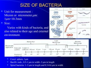
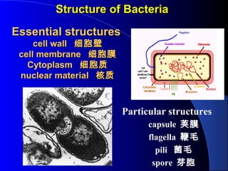
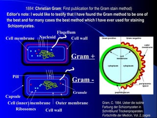
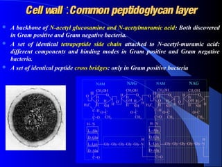
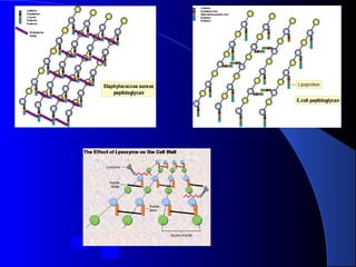
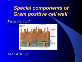
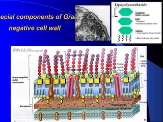
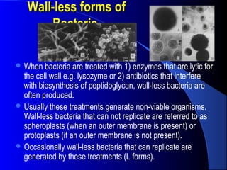
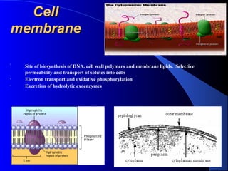
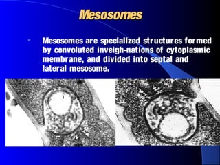
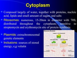
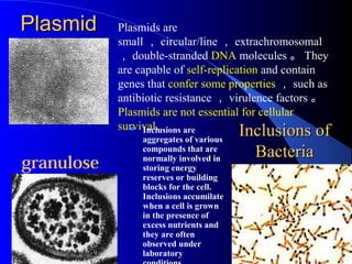
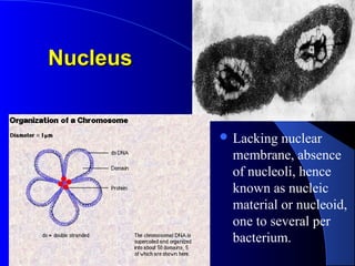
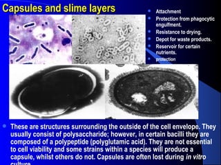
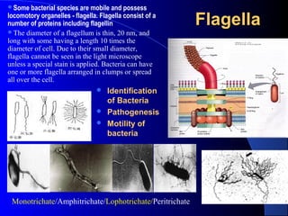
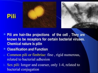
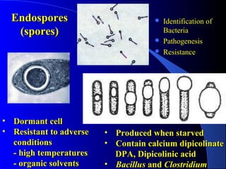
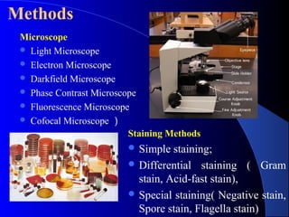
Bacterial Morphology and StructureBacterial Morphology and Structure
Xiao-Kui Guo PhD
http://basic.shsmu.edu.cn/passw/micro2/index.asp
SIZE OF BACTERIASIZE OF BACTERIA
 Unit for measurement :
Micron or micrometer,μm:
1μm=10-3mm
 Size:
Varies with kinds of bacteria, and
also related to their age and external
environment.
 Cocci: sphere, 1μm
 Bacilli: rods , 0.5-1 μm in width -3 μm in length
 Spiral bacteria: 1~3 μm in length and 0.3-0.6 μm in width
Structure of BacteriaStructure of Bacteria
Particular structures
capsule 荚膜
flagella 鞭毛
pili 菌毛
spore 芽胞
Essential structuresEssential structures
cell wallcell wall 细胞壁细胞壁
cell membranecell membrane 细胞膜细胞膜
CytoplasmCytoplasm 细胞质细胞质
nuclear materialnuclear material 核质核质
Gram +
Gram -
Cell wall
Cell (inner) membrane Outer membrane
Ribosomes
Granule
Cell wall
NucleoidCell membrane
Capsule
Flagellum
Pili
Gram, C. 1884. Ueber die isolirteGram, C. 1884. Ueber die isolirte
Farbung der Schizomyceten inFarbung der Schizomyceten in
SchnittÄund Trockenpraparaten.SchnittÄund Trockenpraparaten.
Fortschritte der MedicinFortschritte der Medicin, Vol. 2, pages, Vol. 2, pages
1884:1884: Christian GramChristian Gram: First publication for the Gram stain method): First publication for the Gram stain method)
Editor's note: I would like to testify that I have found the Gram method to be one ofEditor's note: I would like to testify that I have found the Gram method to be one of
the best and for many cases the best method which I have ever used for stainingthe best and for many cases the best method which I have ever used for staining
Schizomycetes.Schizomycetes.
Cell wallCell wall
 Situation:
outmost portion.
15-30nm in
thickness, 10%-
25% of dry
weight.
Cell wallCell wall ::Common peptidoglycan layerCommon peptidoglycan layer
 A backbone of N-acetyl glucosamine and N-acetylmuramic acid: Both discovered
in Gram positive and Gram negative bacteria.
 A set of identical tetrapeptide side chain attached to N-acetyl-muramic acid:
different components and binding modes in Gram positive and Gram negative
bacteria.
 A set of identical peptide cross bridges: only in Gram positive bacteria
Special components ofSpecial components of
Gram positive cell wallGram positive cell wall
Teichoic acid
SPA / M POTEIN
pecial components of Grampecial components of Gram
negative cell wallnegative cell wall
FunctionsFunctions of Cell Wallof Cell Wall
 Maintaining the cell's characteristic shape- the rigid
wall compensates for the flexibility of the
phospholipid membrane and keeps the cell from
assuming a spherical shape
 Countering the effects of osmotic pressure
 Providing attachment sites for bacteriophages
 Providing a rigid platform for surface appendages-
flagella, fimbriae, and pili all emanate from the
wall and extend beyond it
 Play an essential role in cell division
 Be the sites of major antigenic determinants of
the cell surface 。
 Resistance of Antibiotics
Wall-less forms ofWall-less forms of
BacteriaBacteria..
 When bacteria are treated with 1) enzymes that are lytic for
the cell wall e.g. lysozyme or 2) antibiotics that interfere
with biosynthesis of peptidoglycan, wall-less bacteria are
often produced.
 Usually these treatments generate non-viable organisms.
Wall-less bacteria that can not replicate are referred to as
spheroplasts (when an outer membrane is present) or
protoplasts (if an outer membrane is not present).
 Occasionally wall-less bacteria that can replicate are
generated by these treatments (L forms).
CellCell
membranemembrane
•
Site of biosynthesis of DNA, cell wall polymers and membrane lipids. Selective
permeability and transport of solutes into cells
• Electron transport and oxidative phosphorylation
• Excretion of hydrolytic exoenzymes
MesosomesMesosomes
• Mesosomes are specialized structures formed
by convoluted inveigh-nations of cytoplasmic
membrane, and divided into septal and
lateral mesosome.
CytoplasmCytoplasm
 Composed largely of water, together with proteins, nucleic
acid, lipids and small amount of sugars and salts
 Ribosomes: numerous, 15-20nm in diameter with 70S;
distributed throughout the cytoplasm; sensitive to
streptomycin and erythromycin site of protein synthesis
 Plasmids: extrachromosomal
genetic elements
 Inclusions: sources of stored
energy, e,g volutin
PlasmidPlasmid Plasmids are
small , circular/line , extrachromosomal
, double-stranded DNA molecules 。 They
are capable of self-replication and contain
genes that confer some properties , such as
antibiotic resistance , virulence factors 。
Plasmids are not essential for cellular
survival.
Inclusions ofInclusions of
BacteriaBacteria
 Inclusions are
aggregates of various
compounds that are
normally involved in
storing energy
reserves or building
blocks for the cell.
Inclusions accumilate
when a cell is grown
in the presence of
excess nutrients and
they are often
observed under
laboratory
granulose
NucleusNucleus
 Lacking nuclear
membrane, absence
of nucleoli, hence
known as nucleic
material or nucleoid,
one to several per
bacterium.
Capsules and slime layersCapsules and slime layers
 These are structures surrounding the outside of the cell envelope. They
usually consist of polysaccharide; however, in certain bacilli they are
composed of a polypeptide (polyglutamic acid). They are not essential
to cell viability and some strains within a species will produce a
capsule, whilst others do not. Capsules are often lost during in vitro
 Attachment
 Protection from phagocytic
engulfment.
 Resistance to drying.
 Depot for waste products.
 Reservoir for certain
nutrients.
 protection
FlagellaFlagella
Monotrichate/Amphitrichate/Lophotrichate/Peritrichate
 Identification
of Bacteria
 Pathogenesis
 Motility of
bacteria
Some bacterial species are mobile and possess
locomotory organelles - flagella. Flagella consist of a
number of proteins including flagellin
The diameter of a flagellum is thin, 20 nm, and
long with some having a length 10 times the
diameter of cell. Due to their small diameter,
flagella cannot be seen in the light microscope
unless a special stain is applied. Bacteria can have
one or more flagella arranged in clumps or spread
all over the cell.
PiliPili
 Pili are hair-like projections of the cell , They are
known to be receptors for certain bacterial viruses.
Chemical nature is pilin
 Classification and Function
a. Common pili or fimbriae: fine , rigid numerous,
related to bacterial adhesion
b. Sex pili: longer and coarser, only 1-4, related to
bacterial conjugation
EndosporesEndospores
(spores)(spores)
• Dormant cellDormant cell
• Resistant to adverseResistant to adverse
conditionsconditions
- high temperatures- high temperatures
- organic solvents- organic solvents
• Produced when starvedProduced when starved
• Contain calcium dipicolinateContain calcium dipicolinate
DPA,DPA, DDipicolinic acidipicolinic acid
• BacillusBacillus andand ClostridiumClostridium
 Identification of
Bacteria
 Pathogenesis
 Resistance
Microscope
 Light Microscope
 Electron Microscope
 Darkfield Microscope
 Phase Contrast Microscope
 Fluorescence Microscope
 Cofocal Microscope )
Methods
Staining Methods
 Simple staining;
 Differential staining ( Gram
stain, Acid-fast stain),
 Special staining( Negative stain,
Spore stain, Flagella stain)

2

  • 1.
    Bacterial Morphology andStructureBacterial Morphology and Structure Xiao-Kui Guo PhD http://basic.shsmu.edu.cn/passw/micro2/index.asp
  • 2.
    SIZE OF BACTERIASIZEOF BACTERIA  Unit for measurement : Micron or micrometer,μm: 1μm=10-3mm  Size: Varies with kinds of bacteria, and also related to their age and external environment.  Cocci: sphere, 1μm  Bacilli: rods , 0.5-1 μm in width -3 μm in length  Spiral bacteria: 1~3 μm in length and 0.3-0.6 μm in width
  • 3.
    Structure of BacteriaStructureof Bacteria Particular structures capsule 荚膜 flagella 鞭毛 pili 菌毛 spore 芽胞 Essential structuresEssential structures cell wallcell wall 细胞壁细胞壁 cell membranecell membrane 细胞膜细胞膜 CytoplasmCytoplasm 细胞质细胞质 nuclear materialnuclear material 核质核质
  • 4.
    Gram + Gram - Cellwall Cell (inner) membrane Outer membrane Ribosomes Granule Cell wall NucleoidCell membrane Capsule Flagellum Pili Gram, C. 1884. Ueber die isolirteGram, C. 1884. Ueber die isolirte Farbung der Schizomyceten inFarbung der Schizomyceten in SchnittÄund Trockenpraparaten.SchnittÄund Trockenpraparaten. Fortschritte der MedicinFortschritte der Medicin, Vol. 2, pages, Vol. 2, pages 1884:1884: Christian GramChristian Gram: First publication for the Gram stain method): First publication for the Gram stain method) Editor's note: I would like to testify that I have found the Gram method to be one ofEditor's note: I would like to testify that I have found the Gram method to be one of the best and for many cases the best method which I have ever used for stainingthe best and for many cases the best method which I have ever used for staining Schizomycetes.Schizomycetes.
  • 5.
    Cell wallCell wall Situation: outmost portion. 15-30nm in thickness, 10%- 25% of dry weight.
  • 6.
    Cell wallCell wall::Common peptidoglycan layerCommon peptidoglycan layer  A backbone of N-acetyl glucosamine and N-acetylmuramic acid: Both discovered in Gram positive and Gram negative bacteria.  A set of identical tetrapeptide side chain attached to N-acetyl-muramic acid: different components and binding modes in Gram positive and Gram negative bacteria.  A set of identical peptide cross bridges: only in Gram positive bacteria
  • 8.
    Special components ofSpecialcomponents of Gram positive cell wallGram positive cell wall Teichoic acid SPA / M POTEIN
  • 9.
    pecial components ofGrampecial components of Gram negative cell wallnegative cell wall
  • 10.
    FunctionsFunctions of CellWallof Cell Wall  Maintaining the cell's characteristic shape- the rigid wall compensates for the flexibility of the phospholipid membrane and keeps the cell from assuming a spherical shape  Countering the effects of osmotic pressure  Providing attachment sites for bacteriophages  Providing a rigid platform for surface appendages- flagella, fimbriae, and pili all emanate from the wall and extend beyond it  Play an essential role in cell division  Be the sites of major antigenic determinants of the cell surface 。  Resistance of Antibiotics
  • 11.
    Wall-less forms ofWall-lessforms of BacteriaBacteria..  When bacteria are treated with 1) enzymes that are lytic for the cell wall e.g. lysozyme or 2) antibiotics that interfere with biosynthesis of peptidoglycan, wall-less bacteria are often produced.  Usually these treatments generate non-viable organisms. Wall-less bacteria that can not replicate are referred to as spheroplasts (when an outer membrane is present) or protoplasts (if an outer membrane is not present).  Occasionally wall-less bacteria that can replicate are generated by these treatments (L forms).
  • 12.
    CellCell membranemembrane • Site of biosynthesisof DNA, cell wall polymers and membrane lipids. Selective permeability and transport of solutes into cells • Electron transport and oxidative phosphorylation • Excretion of hydrolytic exoenzymes
  • 13.
    MesosomesMesosomes • Mesosomes arespecialized structures formed by convoluted inveigh-nations of cytoplasmic membrane, and divided into septal and lateral mesosome.
  • 14.
    CytoplasmCytoplasm  Composed largelyof water, together with proteins, nucleic acid, lipids and small amount of sugars and salts  Ribosomes: numerous, 15-20nm in diameter with 70S; distributed throughout the cytoplasm; sensitive to streptomycin and erythromycin site of protein synthesis  Plasmids: extrachromosomal genetic elements  Inclusions: sources of stored energy, e,g volutin
  • 15.
    PlasmidPlasmid Plasmids are small, circular/line , extrachromosomal , double-stranded DNA molecules 。 They are capable of self-replication and contain genes that confer some properties , such as antibiotic resistance , virulence factors 。 Plasmids are not essential for cellular survival. Inclusions ofInclusions of BacteriaBacteria  Inclusions are aggregates of various compounds that are normally involved in storing energy reserves or building blocks for the cell. Inclusions accumilate when a cell is grown in the presence of excess nutrients and they are often observed under laboratory granulose
  • 16.
    NucleusNucleus  Lacking nuclear membrane,absence of nucleoli, hence known as nucleic material or nucleoid, one to several per bacterium.
  • 17.
    Capsules and slimelayersCapsules and slime layers  These are structures surrounding the outside of the cell envelope. They usually consist of polysaccharide; however, in certain bacilli they are composed of a polypeptide (polyglutamic acid). They are not essential to cell viability and some strains within a species will produce a capsule, whilst others do not. Capsules are often lost during in vitro  Attachment  Protection from phagocytic engulfment.  Resistance to drying.  Depot for waste products.  Reservoir for certain nutrients.  protection
  • 18.
    FlagellaFlagella Monotrichate/Amphitrichate/Lophotrichate/Peritrichate  Identification of Bacteria Pathogenesis  Motility of bacteria Some bacterial species are mobile and possess locomotory organelles - flagella. Flagella consist of a number of proteins including flagellin The diameter of a flagellum is thin, 20 nm, and long with some having a length 10 times the diameter of cell. Due to their small diameter, flagella cannot be seen in the light microscope unless a special stain is applied. Bacteria can have one or more flagella arranged in clumps or spread all over the cell.
  • 19.
    PiliPili  Pili arehair-like projections of the cell , They are known to be receptors for certain bacterial viruses. Chemical nature is pilin  Classification and Function a. Common pili or fimbriae: fine , rigid numerous, related to bacterial adhesion b. Sex pili: longer and coarser, only 1-4, related to bacterial conjugation
  • 20.
    EndosporesEndospores (spores)(spores) • Dormant cellDormantcell • Resistant to adverseResistant to adverse conditionsconditions - high temperatures- high temperatures - organic solvents- organic solvents • Produced when starvedProduced when starved • Contain calcium dipicolinateContain calcium dipicolinate DPA,DPA, DDipicolinic acidipicolinic acid • BacillusBacillus andand ClostridiumClostridium  Identification of Bacteria  Pathogenesis  Resistance
  • 21.
    Microscope  Light Microscope Electron Microscope  Darkfield Microscope  Phase Contrast Microscope  Fluorescence Microscope  Cofocal Microscope ) Methods Staining Methods  Simple staining;  Differential staining ( Gram stain, Acid-fast stain),  Special staining( Negative stain, Spore stain, Flagella stain)