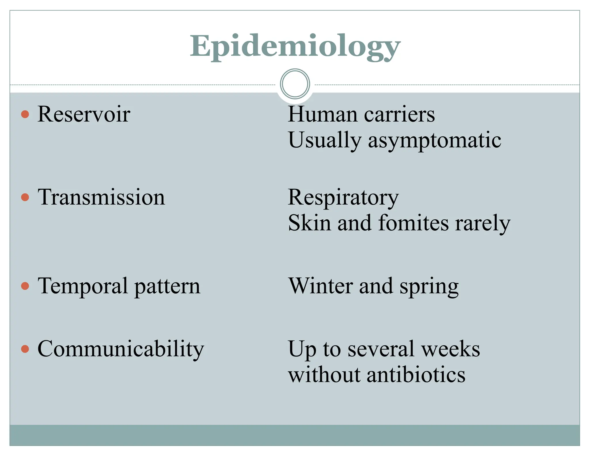
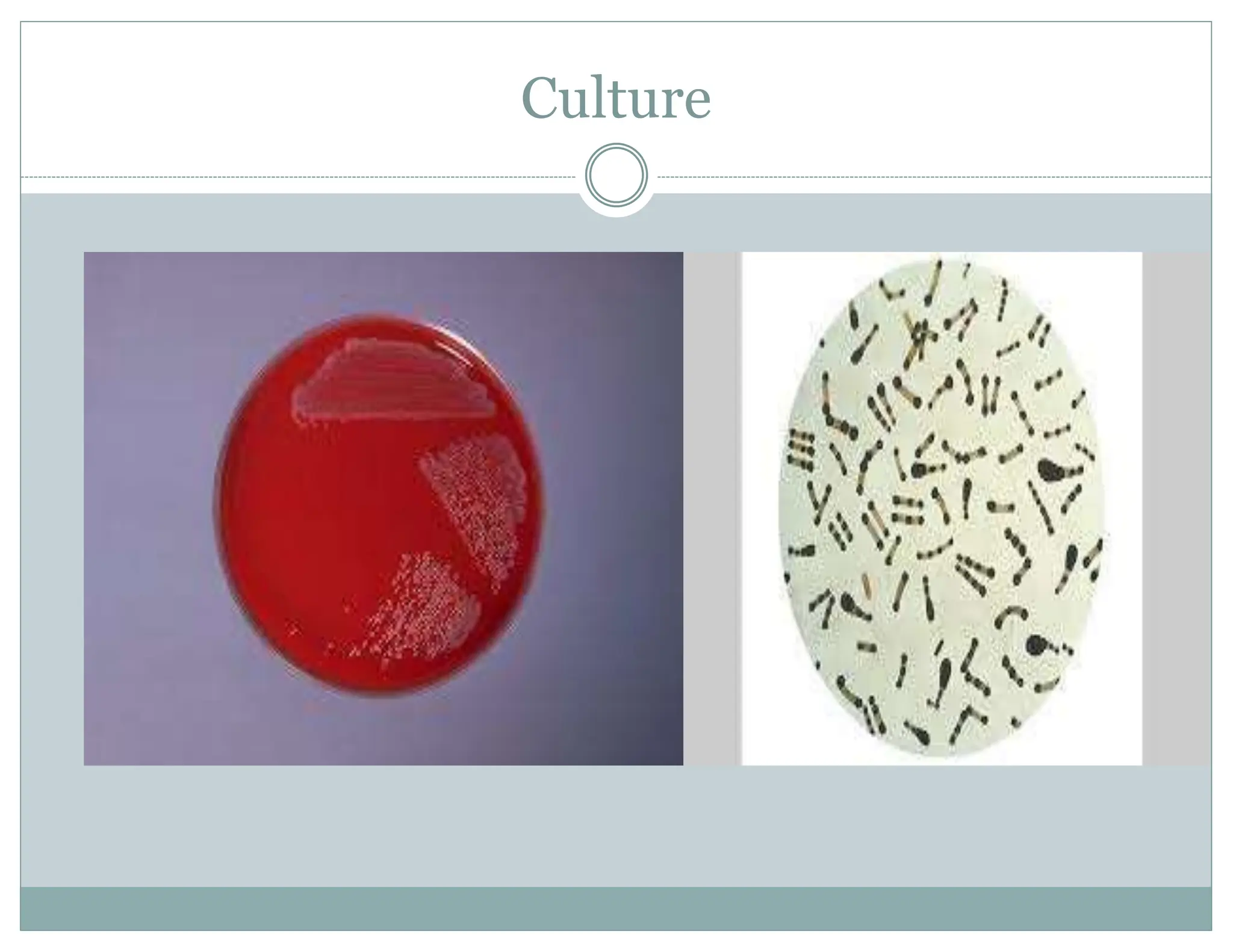
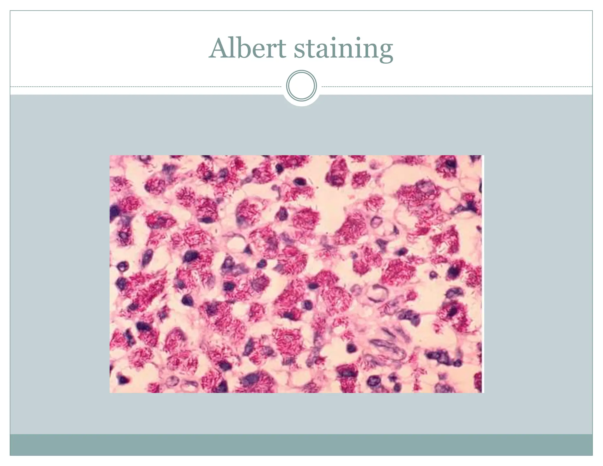
Diphtheria
Dr. Rachana Choudhary
Department of Microbiology
Shri Shankaracharya Mahavidyalaya Junwani
Bhilai
Introduction
 It is Caused by Aerobic Gram +ve rods
Cornybacterium diphtheria.
 Exotoxin production only if infected by virus
phage infected carrying toxin gene.
Gram +ve Bacilli and Colonies
Epidemiology
 Reservoir Human carriers
Usually asymptomatic
 Transmission Respiratory
Skin and fomites rarely
 Temporal pattern Winter and spring
 Communicability Up to several weeks
without antibiotics
Clinical Features
 Incubation period 2-5 days
(range, 1-10 days)
 May involve any mucous membrane
 Classified based on site of infection
Anterior nasal
Pharyngeal
Tonsillar
Laryngeal
Anterior Nasal
Thick Membrane
Pseudo membrane
‘Bull Neck’
Skin Lesions
Symptoms
Diagnosis
 Culture
 Fluorescent antibody technique
 Albert`s staining
Culture
Fluorescent Antibody Techniques
Figure 18.10a
Albert staining
Treatment
Penicillin :
 Procaine penicillin ( 3 – 6 units IM at 12 hourly)
 Oral penicillin ( 125 – 250 mg units)
 Erythromycin ( 25 – 30 mg / kg / day ) for 14 days
 Three negative cultures at 24 hours
intervals should be obtained before the
patient is declared free of the organism
Retention of Penicillin G
Figure 20.7
Prevention
Vaccination: Immunisation with diphtheria
toxoid, combined with tetanus and pertussis
toxoid (DTP vaccine), should be given to all
children at two, three and four months of age.
The child is given a further booster vaccine before
leaving school and is then considered to be
protected for a further 10 years (16 – 18 years).
Reference
Book Writer
 Medical Microbiology P. Chakraverti
 Microbiology Dubey and Maheshwari
 Immunology Kubey

DIPTHERIA.ppt