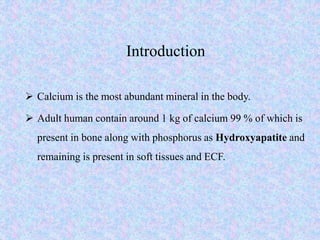
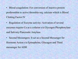
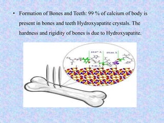
Calcium and phosphorus are important minerals that make up bones and teeth and are involved in many metabolic processes. Calcium homeostasis is regulated by parathyroid hormone, calcitonin, and vitamin D which act on the intestines, kidneys, and bones. Hypocalcemia can result from hypoparathyroidism, vitamin D deficiency, or renal disease and causes tetany and muscle spasms. Hypercalcemia has causes like hyperparathyroidism or cancer and symptoms of nausea, constipation, and renal problems. Phosphorus is found in bones and tissues and is needed for energy metabolism and cell signaling. Its absorption is regulated similarly to calcium. Hypophosphatemia can be caused by
Calcium,magnesium,phosphate and chloride imbalances Jyothi Swaroop
Calcium,magnesium,phosphate and chloride imbalances
Their treatment,my main reference is Eric strong's lectures in youtube,and some of the websites.Hope everyone finding Serum electrolytes find atleast some use of it .
Thank you
This document summarizes mineral metabolism, focusing on calcium. It describes calcium as the most abundant mineral, mainly stored in bones. It discusses calcium absorption in the small intestine and regulation of blood calcium levels by the bones, kidneys, intestine, parathyroid hormone, calcitriol, and calcitonin working together. Hypercalcemia occurs when blood calcium levels exceed 11 mg/dL and can be caused by hyperparathyroidism.
Calcium is essential for many bodily functions like bone formation, muscle contraction, nerve signaling etc. 99% of calcium is stored in bones and remaining 1% is present in extracellular fluids. Calcium level is tightly regulated by parathyroid hormone, calcitriol (active form of vitamin D), and calcitonin. These hormones work to maintain calcium between 9-11 mg/dL by mobilizing calcium from bones and kidneys and absorbing it from intestines. An imbalance in these regulatory hormones can lead to conditions like osteoporosis and rickets.
This document discusses calcium metabolism and disorders. It defines calcium and its daily requirements. Calcium levels are regulated by parathyroid hormone, vitamin D, and calcitonin. Disorders include hypercalcemia caused by overactive parathyroids, and hypocalcemia caused by deficiencies. Hyperparathyroidism has primary, secondary, and tertiary forms caused by changes in calcium levels. The case discusses an older patient with hypercalcemia, high PTH, and symptoms of fatigue from possible primary hyperparathyroidism.
Hypocalcemia is a low level of calcium in the blood. Calcium is essential for nerve impulse conduction, muscle contraction, and other cellular functions. Hypocalcemia can be caused by low albumin, low parathyroid hormone, vitamin D deficiency, or other factors. Symptoms include neuromuscular irritability, tetany, seizures, and EKG changes. Treatment involves oral calcium and vitamin D supplements to replace calcium and maintain adequate blood levels. Intravenous calcium may be needed for severe acute hypocalcemia.
This document discusses vitamin D, calcium, and phosphate metabolism. It covers the roles and regulation of vitamin D, calcium, and phosphate in the body. Vitamin D helps regulate calcium and phosphate levels and is required for bone mineralization. Deficiencies can lead to conditions like rickets, osteomalacia, and osteoporosis. The document also discusses oral implications of these nutritional deficiencies and metabolic bone diseases. Maintaining proper levels of vitamin D, calcium, and phosphate is important for overall health and bone health.
Calcium metabolism can be disrupted, resulting in hypercalcemia or hypocalcemia. Hypercalcemia is caused by increased bone resorption, GI absorption, or decreased renal excretion and may be due to primary hyperparathyroidism, certain cancers, or excessive vitamin D intake. Hypocalcemia occurs when ionized calcium levels decrease below normal due to hypoparathyroidism, vitamin D deficiency, or other conditions. Symptoms of hypercalcemia include fatigue, nausea, and renal problems while hypocalcemia causes muscle spasms, tingling, and seizures. Treatment depends on the underlying cause and severity but aims to restore normal calcium levels through rehydration, diuretics, bisphosphonates
calcium metabolism and hypocalcimia / dental implant courses by Indian dental...Indian dental academy
The Indian Dental Academy is the Leader in continuing dental education , training dentists in all aspects of dentistry and
offering a wide range of dental certified courses in different formats.for more details please visit
www.indiandentalacademy.com
1. Calcium homeostasis is regulated by parathyroid hormone (PTH), calcitonin, vitamin D, and the kidneys and bones. Abnormalities can cause hypo- or hypercalcemia.
2. Hypocalcemia presents with symptoms like numbness, cramps, and seizures. It is diagnosed through blood tests and may be treated with oral calcium or IV calcium gluconate.
3. Hypercalcemia has various causes like primary hyperparathyroidism and can be asymptomatic or cause polyuria, nausea, and kidney stones. Treatment involves rehydration, calcitonin, and bisphosphonates.
Calcium, phosphorus, and potassium are essential minerals that play important roles in bone health, cell signaling, energy production, and other physiological processes. Calcium is critical for bone structure and strength. Phosphorus is also structural and involved in energy production. Potassium helps nerves and muscles function properly. Deficiencies can lead to bone diseases like rickets or osteomalacia. Maintaining adequate intake through diet or supplements is important for prevention of diseases like osteoporosis and kidney stones.
Hypercalcaemia is a condition caused by excessive calcium in the blood, which can result from increased skeletal calcium release, intestinal calcium absorption, or decreased renal calcium excretion. Symptoms include constipation, kidney stones, depression, confusion, and at higher levels coma and cardiac arrest. Treatment involves hydration, increasing salt intake, and forced diuresis.
This document summarizes calcium, phosphate, and magnesium metabolism. It discusses that these minerals have important intracellular and extracellular functions regulated by hormones like PTH and vitamin D. The majority of calcium is stored in bone, while phosphate and magnesium also have roles in cell structures and energy processes. Tight homeostasis maintains mineral levels in the blood and body through balances of absorption, excretion, and bone remodeling. Disorders can develop if these processes are disrupted.
This document discusses calcium imbalance and hypocalcemia. It defines hypocalcemia and describes its various causes including prematurity, birth asphyxia, infants of diabetic mothers, vitamin D deficiency, hypoparathyroidism, and renal failure. The roles of parathyroid hormone, vitamin D, and calcitonin in regulating calcium levels are explained. Symptoms of hypocalcemia include neuromuscular irritability, cardiac involvement, and dermatological manifestations. The pathophysiology and various factors affecting calcium absorption and homeostasis are also summarized.
Three hormones - parathyroid hormone, vitamin D, and calcitonin - regulate serum calcium levels by altering their secretion in response to changes in ionized calcium. About 99% of calcium in the body is stored in bones, while the remaining 1% circulates in blood and other tissues. Calcium in blood is distributed between free ions, protein-bound forms, and forms bound to anions. Hypocalcemia can cause neuromuscular and cardiac symptoms, while hypercalcemia affects the neurological, gastrointestinal, and renal systems. Total calcium is measured using assays involving complexation with dyes like ortho-cresolphthalein or arsenazo, while ionized calcium is measured using ion-
This document discusses magnesium disorders and hypomagnesemia. It begins by providing background on magnesium, including where it is stored in the body and its many functions. It then discusses the causes, clinical manifestations, diagnosis, and treatment of hypomagnesemia. Hypomagnesemia can be caused by renal magnesium wasting due to drugs, diseases, or genetic disorders, or by extrarenal losses from poor nutrition, malabsorption, diarrhea, or sweat losses. Symptoms affect the cardiovascular, neuromuscular, skeletal, and central nervous systems. Treatment involves identifying and addressing the underlying cause, as well as oral or parenteral magnesium supplementation.
Calcium is essential for many bodily functions and is mainly stored in bones. Calcium levels are tightly regulated by parathyroid hormone, vitamin D, and calcitonin. Hypocalcemia can result from hypoparathyroidism, vitamin D deficiency, or kidney disease and causes neuromuscular symptoms. Hypercalcemia generally comes from excessive bone resorption due to cancers or hyperparathyroidism and can lead to gastrointestinal, renal, and neurological issues.
Calcium homeostasis is tightly regulated to keep plasma calcium levels within a narrow range. Three hormones - parathyroid hormone (PTH), calcitriol, and calcitonin - work together to regulate calcium levels by moving calcium between bones and extracellular fluid. The kidneys also play an important role by reabsorbing around 80% of filtered calcium in the proximal tubule and regulating further calcium reabsorption in other parts of the nephron in response to these hormones and other factors like calcium levels and pH. Disruptions to this regulatory system can lead to hypocalcemia or hypercalcemia.
Calcium homeostasis is tightly regulated by the parathyroid hormone (PTH), vitamin D, and calcitonin. PTH increases calcium absorption from the intestine and resorption from bone. Vitamin D increases intestinal calcium absorption and bone resorption. Calcitonin decreases calcium levels by inhibiting bone resorption. Together, these hormones maintain calcium levels in a narrow range.
Calcium homeostasis is tightly regulated by the interaction of the parathyroid gland, kidneys, bone, intestine, and hormones. Parathyroid hormone (PTH) increases blood calcium levels by promoting bone resorption and renal calcium reabsorption. Vitamin D assists PTH by increasing intestinal calcium absorption. Disorders occur when calcium levels decrease or increase outside the normal range, which can cause symptoms like numbness, seizures, or kidney stones. Maintaining balanced calcium levels requires the coordinated functions of multiple organ systems and hormones.
Hypercalcaemia certainly possesses some diagnostic challenges
Cases are too different in ways of presentation and management do need a lot of things to be checked out. This is merely an approach for such patients.
Calcium homeostasis involves absorption of calcium from the intestine, regulation by parathyroid hormone (PTH), vitamin D, and calcitonin, and storage in bone. PTH increases calcium levels by stimulating bone resorption and renal reabsorption and vitamin D absorption. Vitamin D increases intestinal calcium absorption. Calcitonin decreases calcium by inhibiting bone resorption. Hypocalcemia causes neurological symptoms and hypercalcemia causes gastrointestinal and renal issues. Conditions are diagnosed by calcium levels and other tests and treated by calcium supplementation or intravenous calcium for hypocalcemia and hydration and medications for hypercalcemia.
Calcium is an important cation found mostly in bone but also in soft tissues. Serum calcium levels are tightly regulated by parathyroid hormone (PTH) and calcitriol. Hypocalcemia can be caused by low PTH levels due to various conditions or high PTH levels due to vitamin D deficiency or resistance. Symptoms range from mild tingling to seizures. Diagnosis involves measuring serum calcium, PTH, and other electrolytes. Treatment depends on severity but may include calcium supplementation as well as addressing the underlying cause.
This document discusses calcium homeostasis and disorders, including hypercalcemia and hyperparathyroidism. It provides details on the roles of parathyroid hormone (PTH), vitamin D, and calcitonin in regulating calcium levels. It examines causes of hypercalcemia such as primary hyperparathyroidism, malignancy, and vitamin D toxicity. Guidelines for treatment of asymptomatic primary hyperparathyroidism with surgery or medication are reviewed based on calcium levels, bone mineral density, kidney stones, and age. Non-surgical treatments including vitamin D supplementation and cinacalcet are also discussed.
This document provides an overview of minerals, including their sources, daily requirements, absorption, functions, regulation, and clinical manifestations of deficiencies and toxicities. It discusses the key macro minerals sodium, potassium, chloride, calcium, phosphorus, and magnesium, as well as the trace minerals iron, iodine, zinc, copper, molybdenum, fluorine, selenium, cobalt, chromium, and manganese. For each mineral, it describes its major roles and implications of insufficient or excessive levels on human health. The document is intended as a seminar on minerals and their importance for various metabolic processes and maintaining overall health.
The document discusses calcium's biochemical functions including roles in bone and teeth formation, muscle contraction, blood coagulation, nerve transmission, and enzyme activation. It describes calcium requirements, sources, absorption factors, homeostatic regulation by parathyroid hormone, calcitonin, and vitamin D, and excretion. It also examines diseases like hypercalcemia, hypocalcemia, rickets, osteoporosis, and osteopetrosis.
Calcium homeostasis is regulated by calcitonin, parathyroid hormone, and vitamin D. Calcium is primarily stored in bones and is present in small amounts in extracellular fluid. The majority of calcium in blood is ionized and plays important roles in cell excitability, muscle contraction, and hormone secretion. Dietary calcium absorption occurs mainly in the duodenum and is regulated by hormones and vitamin D. Hypocalcemia and hypercalcemia can result from disorders of the parathyroid gland, kidneys, bone, or vitamin D metabolism and cause neuromuscular and cardiac symptoms. Treatment involves addressing the underlying cause and correcting calcium levels.
Calcium is essential for many physiological processes in the body. It makes up 1-1.5% of total body weight, with 99% located in bones and teeth. Dietary sources include dairy products, eggs, fish, and leafy greens. The recommended daily intake is 500 mg for adults and 1200 mg for children. Calcium is absorbed in the duodenum and jejunum through an active transport process requiring energy and carrier proteins. Homeostasis is maintained by calcitriol, parathyroid hormone, and calcitonin which regulate absorption from the intestine and resorption from bones. Imbalances can cause hypercalcemia with symptoms like confusion and arrhythmias, or hypocalcemia/
Magnesium is an important intracellular cation that plays a key role in many cellular processes. Hypomagnesemia can result from reduced intake, malabsorption, renal losses due to drugs or conditions like Gitelman syndrome, while hypermagnesemia commonly occurs in renal failure or with magnesium-containing drugs. Both conditions can impact neuromuscular and cardiac function. Treatment of hypomagnesemia involves oral or IV magnesium supplementation while hypermagnesemia may require calcium, diuretics, or dialysis. Magnesium levels also influence PTH secretion and activity.
This document discusses calcium and phosphate metabolism. It covers:
1. Calcium is found mainly in bones, soft tissues, and extracellular fluid. The majority is stored in bones.
2. Calcium levels are regulated by parathyroid hormone, vitamin D, and calcitonin which act on bones, kidneys and intestines to increase or decrease calcium absorption and resorption.
3. Hypercalcemia can be caused by primary hyperparathyroidism, cancer, multiple myeloma or excessive vitamin D intake. Hypocalcemia results from vitamin D deficiency or renal failure and causes symptoms like muscle spasms.
Calcium is an essential mineral required for normal growth and maintenance of the body. 99% of calcium in the human body is found in bones. Calcium levels in blood are regulated by parathyroid hormone, vitamin D, and calcitonin. Hypocalcemia and hypercalcemia can result from disorders of these hormones or from other causes like cancer, medications, or kidney disease. Symptoms of hypocalcemia include tetany, seizures, and cardiac issues, while hypercalcemia symptoms include renal damage and gastrointestinal problems. Calcium plays important roles in bone formation, blood clotting, muscle contraction, and nerve transmission.
Calcium metabolism/ oral surgery courses /certified fixed orthodontic courses...Indian dental academy
The Indian Dental Academy is the Leader in continuing dental education , training dentists in all aspects of dentistry and
offering a wide range of dental certified courses in different formats.for more details please visit
www.indiandentalacademy.com
The parathyroid glands regulate calcium levels in the blood. They are located behind the thyroid gland and secrete parathyroid hormone (PTH). PTH increases calcium levels in the blood by releasing calcium from bones, reabsorbing calcium in the kidneys, and enhancing calcium absorption in the intestines. PTH also plays a key role in activating vitamin D, which further aids in intestinal calcium absorption. The level of calcium in the blood provides feedback to regulate PTH secretion, with lower calcium triggering more PTH release.
The parathyroid glands secrete parathyroid hormone (PTH) which regulates blood calcium levels. PTH increases calcium resorption from bones and reabsorption from kidneys to raise calcium levels. It also increases vitamin D activation in kidneys to boost calcium absorption in the gut. PTH secretion is regulated by blood calcium and phosphate levels, increasing when calcium is low or phosphate is high to restore normal calcium concentrations.
This document summarizes key information about calcium homeostasis and metabolism. It discusses how calcium provides structural integrity to bones and is essential for many biochemical processes. It outlines recommended daily calcium intake and factors that influence calcium absorption and excretion. Key regulators of calcium levels like parathyroid hormone, calcitonin, and vitamin D are also described. The document discusses consequences of calcium deficiency and hypercalcemia, and treatments for correcting calcium levels.
Parathyroid hormone (The Guyton and Hall physiology)Maryam Fida
Parathyroid hormone (PTH) regulates calcium levels in the blood and body. It is released by the parathyroid glands when calcium levels drop. PTH acts on bone, intestines, and kidneys to raise blood calcium levels. It stimulates bone cells to release calcium into the blood, increases intestinal calcium absorption, and reduces calcium excretion by the kidneys. Calcitonin produced by the thyroid acts opposing PTH to lower blood calcium and deposit calcium in bone. The body tightly controls calcium levels to prevent hypocalcemia and hypercalcemia through the actions of PTH, calcitonin, kidneys, gastrointestinal tract, and skeleton.
This document discusses calcium disorders including an overview of calcium, hypercalcemia, hypocalcemia, and inherited calcium disorders. It defines calcium and its roles in the body. Hypercalcemia is defined as a serum calcium level >10.5 mg/dL and can be caused by increased bone resorption, gastrointestinal absorption, or decreased renal excretion. It discusses the etiology, clinical exam, differential diagnosis, investigations, management, and complications of hypercalcemia. Hypocalcemia is defined as a total calcium <8.5 mg/dL and discusses its etiology including hypoparathyroidism, renal insufficiency, hypomagnesemia, vitamin D deficiency, and others. It also discusses the
Calcium is essential for bone health, muscle function, and other bodily processes. Its levels are tightly regulated by parathyroid hormone (PTH), vitamin D, and calcitonin. Disorders can result from too little or too much calcium and include rickets, osteomalacia, osteoporosis, hypercalcemia, and hypocalcemia. Hyperparathyroidism is a common cause of hypercalcemia due to excessive PTH levels and can result from primary or secondary hyperfunction of the parathyroid glands. A case example is presented of a woman with chronic fatigue and mood changes found to have hypercalcemia due to long term thiazide use increasing calcium resorption.
This document discusses electrolyte concentrations in the three body fluid compartments - intracellular fluid, interstitial fluid, and plasma. It focuses on the major cations sodium, potassium, and calcium, their roles, regulation and what causes hyponatremia/hypernatremia, hypopotassemia/hyperpotassemia, and hypocalcemia/hypercalcemia. Treatment methods for electrolyte imbalances are also covered such as sodium replacement and ways to reduce intestinal calcium absorption.
This document discusses calcium metabolism and provides details on calcium homeostasis, absorption, excretion, and the roles of parathyroid hormone and vitamin D. It also covers hypocalcemia and hypercalcemia, defining each condition and describing causes, clinical manifestations, diagnostic workup, and treatment approaches. Hypocalcemia can result from neonatal issues, vitamin D deficiency, hypoparathyroidism, or other causes. Hypercalcemia has causes including parathyroid hormone excess, malignancy, vitamin D excess, and genetic conditions.
This document discusses calcium homeostasis and hypercalcemia. It notes that calcium is critical for many physiological functions and is mainly stored in bones. Hypercalcemia can be caused by primary hyperparathyroidism, vitamin D excess, certain malignancies, and other conditions. The diagnostic approach involves distinguishing between hyperparathyroidism and hypercalcemia of malignancy based on lab tests. Treatment focuses on rehydration, increasing calciuresis, and decreasing bone resorption or intestinal calcium absorption using medications like calcitonin, bisphosphonates, glucocorticoids, or dialysis depending on the severity of hypercalcemia.
This document discusses the physiologic and biochemical functions of various minerals in the human body. It covers major minerals like calcium, phosphorus, magnesium, sodium, potassium, and chloride. It describes their roles, absorption, transport in the blood, homeostasis, and impact on various diseases when levels are too high or too low. The minerals are essential for many metabolic processes and helping maintain acid-base balance, fluid balance, nerve transmission, muscle contraction and more.
Approach to patient with hypo/hyper calcaemiaNassr ALBarhi
This document discusses calcium homeostasis and disorders of calcium metabolism. It begins by describing the functions of calcium in the body and where calcium is stored. It then discusses calcium regulation by parathyroid hormone, vitamin D, and calcitonin. Causes, signs and symptoms, and treatment approaches for hypercalcemia and hypocalcemia are reviewed. The key points are that calcium levels are tightly controlled by hormones to maintain levels between 2.25-2.62 mmol/L and that disorders can result from excess or deficiencies of these regulating hormones.
Calcium is an essential mineral that makes up 2% of body weight. Over 99% is stored in bones, with the rest in tissues and plasma. Calcium levels are tightly regulated by parathyroid hormone (PTH), calcitonin, and calcitriol (active vitamin D). PTH increases calcium levels by promoting bone resorption, while calcitonin and calcitriol decrease calcium levels by reducing resorption. Bisphosphonates are used to treat osteoporosis and Paget's disease by inhibiting bone resorption. They decrease osteoclast activity and survival. Calcium supplements are used to treat deficiencies and osteoporosis, while bisphosphonates and calcim
This document discusses calcium and phosphate metabolism and related disorders. It covers the distribution, functions, absorption and excretion of calcium and phosphate. It describes the roles of parathyroid hormone, vitamin D, and calcitonin in regulating calcium and phosphate levels. Disorders resulting from calcium and phosphate imbalance like hypercalcemia, hypocalcemia, rickets, and osteomalacia/osteoporosis are explained. Factors affecting calcium and phosphate levels and their clinical importance are also summarized.
The document discusses calcium metabolism. It states that 99% of calcium in the body is found in bones. Dietary sources of calcium include milk, cheese, fish and vegetables. The daily calcium requirement is 500mg for adults, 1200mg for children, and 1300mg for pregnant/lactating individuals. Calcium is absorbed in the duodenum and regulated by parathyroid hormone, vitamin D, and calcitonin. Disorders of calcium metabolism include hypercalcemia, hypocalcemia, hyperparathyroidism, and hypoparathyroidism.
Agents that affect bone mineral homeostasis paulPaul Ndung'u
This document discusses various agents that affect bone mineral homeostasis. Parathyroid hormone (PTH) and vitamin D principally regulate calcium and phosphate levels. PTH stimulates vitamin D production and bone resorption, while vitamin D promotes intestinal absorption of calcium and phosphate. Other agents discussed include calcitonin, bisphosphonates, estrogens, glucocorticoids, thiazides, fluoride, and phosphate binders, which all act on bone formation, resorption, or mineral levels in various ways to maintain bone mineral homeostasis.
The document discusses the four main types of DNA damage and the mechanisms cells use to repair DNA damage. It describes the key steps of DNA repair as recognition of damaged DNA, removal of the damaged region, replacing the excised region using DNA polymerase, and sealing the repaired DNA strand. The main DNA repair pathways discussed are mismatch repair, base excision repair, nucleotide excision repair, direct repair, and homologous recombination and non-homologous end-joining for repairing double-strand breaks. The document also lists some diseases associated with defects in specific DNA repair mechanisms.
Mutations are permanent changes in the DNA sequence that can be caused by errors during DNA replication, environmental factors like radiation and chemicals, or spontaneous changes. Mutations in germ cells can cause inherited diseases while those in somatic cells can lead to cancer. There are several types of mutations, including base substitutions, deletions, insertions, and frameshift mutations. Mutations can have different effects, such as being lethal, silent, beneficial, or carcinogenic by altering regulatory mechanisms and causing uncontrolled cell division.
Polymerase chain reaction (PCR) is a technique used to amplify a single copy of a DNA segment across orders of magnitude, generating thousands to millions of copies. It involves repeated cycles of heating and cooling of the DNA sample to separate and copy the DNA strands. Two primers are used to target the specific segment that will be amplified. During each cycle, the DNA polymerase enzyme adds nucleotides to the primers, duplicating the targeted DNA segment. As the cycles repeat, the copy number increases exponentially. PCR is widely used in clinical diagnostics and research for applications such as disease diagnosis, genetic testing, and forensic analysis.
Cancer arises from uncontrolled cell growth and spread. It was named for swollen veins around tumors resembling a crab. Indian medicine previously called it "arbuda". Cancer is characterized by loss of control over cellular growth and proliferation beyond physiological needs. The main types are carcinomas of epithelial cells, sarcomas of connective tissues, leukemias of blood stem cells, and lymphomas of lymph nodes. Predisposing factors include age, heredity, lifestyle, diet, occupation and some pre-cancerous diseases. Cancer cells show diminished growth control, invasion of tissues, and metastasis to other parts.
This document discusses iron, an essential trace mineral. It notes that iron is needed in small amounts but is critical for biochemical functions. The document covers where iron is stored and transported in the body, how its absorption is regulated, factors that influence absorption, dietary sources, and clinical conditions related to iron deficiency and overload. It provides details on iron metabolism, functions, normal ranges, deficiency states, causes and treatment of iron deficiency anemia.
ELISA (Enzyme-Linked Immunosorbent Assay) is a sensitive technique used to detect small quantities of antigens, antibodies, or other proteins in biological fluids like blood or urine. There are several types of ELISA including direct, indirect, sandwich, and competitive ELISA. ELISA works by using an enzyme-linked antibody or antigen to detect the presence of a target protein. This allows very small amounts of the target to be detected through the enzyme's catalytic activity.
- Erythrocytes have a lifespan of 120 days in adults before being degraded by macrophages in the spleen and liver.
- Haemoglobin is degraded into its protein (globin) and non-protein (heme) components. About 6 g of haemoglobin is broken down per day in adults.
- Globin can be reutilized for new haemoglobin or degraded into individual amino acids. Heme is broken down by heme oxygenase into iron, carbon monoxide, and biliverdin which is further degraded into bilirubin. Bilirubin is conjugated in the liver and excreted into bile.
Immunoglobulins, also known as antibodies, are glycoproteins produced by plasma cells that recognize and bind to specific antigens. There are five main classes of immunoglobulins - IgG, IgA, IgM, IgD, and IgE - which differ in their structure and function. IgG is the most abundant antibody found in serum and body tissues, while IgA is predominantly found in secretions such as breast milk, tears, and saliva where it provides immune protection of mucosal surfaces. IgM is the first antibody to respond to new antigens and plays a key role in activating the complement system.
Plasma proteins include albumin and globulins. Denaturation involves the loss of a protein's tertiary and quaternary structure, exposing hydrophobic residues. This can be caused by heat, acids, salts, or other chemical or physical factors. Major plasma proteins include albumin, which maintains osmotic pressure, and transports hormones and bilirubin. Globulins include α1-antitrypsin, which inhibits proteases and whose deficiency can cause emphysema, orosomucoid which is an acute phase protein, and ceruloplasmin which transports over 90% of copper in the blood.
This document provides information on fat soluble vitamins A and E. It discusses the isolation and chemical structure of vitamin A, its role in vision through the generation of rhodopsin in rods and cones, and its role in gene expression. Deficiency can cause night blindness, dry eyes, skin lesions and blindness. Vitamin E is an antioxidant that protects membranes from free radical damage and its deficiency impacts fertility. The document details the absorption, transport, functions and deficiency symptoms of both vitamins.
This document discusses acid-base balance and acid-base disorders. It defines the anion gap and explains how to calculate it. It describes the causes and management of different types of acid-base disorders including metabolic acidosis, respiratory acidosis, metabolic alkalosis and respiratory alkalosis. Compensation mechanisms for each disorder are explained. Normal ranges for pH, HCO3, pCO2, pO2 and other electrolytes are provided. Multiple choice questions related to acid-base disorders are included at the end.
This document discusses acid-base balance and the mechanisms that regulate pH levels in the human body. It covers topics like buffers, respiratory regulation, and renal regulation of pH. The three main points are:
1) Buffers like bicarbonate help maintain pH levels by neutralizing excess hydrogen ions. The Henderson-Hasselbalch equation describes the buffering capacity of bicarbonate.
2) The respiratory system regulates pH rapidly through exhalation of carbon dioxide. Hemoglobin transports carbon dioxide as bicarbonate from tissues to the lungs.
3) The kidneys regulate pH over longer periods through reabsorption of bicarbonate, and excretion of fixed acids, ammonium
Glycogen is a highly branched polymer of glucose that serves as the primary storage form of glucose in the body. It is synthesized from glucose through glycogenesis, and broken down to glucose through glycogenolysis. Glycogen synthesis occurs mainly in the liver and muscle, and is regulated by hormones and metabolites to store glucose after meals and release it during fasting or exercise. Glycogen degradation provides glucose for energy and maintains blood glucose levels between meals.
Local Advanced Lung Cancer: Artificial Intelligence, Synergetics, Complex Sys...Oleg Kshivets
Overall life span (LS) was 1671.7±1721.6 days and cumulative 5YS reached 62.4%, 10 years – 50.4%, 20 years – 44.6%. 94 LCP lived more than 5 years without cancer (LS=2958.6±1723.6 days), 22 – more than 10 years (LS=5571±1841.8 days). 67 LCP died because of LC (LS=471.9±344 days). AT significantly improved 5YS (68% vs. 53.7%) (P=0.028 by log-rank test). Cox modeling displayed that 5YS of LCP significantly depended on: N0-N12, T3-4, blood cell circuit, cell ratio factors (ratio between cancer cells-CC and blood cells subpopulations), LC cell dynamics, recalcification time, heparin tolerance, prothrombin index, protein, AT, procedure type (P=0.000-0.031). Neural networks, genetic algorithm selection and bootstrap simulation revealed relationships between 5YS and N0-12 (rank=1), thrombocytes/CC (rank=2), segmented neutrophils/CC (3), eosinophils/CC (4), erythrocytes/CC (5), healthy cells/CC (6), lymphocytes/CC (7), stick neutrophils/CC (8), leucocytes/CC (9), monocytes/CC (10). Correct prediction of 5YS was 100% by neural networks computing (error=0.000; area under ROC curve=1.0).
Osteoporosis - Definition , Evaluation and Management .pdfJim Jacob Roy
Osteoporosis is an increasing cause of morbidity among the elderly.
In this document , a brief outline of osteoporosis is given , including the risk factors of osteoporosis fractures , the indications for testing bone mineral density and the management of osteoporosis
Promoting Wellbeing - Applied Social Psychology - Psychology SuperNotesPsychoTech Services
A proprietary approach developed by bringing together the best of learning theories from Psychology, design principles from the world of visualization, and pedagogical methods from over a decade of training experience, that enables you to: Learn better, faster!
Here is the updated list of Top Best Ayurvedic medicine for Gas and Indigestion and those are Gas-O-Go Syp for Dyspepsia | Lavizyme Syrup for Acidity | Yumzyme Hepatoprotective Capsules etc
Basavarajeeyam is a Sreshta Sangraha grantha (Compiled book ), written by Neelkanta kotturu Basavaraja Virachita. It contains 25 Prakaranas, First 24 Chapters related to Rogas& 25th to Rasadravyas.
8 Surprising Reasons To Meditate 40 Minutes A Day That Can Change Your Life.pptxHolistified Wellness
We’re talking about Vedic Meditation, a form of meditation that has been around for at least 5,000 years. Back then, the people who lived in the Indus Valley, now known as India and Pakistan, practised meditation as a fundamental part of daily life. This knowledge that has given us yoga and Ayurveda, was known as Veda, hence the name Vedic. And though there are some written records, the practice has been passed down verbally from generation to generation.
Muktapishti is a traditional Ayurvedic preparation made from Shoditha Mukta (Purified Pearl), is believed to help regulate thyroid function and reduce symptoms of hyperthyroidism due to its cooling and balancing properties. Clinical evidence on its efficacy remains limited, necessitating further research to validate its therapeutic benefits.
Rasamanikya is a excellent preparation in the field of Rasashastra, it is used in various Kushtha Roga, Shwasa, Vicharchika, Bhagandara, Vatarakta, and Phiranga Roga. In this article Preparation& Comparative analytical profile for both Formulationon i.e Rasamanikya prepared by Kushmanda swarasa & Churnodhaka Shodita Haratala. The study aims to provide insights into the comparative efficacy and analytical aspects of these formulations for enhanced therapeutic outcomes.
3. Introduction
• Minerals are inorganic elements required for
variety of functions.
• As per the human requirements minerals can be
grouped as Macrominerals (per day req. more
than 100 mg) and Microminerals (per day req.
less than 100 mg) and Microminerals)
4.
5.
6.
7. Introduction
Calcium is the most abundant mineral in the body.
Adult human contain around 1 kg of calcium 99 % of which is
present in bone along with phosphorus as Hydroxyapatite and
remaining is present in soft tissues and ECF.
8. Important function of Calcium: Calcium(Ca) is required for the
following functions :
Muscle contraction: Muscle contraction is initiated by binding
of calcium to Troponin.
Nerve conduction: Influx of Calcium from ECF to neurons
causes release of Neurotransmitters.
Hormone release: Release of certain hormones s/a parathyroid
hormone and calcitonin req. calcium ions.
9. • Blood coagulation: For conversion of inactive protein
prothrombin to active thrombin req. calcium which is Blood
Clotting Factor IV
• Regulation of Enzyme activity: Activation of several
enzymes require Ca as a cofactor s/a Glycogen Phosphorylase
and Salivary/Pancreatic Amylase.
• Second Messengers: It act as a Second Messenger for
Hormone Action s/a Epinephrine, Glucagon and Third
messenger for ADH
10. • Formation of Bones and Teeth: 99 % of calcium of body is
present in bones and teeth Hydroxyapatite crystals. The
hardness and rigidity of bones is due to Hydroxyapatite.
11. Sources of calcium
• Widely distributed in food substances such as
Milk (Half litre of milk contains 1000
mg of calcium )
Cheese
Egg- yolk
Fish
Beans Lentils Nuts and Cabbage
12.
13. Recommended Dietary Allowance
Per day req. of calcium:
Adults: 800 mg/day
Women's during pregnancy and Lactation and Teenagers:
1200 mg/day
Infants: 300-350 mg/day
14. Calcium Absorption
The absorption of calcium occurs in intestine and depends on
several factors.
Factors favouring calcium absorption:
An acidic pH: Calcium salts are more soluble in acidic pH , the
acidic foods and Organic acids s/a Citric Acid , lactic acid and
pyruvic acid promote calcium absorption.
High protein diet- Lysine and Arginine cause maximal
absorption
15. Vitamin D: stimulates calcium absorption by inducing
synthesis of Calcium binding protein.
Ca : P ratio- A ratio of dietary Ca: P not more than 2:1 is
adequate for optimal absorption, ratio of less than 1:2 reduces
absorption
State of health and intact mucosa- A healthy adult absorbs
about 40% of dietary calcium.
PTH (Parathormone) stimulates the activation of vitamin
D, thus indirectly increases absorption of vitamin D
16. Factors inhibiting absorption of calcium
Alkaline pH
High fat diet- High amount of Fatty acids form calcium
soaps that can not be absorbed
Presence of Phytates and oxalates- Insoluble calcium salts
are formed which can not be absorbed
Dietary fiber in excess inhibits absorption
Excess phosphates form Insoluble Calcium-Phosphate.
17. Calcitonin reduces calcium absorption indirectly by
inhibiting the activation of vitamin D
Advancing age and intestinal inflammatory disorders
inhibit absorption of calcium
Excretion
The excretion of Calcium occurs partially through kidney
and mostly by the way of Intestine through Feces
18. Distribution of Body calcium
Of the total amount, 50% is free ionized calcium, 10% is
combined with various anions (including bicarbonate,
citrate, phosphate, lactate and sulphate) and the remaining
40% is bound to serum proteins mainly albumin.
Free ionized calcium is the physiologically
important component of the total calcium.
In plasma, the ionized calcium concentration is
normally maintained within a tight range (1.0-
1.25mmol/l).
20. • The plasma Calcium concentration of Normal Individual is
9-11 mg/dl
Regulation of calcium homeostasis
Three principal hormones are involved in calcium homeostasis
• Vitamin D
• Parathormone and
• Calcitonin
21. Which act on three target organs:
• Intestine,
• Bone and
• Kidneys
22. The four major processes are:
Absorption of calcium from Intestine through Vitamin D
Reabsorption of Calcium from Kidney through Vitamin D
and PTH
Demineralization of Bones mainly through the action of
PTH and supported by Vitamin D
Mineralization of bone through Calcitonin.
23. Role of vitamin D in calcium
homeostasis
The actions of Vitamin D(Calcitriol) are as follows:
The main role of Vitamin D is to increase Serum Calcium by
following Mechanism:
Enhances calcium absorption from the intestine
Facilitates calcium re-absorption from the kidney
Mobilizes calcium and phosphate from
Bones(Demineralization)
24. Role of Parathyroid hormone (PTH)
• Parathyroid hormone is releases in response to Low Blood
calcium level which is a linear polypeptide containing 84
amino acid residues.
• It is secreted by the chief cells in the four parathyroid
glands.
• It mainly acts on two main Target organs i.e. Bones and
Kidney and indirectly on Intestine by activation of
Vitamin D.
25. Action on Bones: PTH stimulates bone
demineralization by moving calcium and phosphates
from bones to plasma.
Hence increases Osteoclastic activity.
It also decreases uptake of Calcium and Phosphates from
bones.
26. Action on Kidney: PTH stimulates renal reabsorption
and decreases excretion of calcium to maintain blood calcium
level. It also increases excretion of Phosphates.
Action on Intestine: action of PTH on Intestine is
indirect via Vitamin D
27. Role of Calcitonin
Calcitonin is a 32 amino acid polypeptide secreted by the
parafollicular cells in the thyroid gland .
It tends to decrease serum calcium concentration and, in
general, has effects opposite to those of PTH.
28. The actions of calcitonin are as follows:
Inhibits bone resorption
Increases renal calcium excretion
The exact physiological role of calcitonin in calcium
homeostasis is uncertain.
The effects of calcitonin on bone metabolism are much
weaker than those of either PTH or vitamin D.
29.
30.
31. Hypocalcaemia
Hypocalcemia is Total Serum Ca concentration < 8.8 mg/dL (<
2.20 mmol/L) or a serum ionized Ca concentration < 4.7 mg/dL (<
1.17 mmol/L).
Causes Include: Hypoparathyroidism(Surgical Removal of
Gland or d/t Mg Deficiency), Vitamin D deficiency(Dietary
Insufficiency, Malabsorption or low exposure to sunlight), and
Renal disease(Failure to synthesize Calcitriol)
32. Acute hypocalcaemia can also occur in the immediate
post-operative period, following removal of the thyroid or
parathyroid glands.
Hypocalcaemia can occur following rapid administration of
citrated blood or large volumes of albumin.
Drugs including anticonvulsants (e.g., phenytoin ,
phenobarbital and rifampcin which alter vitamin D
metabolism)
33. Clinical manifestations of Hypocalcaemia
Hypocalcemia is frequently asymptomatic. Major clinical
manifestations of hypocalcemia are due to disturbances in
cellular membrane potential, resulting in neuromuscular
irritability.
Clinical signs include: tetany, carpopedal spasm and
Sensory symptoms consisting of paresthesias of the lips,
tongue, fingers, and feet
Generalized muscle aching and spasm of facial
musculature are also there
34. Clinical manifestations of Hypocalcaemia
• Hypocalcaemia may lead to Cardiac Dysrhythmias
• Decreased cardiac contractility
• Neuromascular Irritibality
• Neurological features s/a Tingling, Tetany Numbness(Finger and
Toes)
• Mascular Cramps
• Chronic hypocalcemia, such as dry and scaly skin, brittle
nails, and coarse hair.
35.
36. Diagnosis of Hypocalcaemia
Estimation of ionized Ca
Biochemical Analysis of Phosphorus Vitamin D and Magnesium
Electrocardiographic changes
37. Treatment of Hypocalcaemia
• IV Ca Gluconate for tetany
• Oral Ca for postoperative hypoparathyroidism
• Oral Ca and vitamin D
• In patients without renal failure, vitamin D is
given as a standard oral supplement (e.g.,
Cholecalciferol 800 IU once/day).
38. Hypercalcaemia
Hypercalcemia is total serum Ca concentration > 10.4 mg/dL
(> 2.60 mmol/L) or ionized serum Ca > 5.2 mg/dL (> 1.30
mmol/L).
Principal Causes of Hypercalcemia:
Hypercalcemia usually results from excessive bone
resorption. There are many causes of hypercalcemia
Any Malignancy related to Bones
Hyperparathyroidism
39. Clinical manifestations of Hypercalcaemia
In mild hypercalcemia, many patients are asymptomatic.Clinical
manifestations of hypercalcemia include
GI problems s/a constipation, anorexia, nausea and vomiting,
abdominal pain
Renal features s/a polyuria, nocturia, and polydipsia.
Muscle Weakness
Neurological Symptoms s/a Depression confusion and lack of
concentration
40. Elevation of serum Ca > 12 mg/dL (> 3.00 mmol/L) can
cause emotional lability, confusion, delirium, psychosis, and
coma.
Hypercalciuria with nephrolithiasis is common(Renal Calculi)
Hypercalcemia > 18 mg/dL (> 4.50 mmol/L) may cause shock,
renal failure, and death.
41. Diagnosis of Hypercalcaemia
Total serum Ca concentration
ionized Ca, PO4, alkaline phosphatase
Measurement of PTH
42. Treatment of Hypercalcaemia
There are 4 main strategies for lowering serum Ca:
Decrease intestinal Ca absorption
Increase urinary Ca excretion
Decrease bone resorption
Remove excess Ca through dialysis
43.
44. Ca is required for the proper functioning of muscle
contraction, nerve conduction, hormone release, blood
coagulation and for various other metabolic processes.
Maintenance of body Ca stores depends on Dietary
Ca intake
The regulation of both Ca and PO4balance is greatly
influenced by concentrations of circulating PTH, vitamin D,
and, to a lesser extent, Calcitonin.
45. Hypocalcemia is total serum Ca concentration < 8.8 mg/dL (<
2.20 mmol/L) or a serum ionized Ca concentration < 4.7 mg/dL
(< 1.17 mmol/L).
Causes include hypoparathyroidism, vitamin D
deficiency, and renal disease.
Manifestations include paresthesias, tetany, and, when severe,
seizures and heart failure.
Diagnosis involves measurement of serum Ca
Treatment is administration of Ca, sometimes with
vitamin D.
46. • Hypercalcemia is total serum Ca concentration > 10.4 mg/dL
(> 2.60 mmol/L) or ionized serum Ca > 5.2 mg/dL (> 1.30
mmol/L).
Principal causes include hyperparathyroidism, vitamin D
toxicity, and cancer.
Clinical features include polyuria, constipation, muscle
weakness, confusion, and coma.
Diagnosis is by serum ionized Ca and parathyroid
hormone concentrations.
47.
48. Phosphorous isa widely distributed in thebody
Thehuman body contains about 1kg of phosphorous out of which
about 80%of phosphorous isfound in bones &teeth in combination
with calcium
About 10 % of phosphorous ispresent in Muscles and Blood
Circulation in the form of component of phospholipids,
phosphoproteins, nucleic acids & nucleoproteins.
Remaining 10 % is occurs as a Chemical Compounds.
49. Requirement and Sources
Thefood rich in calciumarealso rich in phosphorous, i.e. milk,
cheese,beans, eggs, cereals, fish and meat
Milk isgood source of phosphorous, which contains about100
mg/dl of phosphorous
Thedaily requirement of phosphorous isabout 800mg/day
During pregnancy and lactation 1200mg/day is required
50. Biochemical Function of Phosphorus
Phosphorous isessential for formation of bones &teeth.
Inorganic phosphate isconstituent of hydroxyapatite in bone
It provides structural support
The formation and utilization of high energy
phosphate compounds like ATP, ADP, GTP, Creatine
phosphate, etc. contains phosphorous
Essentialfor the formation of phospholipids, phosphoproteins,
nucleicacids,nucleotides (NAD, NADP, cAMP, c-GMP)
51. Phosphate present in nucleotides, some of which function as
coenzymes, PLP,TPP,NADP and flavincoenzymes
Several enzymes and proteins are activated by
phosphorylation (Phosphorylation & Dephosphorylation)
Mixture of HPO4
-- and H2PO4
-constitutes the phosphate
buffer which plays a role in maintaining the pH of body
fluid
Formation of phosphate esters, suchasglucose-6-phosphatase
52. Absorptionand regulation
About 90%of dietary phosphorous isabsorbed
Phosphorous isabsorbed from small intestine
Theabsorption isstimulated by both PTHandcalcitriol
TheCa:Pratio in diet affects the absorption &excretion of
phosphorous
Regulation of Ca &Pisunder the similar control mechanisms
by kidney with respect to PTHandcalcitriol
53. PTHincreases calcium &phosphate release from the bone
& decreases lossof calcium&increases lossof phosphate in
urine
Excretion
500mg of phosphate isexcreted through urine per day
Phosphate excretion isinfluenced by many factors
including musclemass,renal function &age
Phosphates are mainly excreted by kidneys as NaH2PO4
through the urine
54. About 90%of the phosphate filtered at the
glomeruli is reabsorbed by the tubules
PTHdecreases the reabsorption of phosphorous from the
proximal as well as distal convoluted tubules &cause increased
excretion of phosphorous in urine
Only small amounts are excreted infaeces
55. Normal Range
Plasma phosphorous is3 - 4mg/dl inadults
In children’s it isabout 5.0mg/dl - 6.0mg/dl
56. Hypophosphataemia
• Serum inorganic phosphate concentration < 2.5mg/dl iscalled as
Hypophosphataemia
• Commonly seen in conditionslike:
Hyperparathyroidism: High PTHincreases phosphate excretion by the
kidney &this leads to low serum concentration of phosphate
In the treatment of Diabetes the effect of insulin in causing the
shift of glucose into cellsalso enhances the transport of phosphate
into cells,which may result into hypophosphataemia
57. Renal rickets isassociateswith low phosphate &increased ALP
concentration
Congenital defect of tubular phosphate reabsorption.
Symptoms
Cellular function is impaired
Muscle pain, weakness with respiratory failure and
decreased myocardial output
Ricketsin children’s &Osteomalacia in adults may develop
58. Hyperphosphataemia
Increase in serum inorganic phosphate levels than the normal
levels iscalled as hyerphosphataemia
• Seen in conditionslike:
Renalfailure: In renal failure, excretion of phosphorous is
impaired, leads to increased serum phosphate levels
Hypoparathyroidism: Low PTHdecreases phosphate excretion
by the kidney and leads to high serum concentration
59. Symptoms:
Increased serum phosphate levels causesdecrease in serum
calciumconcentration; therefore tetany & seizures may be the
presenting symptoms
60. • Questions:
A 10 year old boy appears with Muscle
pain, stiffness cramps and spasm in both
hands and feet. He was a strict vegetarian
and did not even consume milk and milk
products. On examination no signs of
rickets were found and he had positive
Trosseau’s and Chcostek’s sign. Serum
calcium level were as low as 4 mg/dl. How
will you investigate the case and what is
your probable diagnosis?
Overuse of Anta-acids leads to hypo-
phosphatemia why??
62. Sodium is the principal cation of ECF. Total body content of
sodium is about 70 gm. About 50 % of which occurs in bones
and 40% in ECF and remaining 10 % in soft organs.
Dietary sources: common salt (Nacl) used in cooking medium
is the major source of sodium. Whole grains, nuts, eggs, leafy
vegetables, milk and bread are good source of Na
Absorption: sodium is readily absorbed from GIT and very
little is excreted.
63. RDA: mostly sodium is ingested as common salt. The RDA for
sodium is 5-10 gm/day. This should be low in cases of persons
with family history of hypertension (5 gm/day) and 1 gm/day is
recommended for patients of HTN
10 gm of NACL contain 4 gm of Na
64. Sodium in ECF: the normal concentration of sodium in
plasma/serum is 135-145 mEq/L.
Sodium metabolism is largely monitored by Aldosterone
Excretion: kidney is the major route for the excretion of sodium
from the body. Sweating also causes considerable amount of
sodium loss from the body.
65. • Biochemical Functions:
Sodium regulate Acid-base balance of body along with chloride
and bicarbonate. It is involved in forming bicarbonate buffer
system and phosphate buffer system.
Plays important role in maintaining osmotic pressure and fluid
balance
Sodium is important for muscle excitability and necessary for
initiating and maintenance of heart beat.
Important role in cellular permeability
66. Absorption of glucose galactose and amino acids are done by
sodium
Major inorganic component of saliva, gastric juice, pancreatic
and intestinal juices
Na-K pump maintains electrical neutrality
Involved in formation of bile salts
67. Clinical Importance:
Hyponatremia: low sodium than normal range is k/a hyponatremia.
The major causes are:
• Vomiting and Diarrhoea
• Burns
• Addison’s disease
• Renal tubular acidosis
• Severe sweating
Symptoms include: muscle cramps, headache, nausea,
• Chronic hyponatremia leads to low BP and cardiac failure
68. Hypernatremia: this condition is marked by elevation in plasma
sodium level. Less common than hyponatremia and occurs in
low body water content.
The major causes are:
• Cushing syndrome
• Prolonged cortisol therapy
• Pregnancy(steroid hormones causes sodium retention)
• Dehydration
Symptoms include : increase in blood volume and blood pressure
70. Potassium is the major intracellular cation.
Total body potassium is about 3500 mEq (150 grams) out of
which 75 % is in skeletal muscle and remaining 25 % is
distributed in all over body.
Sources: Banana, orange, pine-apple, potato, beans, meat, are
good sources of Potassium. Coconut water is best source of
potassium.
RDA: 3-4 gm/day.
71. Absorption : Potassium is readily absorbed by passive diffusion
from gastrointestinal tract. Almost 90 % of potassium is absorbed
and very little is lost.
The amount of potassium in the body depends on the balance
between potassium intake and output.
72. Excretion
Potassium output occurs through three primary routes, the
gastrointestinal tract, the skin and the urine.
Under the normal conditions loss of potassium through
gastrointestinal tract and skin is very small.
The major means of potassium excretion is by the kidney
through Urine.
Aldosterone increases excretion of potassium.
73. Biochemical Functions:
Many functions of potassium and sodium are carried out in
coordination with each other and are common.
1. Potassium influences the muscular activity.
2. Involved in acid-base balance and water balance in cells.
3. It has an important role in cardiac function.
4. Certain enzymes such as pyruvate kinase require K+ as cofactor.
5. Involved in neuromuscular irritability and nerve conduction
process.
6. Potassium is required for proper biosynthesis of proteins by
ribosomes.
74. 7. Potassium maintains osmotic pressure: movement of water
across the biological membrane is dependent on osmotic
pressure differences between ICF and ECF . In healthy state
the osmotic pressure of ECF (mainly due to sodium) is equal to
osmotic pressure of ICF(due to potassium)
75. Normal Serum/Plasma Potassium concentration is 3.5 to 5.0
mEq/L.
Clinical Importance
Hypokalemia:
Plasma potassium level below 3 mEq/L is considered as
Hypokalemia
It is clinical condition associated with low plasma potassium
concentration than the normal level. Low serum K results from
depletion of total body K. Nearly all food contains K in sufficient
amount hence dietary deficiency is uncommon.
76. Common causes of K loss are mentioned below:
Gastrointestinal losses: both prolonged vomiting and severe
diarrhoea
Excessive loss of Fluids
Habitual users of Laxatives eventually develop potassium loss
Loss in Urine: many diuretics leads to potassium depletion along
with sodium loss.
Conn’s Tumour also causes loss of K in urine
77. Cushing syndrome also leads to hypokalemia
Loss of Extracellular potassium into Intracellular spaces: in
Diabetic Ketoacidosis and Alkalosis(Redistribution of potassium
occurs in exchange of Hydrogen ions)
In renal tubular acidosis low serum potassium is seen.
78. Symptoms of Hypokalemia: The symptoms includes
anorexia,
nausea,
vomiting,
muscle cramps, or tender ness,
electrocardiographic changes,
polyuria, polydipsia, lethargy and confusion
79. Hyperkalaemia: Plasma level above 5.5 mEq/L is considered as
hyperkalaemia.
The mechanism of excretion of potassium is so effective in normal
person that it is difficult to produce Hyperkalaemia by just high
oral intake. In clinical practice hyperkalaemia may be d/t Kidney
failure with decreased excretion or Sudden release of potassium
from ICF
Anuria: complete shut down of kidney function(Renal Failure)
80. Tissue Damage: Sudden Trauma/Muscle Injury /Massive
haemolysis leads to movement of potassium into ECF
Vigorous muscle exercise leads to temporary hyperkalaemia due
to movement of potassium into ECF.
Addison’s Disease: In absence of Aldosterone exchange of
Sodium and Potassium is disturbed hence Increased excretion of
sodium occurs and retention of potassium occurs.
81. Diabetes Mellitus: in ketoacidosis there is substantial loss of
Intracellular K in ECF. This is due to overactivity of Na-K
ATPase which is d/t impairment in Glucose metabolism. If
ketoacidosis persist for long time then major depletion of body K
occurs.
Symptoms: in hyperkalaemia there is increased membrane
excitability occurs which leads to ventricular arrhythmia and
ventricular fibrillation, bradycardia and may lead to cardiac
arrest.
82. Pseudohyperkalemia: it is seen in haemolysis, thrombocytosis,
leucocytosis, and polycythaemias. In these cases while sample
collection potassium leaks into plasma which give false result of
potassium level increased.
84. Chloride
Chlorine is a constituent of sodium chloride hence metabolism of
sodium and chlorine are closely related.
RDA: 5-10 g/day
Sources: common salt, leafy vegetables, eggs and milk.
Absorption: chloride is totally absorbed from GIT
Plasma chloride: 95-105 mEq/L
CSF Chloride: 125 mEq/L
Renal threshold for chloride is 110 mEq/L
85. Biochemical Functions:
Chloride is involved in regulation of acid base equilibrium, fluid
balance and osmotic pressure. These functions are carried out by
interaction of chloride with Na and K
Chloride is necessary for the formation of HCL in gastric juice.
Chloride shift involves participation of chloride.
The enzyme salivary amylase is activated by Chloride
86. Hypochloremia: Reduction in serum chloride occurs may be d/t
Vomiting removes hydrochloric acid from the stomach. Frequent
vomiting can cause a chloride deficiency.
Addison’s disease
Excessive sweating
Hyperchloremia: Increased concentration of chloride is may be d/t
Dehydration,
Respiratory Acidosis And
Cushing’s Syndrome
88. MAGNESIUM (Mg++)
Magnesium is the fourth most abundant cation in the body and
second most prevalent intracellular cation.
Magnesium is mainly seen in intracellular fluid. Total body
magnesium is about 25 g, 60% of which is complexed with
calcium in bone.
One-third of skeletal magnesium is exchangeable with serum.
Magnesium orally produces diarrhea; but intravenously it
produces CNS depression.
89. Requirement:
The requirement is about 400 mg/day for men and 300 mg/day
for women.
Doses above 600 mg may cause diarrhea.
Major sources are cereals, beans, leafy vegetables and fish.
Normal Serum Level of Magnesium Normal serum level Mg++ is
1.8-2.2 mg/dl. Inside the RBC, the magnesium content is 5
mEq/L. In muscle tissue Mg++ is 20 mEq/L.
90. About 70% of magnesium exists in free state and remaining
30% is protein-bound (25% to albumin and 5% to globulin).
Homeostasis is maintained by intestinal absorption as well as
by excretion by kidney.
Magnesium is reabsorbed from loop of Henle and not from
proximal tubules.
91. Functions of Magnesium
1. Mg++ is the activator of many enzymes requiring ATP. Alkaline
phosphatase, hexokinase, fructokinase, phosphofructokinase,
adenyl cyclase, cAMP dependent kinases, etc. need magnesium.
2. Neuromuscular irritability is lowered by magnesium.
3. Insulin-dependent uptake of glucose is reduced in magnesium
deficiency. Magnesium supplementation improves glucose
tolerance.
92. Hypomagnesemia
It is commonly seen in hospital patients.
Conditions which require magnesium estimation are mentioned
below:
1. Increased urinary loss (Tubular necrosis)
2. Hyperaldosteronism, volume expansion
3. Familial hypomagnesemia
4. Increased intestinal loss Diarrhea, laxatives, ulcerative colitis
Nasogastric suction, vomiting
94. Hypomagnesemia
When serum magnesium level falls below 1.7 mg/dl, it is called
hypomagnesemia.
Vomiting, nasogastric suction, diarrhea, liver cirrhosis, protein-
calorie malnutrition and diuretic therapy are the common causes
Serum magnesium levels need not always reflect body content.
Measurement of urinary magnesium excretion will distinguish
between renal and gastrointestinal losses.
95. Deficiency of magnesium leads to neuromuscular
hyperirritability and cardiac arrhythmias. The magnesium
deficiency symptoms are similar to those of calcium deficiency;
but symptoms will be relieved only when magnesium is given.
Oral therapy may lead to diarrhea, hence intravenous
magnesium sulfate is given.
96. Hypermagnesemia It is uncommon and always due to excessive
intake either orally (antacids), rectally (enema) or parenterally.
Causes of hypermagnesemia are listed below:
1. Excess intake orally or parenterally
2. Renal failure
3. Hyperparathyroidism
4. Oxalate poisoning
5. Rickets
6. Multiple myeloma
7. Dehydration
8. Drugs: Aminoglycosides Antacids Calcitriol Tacrolimus
97. Magnesium intoxication causes depression of neuromuscular
system, causing lethargy, hypotension, respiratory depression,
bradycardia and weak tendon reflexes
In severe conditions, acute rhabdomyolysis results.
Hypermagnesemia induces decrease in serum calcium by
inhibiting PTH secretion, which in turn will have deleterious
effects.
99. SULFUR
Source of sulfates is mainly amino acids
cysteine and methionine. Proteins contain
about 1% sulfur by weight.
Inorganic sulfates of Na+, K+ and Mg++,
though available in food, are not utilized.
100. Functions of Sulfur
Sulfur containing amino acids are important constituents of
body proteins. The disulfide bridges keep polypeptide units
together, e.g. insulin, immunoglobulins.
Chondroitin sulfates are seen in cartilage and bone.
Keratin is rich in sulfur, and is present in hair and nail.
Many enzymes and peptides contain -SH group at the active
site, e.g. glutathione.
Co-enzymes derived from thiamine, biotin, pantothenic acid
and lipoic acid also contain sulfur.
101. If sulfate is to be introduced in glycosaminoglycans or in
phenols for detoxification, it can be done only by
phosphoadenosine phosphosulfate (PAPS).
Sulfates are also important in detoxification mechanisms,
e.g. production of indoxyl sulphate.
102. Excretion
All the sulfur groups are ultimately oxidized in liver to sulfate
(SO4) group and excreted in urine.
The total quantity of sulfur in urine is about 1 gm/day. This
contains 3 categories. i. Inorganic sulfates: It is about 80% of the
total excretion. This is proportional to the protein intakeii. Organic
sulfate or ethereal sulfate: It is also called conjugated sulfate. It
constitutes 10% of urinary sulfates. This part is also proportional
to protein intake.
103. iii. Neutral sulfur or unoxidized sulfur: This fraction constitutes
10% of total sulfates. Sulfur containing organic compounds
such as amino acids, thiocyanates and urochrome constitute
this fraction. This will not vary with diet.