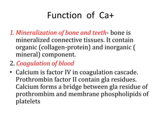
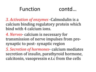
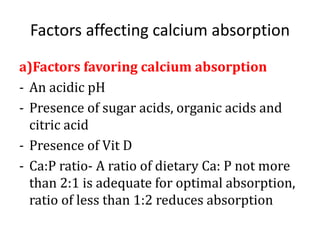
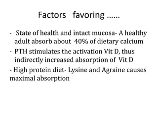
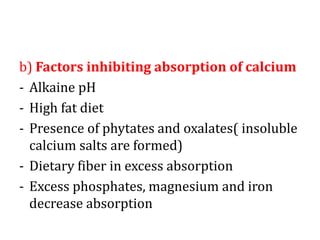
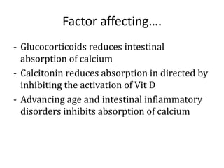
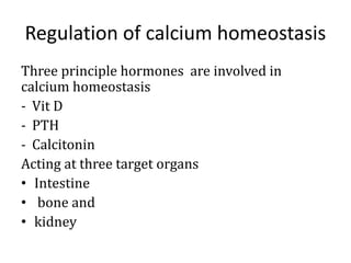
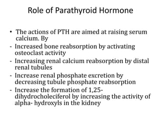
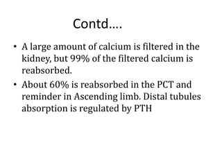
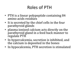
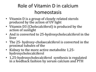
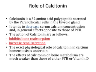
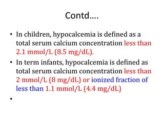
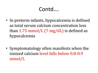
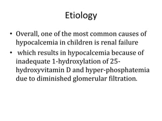
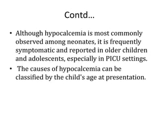
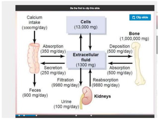
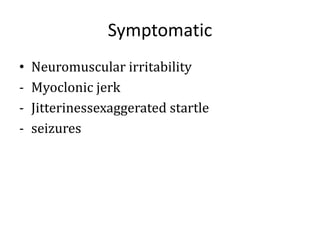
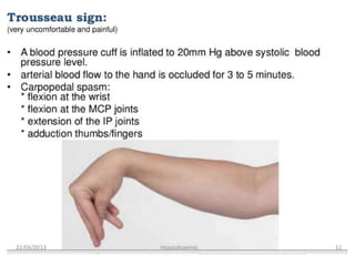
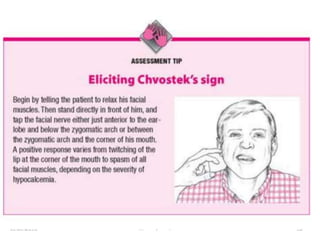
2. Function of Ca+
1. Mineralization of bone and teeth- bone is
mineralized connective tissues. It contain
organic (collagen-protein) and inorganic (
mineral) component.
2. Coagulation of blood
• Calcium is factor IV in coagulation cascade.
Prothrombin factor II contain gla residues.
Calcium forms a bridge between gla residue of
prothrombim and membrane phospholipids of
platelets
3. Function contd…
3. Activation of enzymes -Calmodulin is a
calcium binding regulatory protein which
bind with 4 calcium ions.
4. Nerves- calcium is necessary for
transmission of nerve impulses from pre-
synaptic to post- synaptic region
5. Secretion of hormones- calcium mediates
secretion of insulin, parathyroid hormone,
calcitonin, vasopressin e.t.c from the cells
5. Factors affecting calcium absorption
a)Factors favoring calcium absorption
- An acidic pH
- Presence of sugar acids, organic acids and
citric acid
- Presence of Vit D
- Ca:P ratio- A ratio of dietary Ca: P not more
than 2:1 is adequate for optimal absorption,
ratio of less than 1:2 reduces absorption
6. Factors favoring ……
- State of health and intact mucosa- A healthy
adult absorb about 40% of dietary calcium
- PTH stimulates the activation Vit D, thus
indirectly increased absorption of Vit D
- High protein diet- Lysine and Agraine causes
maximal absorption
7. b) Factors inhibiting absorption of calcium
- Alkaine pH
- High fat diet
- Presence of phytates and oxalates( insoluble
calcium salts are formed)
- Dietary fiber in excess absorption
- Excess phosphates, magnesium and iron
decrease absorption
8. Factor affecting….
- Glucocorticoids reduces intestinal
absorption of calcium
- Calcitonin reduces absorption in directed by
inhibiting the activation of Vit D
- Advancing age and intestinal inflammatory
disorders inhibits absorption of calcium
9. Influences on calcium concentrations
• The plasma pH and the total plasma protein
concentration influences the total calcium
level
• Since a significant proportion of calcium in
the blood is bound to albumin, it is
important to know the Plasma albumin
concentration when evaluating the total
plasma calcium
10. Influences contd…
• In general, 0.2 mmol/l must be added to the
total calcium concentration for each 1gm/dl
decreases in albumin concentration
• Ionized calcium increases with acidosis, and
decreases with alkalosis
11. Contd….
• Calcium is the most abundant mineral in the
body. Of the body's total calcium, 99% is stored
in bone, and serum levels constitute less than
1%.
• Various factors regulate the homeostasis of
calcium and maintain serum calcium within a
narrow range.
• These include parathormone (PTH), vitamin D,
hepatic and renal function (for conversion of
vitamin D to active metabolites), and serum
phosphate and magnesium levels.
12. • Serum calcium is present in two forms: the
free (ionized) and the bound form.
• Only about 50% of circulating calcium is
present in the physiologically free form.
• The rest is either bound to proteins (40%)
or complexes (10%) with bicarbonate,
citrate, and phosphate.
13. • The ionized calcium level varies based on
the level of serum albumin, blood pH, serum
phosphate, magnesium, and bicarbonate
levels, the administration of transfused
blood containing citrate and free fatty acid
content in total parenteral nutrition.
• The normal range for ionized calcium is 1-
1.25 mmol/L (4-5 mg/dL).
14. Regulation of calcium homeostasis
Three principle hormones are involved in
calcium homeostasis
- Vit D
- PTH
- Calcitonin
Acting at three target organs
• Intestine
• bone and
• kidney
15. Role of Parathyroid Hormone
• The actions of PTH are aimed at raising serum
calcium. By
- Increased bone reabsorption by activating
osteoclast activity
- Increasing renal calcium reabsorption by distal
renal tubules
- Increase renal phosphate excretion by
decreasing tubule phosphate reabsorption
- Increase the formation of 1,25-
dihydrocholeciferol by increasing the activity of
alpha- hydroxyls in the kidney
16. • A large amount of calcium is filtered in the
kidney, but 99% of the filtered calcium is
reabsorbed.
• About 60% is reabsorbed in the PCT and
reminder in Ascending limb. Distal tubules
absorption is regulated by PTH
Contd….
17. Roles of PTH
• PTH is a linear polypeptide containing 84
amino acids residues
• It is secreted by the chief cells in the four
parathyroid glands
• plasma ionized calcium acts directly on the
parathyroid gland in a feed back manner to
regulate PTH
• In hypercalcemia, secretion is inhibited, and
the calcium is deposited in the bones
• In hypocalcemia, PTH secretion is stimulated
18. Role of Vitamin D in calcium
homeostasis
• Vitamin D is a group of closely related sterols
produced by the action of UV light
• Vitamin D3 (Cholecalciferol) is produced by the
action of sunlight
• And is converted to 25-hydroxycholecalciferol in the
liver
• The 25- hydroxy-cholaecalciferol is converted in the
proximal tubules of the
• Kidney to the more active metabolite 1,25-
hydroxycholecalciferol
• 1,25-hydroxycholecalciferol synthesis is regulated
in a feedback fashion by serum calcium and PTH
19. Role of Calcitonin
• Calcitonin is a 32 amino acid polypeptide secreted
by the Para follicular cells in the thyroid gland
• It tends to decrease serum calcium concentration
and, in general effects opposite to those of PTH
• The action of Calcitonin are as follows:
- Inhibits bone reabsorption
- Increase renal secretion
• The exact physiological role of calcitonin in calcium
homeostasis is uncertain.
• The effects of calcitonin on bone metabolism are
much weaker than those of either PTH or Vitamin D
20. Glucocorticoids and calcium
homeostasis
• Glucocorticoids lower serum calcium levels by
inhibiting osteoclast formation and activity.
• But over long periods they causes Osteoporosis
by decreasing bone formation and increasing
bone reabsorption.
• They also decreases the absorption of calcium
from the intestine by anti vitamin D action and
increased its renal excretion.
• The decrease in serum calcium concentration
increases the secretion of PTH and bone
reabsorption is facilitated.
21. Growth hormones and calcium levels
• Growth hormone increases calcium
excretion in the urine
• It also increases intestinal absorption of
calcium and this effects may be greater than
• The effects on excretion with a resultant
positive calcium balance
22. Effects of other hormones on calcium
levels
• Thyroids hormones may cause
hypercalcemia, hypercalciuria and in some
instances osteoporosis
• Estrogens prevent osteoporosis, probably
by a direct effects on Osteoblasts.
• Insulin increases bone formation and there
is significant bone loss in untreated diabetes
23. Hypocalcemia
• Hypocalcemia is a laboratory and clinical
abnormality that is observed with relative
frequency, especially in neonatal pediatric
patients.
• Laboratory hypocalcemia is often
asymptomatic, and its treatment in neonates is
controversial.
• However, children with hypocalcemia in
pediatric intensive care units (PICUs) have
mortality rates higher than those of children
with normal calcium levels
24. Definition
• The definition of hypocalcemia is based on
both gestational and postnatal age in
neonates and is different for children.
• Calcium data are presented as both mg/dL
and mmol/L (1 mg/dL = 0.25 mmol/L)
25. Contd….
• In children, hypocalcemia is defined as a
total serum calcium concentration less than
2.1 mmol/L (8.5 mg/dL).
• In term infants, hypocalcemia is defined as
total serum calcium concentration less than
2 mmol/L (8 mg/dL) or ionized fraction of
less than 1.1 mmol/L (4.4 mg/dL)
•
26. Contd….
• In preterm infants, hypocalcemia is defined
as total serum calcium concentration less
than 1.75 mmol/L (7 mg/dL) is defined as
hypocalcemia
• Symptomatology often manifests when the
ionized calcium level falls below 0.8-0.9
mmol/L
27. Etiology
• Overall, one of the most common causes of
hypocalcemia in children is renal failure
• which results in hypocalcemia because of
inadequate 1-hydroxylation of 25-
hydroxyvitamin D and hyper-phosphatemia
due to diminished glomerular filtration.
28. Contd…
• Although hypocalcemia is most commonly
observed among neonates, it is frequently
symptomatic and reported in older children
and adolescents, especially in PICU settings.
• The causes of hypocalcemia can be
classified by the child's age at presentation.
29. Classification
Early onset neonatal hypocalcemia
• Early neonatal hypocalcemia, which occurs
within 48-72 hours of birth, is most commonly
seen in preterm and very low birth weight
infants, infants asphyxiated or depressed at
birth, infants of diabetic mothers, and the
intrauterine growth restricted infants. The
mechanisms underlying hypocalcemia caused
by these conditions are as follows:
30. Contd…
Prematurity:
- Possible mechanisms include inadequate
nutritional intake
- Decreased responsiveness of parathyroid
hormone to vitamin D
- Increased calcitonin level
- Increased urinary losses, and
hypoalbuminemia leading to a decreased
total (but normal ionized) calcium level.
31. Contd…
• Birth asphyxia:
- Delayed introduction of feeds
- Increased calcitonin production
- Increased endogenous phosphate load due
to tissue catabolism
- Renal failure, metabolic acidosis and its
treatment with alkali therapy all may
contribute to hypocalcemia.
32. Contd…
- Infants of a diabetic mother: The degree of
hypocalcemia is associated with the severity
of diabetes in the mother.
Magnesium depletion in mothers with
diabetes mellitus causes a hypomagnesemic
state in the fetus, which induces functional
hypoparathyroidism and hypocalcemia in
the infant.
33. Contd….
In addition, infants of diabetic mothers have
higher serum calcium in utero and this may
also suppress the parathyroid gland.
A high incidence of birth complications due
to microsomal and difficult delivery and, in
some cases, higher incidence of preterm
birth in infants of diabetic mothers are
contributing factors for hypocalcemia.
34. Contd…
Intrauterine growth restriction
- Infants with intrauterine growth restriction
may develop hypocalcemia because of
decreased transplacental passage of calcium.
- In addition, decreased accretion is present if
they are delivered preterm or have
experienced perinatal asphyxia as a result of
placental insufficiency.
35. Contd..
Late-onset neonatal hypocalcemia
• This occurs 3-7 days after birth, although
occasionally it is seen as late as age 6 weeks.
The following are some important causes of
late neonatal hypocalcemia:
-
36. Contd…
Exogenous phosphate load: This is most
commonly seen in developing countries. The
problem results when the neonate is fed with
phosphate-rich formula or cow's milk. Whole
cow's milk has 7 times the phosphate load of
breast milk (956 vs 140 mg/L in breast milk).
This may cause symptomatic hypocalcemia in
neonates.
37. Contd…
- Vitamin D deficiency: In a review of the medical
records of 78 term neonates with hypocalcemia,
moderate-to-severe late-onset neonatal
hypocalcemia developed more often in male infants
and Hispanic infants. It was often a sign of coexistent
vitamin D insufficiency or deficiency and
hypomagnesemia.
The newborns respond well to one or more of the
following: calcium supplements, calcitriol, low
phosphorus formula (PM 60/40), and magnesium
supplements for a limited period of time.
38. Contd…
• Primary immunodeficiency disorder: DiGeorge
Syndrome is the most important immunodeficiency
disorder to be aware of that is associated with
hypocalcemia.
• DiGeorge Syndrome is a primary immunodeficiency,
often but not always, characterized by cellular (T-
cell) deficiency, characteristic faces, congenital heart
disease and hypocalcemia. Hypoparathyroidism
causes hypocalcemia; 90% of infants with the
features of DiGeorge syndrome have a 22q11
chromosomal deletion
39. Contd…
Hypocalcemia in infants and children
• Hypoparathyroidism can result from the
following:
• Aplasia or hypoplasia of parathyroid gland -
DiGeorge syndrome known as velocardiofacial
(Shprintzen) syndrome or 22q11 deletion
syndrome; fetal exposure to retinoic acid;
complex of vertebral defects, anal atresia,
tracheoesophageal fistula with esophageal
atresia, and radial and renal abnormalities
(VATER/VACTERL);
40. Contd…
Parathormone (PTH) receptor defects -
Pseudohypoparathyroidism
• Autoimmune parathyroiditis
• Infiltrative lesions - Hemosiderosis, Wilson
disease, thalassemia
• Activating mutations of the calcium-sensing receptor
leading to inappropriately suppressed PTH secretion
• Idiopathic causes
• Hypoparathyroidism, abnormal vitamin D production or
action, and hyperphosphatemia are among the causes of
hypocalcemia in infants and children.
41. Contd..
• Abnormal vitamin D production or action can be caused
by the following:
• Vitamin D deficiency: Dietary insufficiency and maternal
use of anticonvulsants have been reported.
• Acquired or inherited disorders of vitamin D metabolism
• Resistance to actions of vitamin D
• Liver disease: Liver disease can affect 25-hydroxylation
of vitamin D; certain drugs (eg, phenytoin,
carbamazepine, phenobarbital, isoniazid, and rifampin)
can increase the activity of P-450 enzymes, which can
increase the 25-hydroxylation and also the catabolism of
vitamin D.
• Hyperphosphatemia can result from the following:
42. Contd..
• Excessive phosphate intake from feeding cow
milk or infant formula with improper (low)
calcium to phosphate ratio
• Excessive phosphate intake caused by
inappropriate use of phosphate-containing
enemas
• Excessive phosphate or inappropriate Ca:P
ratio in total parenteral nutrition
• Increased endogenous phosphate load caused
by anoxia, chemotherapy, or rhabdomyolysis
• Renal failure
43. Contd..
Other causes of hypocalcemia in infants and
children include the following:
• Malabsorption syndromes
• Alkalosis: Respiratory alkalosis is caused by
hyperventilation; metabolic alkalosis occurs
with the administration of bicarbonate,
diuretics, or chelating agents, such as the high
doses of citrates taken in during massive blood
transfusions.
• Pancreatitis
44. Contd..
• Pseudohypocalcemia (ie, hypoalbuminemia):
Serum calcium concentration decreases by 0.8
mg/dL for every 1 g/dL fall in concentration of
plasma albumin.
• “Hungry bones syndrome:" Rapid skeletal
mineral deposition is seen in infants with
rickets or hypoparathyroidism after starting
vitamin D therapy.
•
55. Diagnosis
• Serum calcium
• Serum Phosphate
• Serum Albumin
• RFT
• PTH level in serum
• Parathyroid antibodies ( present in idiopahic
hypoparathyrodism)
• Vit D serum level
• Serum magnisium levelX-ray of metacarpals
• Cardiac enzzumes
• ABG analysis
• ECG
56.
57. Management
• Depend on the underlying cause and
severity
• Administration of calcium alone is only
transiently effective
• Mild asymptomatic cases-often adequate to
increase dietary calcium by 1000mg/day
• Symptomatic: treat immediately
59. Contd..
Symptomatic
• Bolus dose of 2ml/kg/dose (diluted)
5% dextrose over 10 min under cardiac
monitoring
• Calcium chloride 20mg/kg may be given
over 10-30 min
60.
61. Contd..
Hypomagnesemia:
• Symptomatic hypocalcemia unresponsive to
adequate doses of IV calcium therapy is
usually due to hypomagnesemia.
• 2 doses of 0.2 mL/kg of 50% MgSO4
injection, 12 hr apart, deep IM followed by a
maintenance dose of 0.2 mL/kg/day of 50%
MgSO4, PO for 3 days.
62. Contd..
High phosphate load:
• These infants have hyperphosphatemia with
near normal calcium levels. Exclusive
breast-feeding should be encouraged and
top feeding with cow’s milk should be
discontinued. Phosphate binding gels
should be avoided.
63. Vitamin D deficiency states:
• These babies have hypocalcemia associated
with hypophosphatemia due to an intact
parathormone response on the kidneys.
They benefit from Vitamin D3
supplementation in a dose of 30-60
ng/kg/day