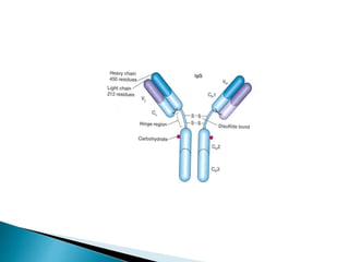
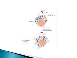
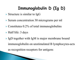
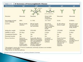
Immunoglobulins
Akanksha Dubey
Immunoglobulin
 Immunoglobulin is a glycoprotein that is made in
response to an antigen and can recognize and bind to
that antigen.
 Abbreviated as Ig
 Are gamma globulins(Mobility in Electric Field)
 Immunoglobulins and Gamma Globulins are synonyms
 Synthesized by plasma cells
 Immunoglobulin is a functional term while
Gamma Globulin is Physical term
 Constitute 25-30 % of total serum proteins
 Antibodies are present in serum, tissue fluids and
mucosal surfaces.
 All antibodies are immunoglobulins, but all
immunoglobulins may not be antibodies
Basic structure
 Composed of 4 polypeptide chains.
 2 identical light and 2 identical heavy chains
 Linked by disulphide bonds
 Light chains similar in all immunoglobulins
 Light chains occur in 2 varieties kappa and lambda
 For eg: IgG may have λ(Lambda) or κ (Kappa) in all
classes
 Light and Heavy chains are subdivided into variable and
constant region as per their amino acid composition.
 VL and CL are terms used for light chain and VH and CH
are used for heavy chain.
 For eg: First 108 aa in light chain and 118 aa in Gamma
Heavy chain constitutes variable region.
 Each heavy and light chain contains amino terminal in
variable region carboxy terminal in constant region
 Heavy chains are structurally and antigenically
distinct for each class
 Each immunoglobulin peptide chain has intra chain
disulphide bonds which form loops
 Each loop is compactly folded to form a globular
structure-domain
 Light chain contains a single variable domain (VL)
and a single constant domain (CL).
 Heavy chain contains one variable domain (VH) and
3 constant domains (CH1, CH2, CH3)
 Hinge region is the segment in heavy chain - between
CH1, CH2
Digestion with proteolytic enzymes
Papain enzyme
 Papain is proteolytic enzyme from papaya cleaves Ig in
2 Fab portions (Fraction Antibody) and 1 Fc(Fraction
Crystallizable) portions.
 Peptide bonds in the hinge region are broken
 Produces 3 fragments
 2 identical fragments called Fab fragments it possess antigen
binding activity.
 Other fragment called Fc fragment (Fraction crystallizable)
Pepsin Digestion
 Another enzyme Pepsin cleaves Ig at other site
Produce a single fragment composed of two Fab like
subunits F(ab)2 binds antigen
 Fc fragment is not recovered- digested to small
numerous peptides.
Classification
 Based on structure and antigenic nature of Heavy
chain the Immunoglobulins are classified into 5
classes.
 Ig G- (gamma)
 Ig A- (alpha)
 Ig M- (mu)
 Ig D- (delta)
 Ig E - (epsilon)
Immunoglobulin G (Ig G)
 Most abundant antibody in serum
 Constitutes 75-80% of total immunoglobulin
 Present in blood, plasma and tissue fluids
 IgG contains two heavy chain and two light chains
 Heavy chain is of Gamma Type
 Contains less carbohydrate than other immunoglobulins
 It has a half life of 23 days: the longest of all of the
immunoglobulin isotypes
 This antibody is seen in Secondary Immune Response.
 It can cross placental barrier hence can protect newborn from
infections.
 Available in neonatal circulation up to 2-4 months
 Acts against bacteria and viruses by opsonizing
 Neutralize toxin
 Activate complement by classical pathway
 Catabolism of IgG is unique in that it varies with
its serum concentration
Sub classes of Ig G
 Ig G1……Ig G2….. Ig G3…..and Ig G4.
Biological Function of Subclasses
 IgG1, IgG3, IgG4 – cross placenta and protect foetus
 IgG3 activates complement
 IgG1 and IgG3 binds to Fc receptor on phagocytic cells,
monocytes and macrophages and mediate opsinization.
 Constitutes 10-15 % of Total Immunoglobulins
 Present in milk, saliva, tears, mucous of Respiratory
Tract, Digestive Tract and Genitourinary Tract.
 In serum exist as monomer
 In external secretions exist as Dimer (4 Heavy chain
and 4 Light Chain) called Secretory Immunoglobulin.
 The J chain connects the dimer. The dimers are stabilized
against proteolytic enzymes by Secretory Piece. This piece is
produced in liver, reaches to intestinal mucosal cells, where it
combines with IgA dimer to form secretory IgA which is then
released.
 Half life: 6-8 days
 The portion of the receptor that remains attached to the Ig A
dimer – secretory component
 Secretory piece protects Ig A from digestive enzymes and
denaturation by bacterial proteases
Functions
 Provides local immunity.
 Secretory Ig A binds to surface antigens of
microorganism and prevent its attachment and invasion of
the mucosal surfaces of respiratory and digestive tract-
immune elimination.
 Secretory IgA provides important line of defense against
salmonella, Vibrio cholerae, N. gonorrhoeae, influenza
virus and poliovirus.
 Secretory IgA present in breast milk protects
newborn during first months of life.
 Activates complement by the alternative pathway
 Promotes phagocytosis and intracellular killing of
microorganisms
Immunoglobulin M (Ig M)
 Accounts for 5-10% of total serum proteins
 Polymer of five monomeric units (pentamer) 10 heavy
chains and 10 light chains joined by disulfide bonds and
‘J’ chain
 It can combine with 5 antigens simultaneously.
 Mol. Wt. of 900,000-10,00,000 (millionaire molecule)
hence they are macroglobulins or 19 S Ig
 Half life: 5 days
 Most of IgM (80%) present intravascularly because of
large molecular weight.
 IgM are predominant class of antibodies in Primary
response. Natural Ab’s are IgM in nature
 Cannot cross placenta
 Presence of IgM antibody in serum of newborn
indicate congenital infection.
 Earliest immunoglobulin to be synthesized by foetus
(20 weeks)
 Relatively short-lived hence it’s demonstration in the
serum indicates recent infection
 Monomeric IgM appears on the surface of
unstimulated B lymphocytes and act as receptors for
antigens
Functions
 It agglutinates bacteria more effectively.
 Activates complement by classical pathway
 Causes opsonization and immune hemolysis
 Believed to be responsible for protection against
blood invasion by microorganisms
Immunoglobulin E (Ig E)
 They are cytophillic antibodies and they mediate allergy,
hypersenstivity and anaphylaxis.
 Structure is similar to Ig G
 Has 4 constant region domains.
 Mol. Wt. 1,90,000
 Half life: 2 days
 Heat labile (inactivated at 560C in 1 hour)
 Normal serum concentration 0.3 ug/ml
 Mostly present extra cellularly
 Does not cross placenta
 Produced in the lining of respiratory and intestinal
tract
 Known as reagin antibody
 Does not activate complement nor agglutinate
antigens
 Binds to the Fc receptors on the membranes of blood
basophils and tissue mast cells
 IgE binds to Fc receptors on the membrane of blood
basophils and tissue mast cells.
 When two IgE molecules on the surface of these cells
are cross linked by binding of the same antigen- cells
degranulates.
 Mediates immediate hypersensitivity reaction.
 Responsible for symptoms of anaphylactic shock, hay
fever and asthma.
 Play a role in immunity against helminthic parasites
and IgE level markedly increased in above condition.
 Release histamine and pharmacological mediators of
anaphylaxis from cell.
 The physiological role of IgE appears to be protection
against pathogens by mast cell degranulation and
release of inflammatory mediators
 Mediates P.K. reaction (PRAUSNITZ & KUSTNER)
 The presence of a serum component responsible for
allergic reaction was first demonstrated by Prausnitz and
Kustner in 1921.
 Kustner was suffering from atopic hypersensitivity to
certain species of fish
 Kustner’s serum was injected intracutaneously in
Prausnitz
 After 24 hrs small quantity of cooked fish antigen
was injected at the same site
 A wheal and flare reaction occurred within minutes.
Immunoglobulin D (Ig D)
 Structure is similar to IgG
 Serum concentration 30 micrograms per ml
 Constitutes 0.2% of total immunoglobulins
 Half life: 3 days
 IgD together with IgM is major membrane bound
immunoglobulin on unstimulated B lymphocytes-acts
as recognition receptors for antigens
Antibody Types
 Polyclonal: When an animal is immunized with complex
antigen some B cells will make antibodies against one Epitope
whereas other cells will make antibodies for other epitope of
an same antigen. An antigen have more than one Epitope
 The antibodies present in blood are actually mixture of
different immunoglobulins molecules. Antibodies are derived
from several B cells they are c/a Polyclonal Antibodies.
 Polyclonal Ab’s are heterogenous mixture
 Monoclonal: They are derived from clones of single B cells.
Clones synthesized one type of antibody specific for just one
epitope.
 Monoclonal antibodies arise from benign or malignant
transformation of single B cell which produces identical
antibodies just for one epitope.
 They are Homogenous made up of single Ig’s
 If clones multiply malignantly s/a in Multiple Myeloma....
Waldenstorm’s Macroglobulinaemia....the serum concentration
of particular Ig’s become very high.
Clinical Importance of Immunoglobulin's:
 Quantitative changes in the amount of Ig’s in plasma and urine
are known in several pathological conditions in human.
Abnormally large amount of Ig’s are found in serum in several
disease. As well as deficiency of Gamma Globulins is found in
rare hereditary disease
Hypergammaglobulinaemia
1. Polyclonal Hypergammaglobulinaemia d/t increased
production of large numbers of different immunoglobulins.
Liver disease s/a hepatitis, cirrhosis, recurrent infections s/a
Rheumatoid Arthritis stimulate B cells for production of
heterogenous antibodies.
In this case Diffuse Discrete pattern is observed on
electrophoresis in contrast to monoclonal where Single band
is observed.
2.monoclonal hypergammaglobulinaemia
Or
Paraproteinaemia
Characterized by abnormal benign or malignant proliferation of
single clone of B lymphocytes or plasma cells that produces
homogenous Ig’s
Discrete Ig’s bands are observed on electrophoresis k/a
paraproteins or monoclonal components d/t production of
single Ig’s
Malignant paraproteinaemia occurs in Multiple Myeloma,
Plasma Cell leukemia, Waldenstorm’s macroglobulinaemia ,
heavy and light chain disease, amyloidosis, and other tumours.
 Multiple Myeloma: Characterized by a malignant proliferation
of plasma cells derived from a single clone.
 Most common primary malignancy of bone (~40%)
 Also known as Plasmocytoma or Monoclonal-gammopathy
Multiple
Myeloma
Bone
Nervous
system
KidneyBlood
Chest
 Multiple myeloma is characterized by excessive numbers of
abnormal plasma cells in the bone marrow and overproduction of
intact monoclonal immunoglobulin usually IgG or IgA, or
Bence-Jones protein (free monoclonal κ and λ light chains).
 BJ Proteins: they are light chain proteins (low molecular weight)
hence they enter the circulation and through glomerulus appear
in urine.
 Multiple myeloma with bence jones proteins is k/a light chain
disease.
Organ damage classified as “CRAB”
 C – calcium elevation (>10 mg/L)
 R – renal dysfunction (creatinine >2 mg/dL)
 A – anemia (hemoglobin <10 g/dL or ≥2 g/dL decrease from
patient’s normal)
 B – bone disease (osteoporosis)
 Waldenstorm’s Macroglobulinaemia
Malignant disease of lymphoid elements characterized by high
serum IgM.
 Amyloid Disease
Amyloidosis is a group of diseases in which abnormal protein,
known as amyloid fibrils, builds up in tissue
Amyloid proteins found in body tissue and fluids. Deposition of
amyloid in tissues is k/a Amyloidosis
Deposition leads to pressure on vital organs.
Generally associated with Multiple Myeloma
 There are about 30 different type of amyloidosis, each due to a
specific protein misfolding
 Diagnosis may be suspected when protein is found in the
urine, organ enlargement is present,
Hypogammaglobulinaemia
Deficiency of Immunoglobulins may be of primary nature
commonly genetic in origin or secondary to some other
disease.
Antibody Diversity
 Human Genome is thought to contain fewer than 105 genes,
yet a human can make at least 1015 different types of
antibodies in terms of Antigen binding specificity.
 One of the important feature of the vertebrate immune system
is its ability to respond to an apparently limitless array of
foreign antigens.
 As immunoglobulin (Ig) sequence data accumulated , virtually
every antibody molecule studied was found to contain a
unique amino acid sequence in its variable region but only one
of a limited number of invariant sequences in its constant
region.
 The genetic basis for this combination of constancy and
tremendous variation in a single protein molecule lies in the
organization of the immunoglobulin genes.
 An Ab combining site is made up of one VL and one VH.
 The specificity of any combining site is determined by its
amino acid sequence.
 There exist at least 106 unique combining sites .
 The formation of a complete variable region of a light or
heavy chain requires the joining of two or three separate
genetic elements by a process of gene rearrangement
Theories
 Germ-Line Model
 Somatic-Variation Model
 Two-Gene Model
 GERM-LINE THEORY –
◦ For every kappa-chain V-region there exists one unique germ-
line gene. A particular antibody-forming cell selects one of
these and expresses it in unmodified form.
 SOMATIC THEORY –
◦ Only a single germ-line gene exists for all kappa-chain V-
regions.
◦ A particular antibody-forming cell expresses this gene
following a process of somatic mutation, which results in each
cell expressing a different version of this gene.
TWO-GENE MODEL
 Dreyer and Bennett proposed the Two-Gene Model.
 In 1965-Proposed that two separate genes encode a single
immunoglobulin heavy or light chain, one gene for the V
region (variable region) and the other for the C region
(constant region).
 They suggested that these two genes must somehow come
together at the DNA level to form a continuous message that
can be transcribed and translated into a single Ig heavy or light
chain.
 Moreover, they proposed that hundreds or thousands of V-
region genes were carried in the germ line, whereas only
single copies of C-region class and subclass genes need exist.
STRUCTURE AND EXPRESSION OF
IMMUNOGLOBULIN GENES
 Three families of immunoglobulin genes exist, each on a
separate chromosome.
◦ kappa genes- chromosome 2
◦ lambda genes- chromosome 22
◦ heavy chain genes- chromosome 14
 Each family consists of a series of V-regions genetically linked
to one or more C-regions.
 Antibody is a protein and antigen binding region is made up of
VL and VH regions of light and heavy polypeptide chain. So
how it is possible to have large variety of antigen specific
antibodies when there are only 30-35000 genes for
polypeptide.
 This is explained by Multi gene theory of Immunoglobulin.
Accordingly each VL region is coded by 2 genes V and J and
each VH region is coded by 3 genes V D and J.
 There are many alleles known for the genes coding VL and
VH regions of light and heavy chains
 For eg: There are 40 VL and 5 JL genes
 There are 51 VH, 27 DH and 6 JH genes
 So by random combination of V and J genes in VL regions or
V D and J genes in VH regions a large variety of different
antigenic specific variables regions are produced. Splicing
produced fully functional antibody molecule.
MECHANISMS FOR GENERATING ANTIBODY
DIVERSITY
 Multiple germ-line gene segments
 Combinatorial V-(D)-J joining
 Junctional flexibility
 P-region nucleotide addition (P-addition)
 N-region nucleotide addition (N-addition)
 Somatic hypermutation
 Combinatorial association of light and heavy chains.
Immunoglobulins
Immunoglobulins

Immunoglobulins

  • 2.
  • 3.
    Immunoglobulin  Immunoglobulin isa glycoprotein that is made in response to an antigen and can recognize and bind to that antigen.  Abbreviated as Ig  Are gamma globulins(Mobility in Electric Field)  Immunoglobulins and Gamma Globulins are synonyms  Synthesized by plasma cells
  • 4.
     Immunoglobulin isa functional term while Gamma Globulin is Physical term  Constitute 25-30 % of total serum proteins  Antibodies are present in serum, tissue fluids and mucosal surfaces.  All antibodies are immunoglobulins, but all immunoglobulins may not be antibodies
  • 6.
    Basic structure  Composedof 4 polypeptide chains.  2 identical light and 2 identical heavy chains  Linked by disulphide bonds  Light chains similar in all immunoglobulins  Light chains occur in 2 varieties kappa and lambda  For eg: IgG may have λ(Lambda) or κ (Kappa) in all classes
  • 7.
     Light andHeavy chains are subdivided into variable and constant region as per their amino acid composition.  VL and CL are terms used for light chain and VH and CH are used for heavy chain.  For eg: First 108 aa in light chain and 118 aa in Gamma Heavy chain constitutes variable region.  Each heavy and light chain contains amino terminal in variable region carboxy terminal in constant region
  • 9.
     Heavy chainsare structurally and antigenically distinct for each class  Each immunoglobulin peptide chain has intra chain disulphide bonds which form loops  Each loop is compactly folded to form a globular structure-domain  Light chain contains a single variable domain (VL) and a single constant domain (CL).
  • 11.
     Heavy chaincontains one variable domain (VH) and 3 constant domains (CH1, CH2, CH3)  Hinge region is the segment in heavy chain - between CH1, CH2
  • 13.
    Digestion with proteolyticenzymes Papain enzyme  Papain is proteolytic enzyme from papaya cleaves Ig in 2 Fab portions (Fraction Antibody) and 1 Fc(Fraction Crystallizable) portions.  Peptide bonds in the hinge region are broken  Produces 3 fragments
  • 14.
     2 identicalfragments called Fab fragments it possess antigen binding activity.  Other fragment called Fc fragment (Fraction crystallizable)
  • 16.
    Pepsin Digestion  Anotherenzyme Pepsin cleaves Ig at other site Produce a single fragment composed of two Fab like subunits F(ab)2 binds antigen  Fc fragment is not recovered- digested to small numerous peptides.
  • 18.
  • 19.
     Based onstructure and antigenic nature of Heavy chain the Immunoglobulins are classified into 5 classes.  Ig G- (gamma)  Ig A- (alpha)  Ig M- (mu)  Ig D- (delta)  Ig E - (epsilon)
  • 20.
    Immunoglobulin G (IgG)  Most abundant antibody in serum  Constitutes 75-80% of total immunoglobulin  Present in blood, plasma and tissue fluids  IgG contains two heavy chain and two light chains  Heavy chain is of Gamma Type
  • 21.
     Contains lesscarbohydrate than other immunoglobulins  It has a half life of 23 days: the longest of all of the immunoglobulin isotypes  This antibody is seen in Secondary Immune Response.  It can cross placental barrier hence can protect newborn from infections.  Available in neonatal circulation up to 2-4 months
  • 23.
     Acts againstbacteria and viruses by opsonizing  Neutralize toxin  Activate complement by classical pathway  Catabolism of IgG is unique in that it varies with its serum concentration
  • 26.
    Sub classes ofIg G  Ig G1……Ig G2….. Ig G3…..and Ig G4.
  • 27.
    Biological Function ofSubclasses  IgG1, IgG3, IgG4 – cross placenta and protect foetus  IgG3 activates complement  IgG1 and IgG3 binds to Fc receptor on phagocytic cells, monocytes and macrophages and mediate opsinization.
  • 28.
     Constitutes 10-15% of Total Immunoglobulins  Present in milk, saliva, tears, mucous of Respiratory Tract, Digestive Tract and Genitourinary Tract.  In serum exist as monomer  In external secretions exist as Dimer (4 Heavy chain and 4 Light Chain) called Secretory Immunoglobulin.
  • 30.
     The Jchain connects the dimer. The dimers are stabilized against proteolytic enzymes by Secretory Piece. This piece is produced in liver, reaches to intestinal mucosal cells, where it combines with IgA dimer to form secretory IgA which is then released.  Half life: 6-8 days
  • 32.
     The portionof the receptor that remains attached to the Ig A dimer – secretory component  Secretory piece protects Ig A from digestive enzymes and denaturation by bacterial proteases
  • 33.
    Functions  Provides localimmunity.  Secretory Ig A binds to surface antigens of microorganism and prevent its attachment and invasion of the mucosal surfaces of respiratory and digestive tract- immune elimination.  Secretory IgA provides important line of defense against salmonella, Vibrio cholerae, N. gonorrhoeae, influenza virus and poliovirus.
  • 34.
     Secretory IgApresent in breast milk protects newborn during first months of life.  Activates complement by the alternative pathway  Promotes phagocytosis and intracellular killing of microorganisms
  • 35.
    Immunoglobulin M (IgM)  Accounts for 5-10% of total serum proteins  Polymer of five monomeric units (pentamer) 10 heavy chains and 10 light chains joined by disulfide bonds and ‘J’ chain  It can combine with 5 antigens simultaneously.  Mol. Wt. of 900,000-10,00,000 (millionaire molecule) hence they are macroglobulins or 19 S Ig  Half life: 5 days
  • 37.
     Most ofIgM (80%) present intravascularly because of large molecular weight.  IgM are predominant class of antibodies in Primary response. Natural Ab’s are IgM in nature  Cannot cross placenta  Presence of IgM antibody in serum of newborn indicate congenital infection.  Earliest immunoglobulin to be synthesized by foetus (20 weeks)
  • 38.
     Relatively short-livedhence it’s demonstration in the serum indicates recent infection  Monomeric IgM appears on the surface of unstimulated B lymphocytes and act as receptors for antigens
  • 39.
    Functions  It agglutinatesbacteria more effectively.  Activates complement by classical pathway  Causes opsonization and immune hemolysis  Believed to be responsible for protection against blood invasion by microorganisms
  • 40.
    Immunoglobulin E (IgE)  They are cytophillic antibodies and they mediate allergy, hypersenstivity and anaphylaxis.  Structure is similar to Ig G  Has 4 constant region domains.  Mol. Wt. 1,90,000  Half life: 2 days  Heat labile (inactivated at 560C in 1 hour)
  • 41.
     Normal serumconcentration 0.3 ug/ml  Mostly present extra cellularly  Does not cross placenta
  • 43.
     Produced inthe lining of respiratory and intestinal tract  Known as reagin antibody  Does not activate complement nor agglutinate antigens  Binds to the Fc receptors on the membranes of blood basophils and tissue mast cells
  • 44.
     IgE bindsto Fc receptors on the membrane of blood basophils and tissue mast cells.  When two IgE molecules on the surface of these cells are cross linked by binding of the same antigen- cells degranulates.
  • 45.
     Mediates immediatehypersensitivity reaction.  Responsible for symptoms of anaphylactic shock, hay fever and asthma.  Play a role in immunity against helminthic parasites and IgE level markedly increased in above condition.
  • 46.
     Release histamineand pharmacological mediators of anaphylaxis from cell.  The physiological role of IgE appears to be protection against pathogens by mast cell degranulation and release of inflammatory mediators
  • 48.
     Mediates P.K.reaction (PRAUSNITZ & KUSTNER)  The presence of a serum component responsible for allergic reaction was first demonstrated by Prausnitz and Kustner in 1921.  Kustner was suffering from atopic hypersensitivity to certain species of fish
  • 49.
     Kustner’s serumwas injected intracutaneously in Prausnitz  After 24 hrs small quantity of cooked fish antigen was injected at the same site  A wheal and flare reaction occurred within minutes.
  • 50.
    Immunoglobulin D (IgD)  Structure is similar to IgG  Serum concentration 30 micrograms per ml  Constitutes 0.2% of total immunoglobulins  Half life: 3 days  IgD together with IgM is major membrane bound immunoglobulin on unstimulated B lymphocytes-acts as recognition receptors for antigens
  • 54.
    Antibody Types  Polyclonal:When an animal is immunized with complex antigen some B cells will make antibodies against one Epitope whereas other cells will make antibodies for other epitope of an same antigen. An antigen have more than one Epitope  The antibodies present in blood are actually mixture of different immunoglobulins molecules. Antibodies are derived from several B cells they are c/a Polyclonal Antibodies.  Polyclonal Ab’s are heterogenous mixture
  • 55.
     Monoclonal: Theyare derived from clones of single B cells. Clones synthesized one type of antibody specific for just one epitope.  Monoclonal antibodies arise from benign or malignant transformation of single B cell which produces identical antibodies just for one epitope.  They are Homogenous made up of single Ig’s  If clones multiply malignantly s/a in Multiple Myeloma.... Waldenstorm’s Macroglobulinaemia....the serum concentration of particular Ig’s become very high.
  • 59.
    Clinical Importance ofImmunoglobulin's:  Quantitative changes in the amount of Ig’s in plasma and urine are known in several pathological conditions in human. Abnormally large amount of Ig’s are found in serum in several disease. As well as deficiency of Gamma Globulins is found in rare hereditary disease
  • 60.
    Hypergammaglobulinaemia 1. Polyclonal Hypergammaglobulinaemiad/t increased production of large numbers of different immunoglobulins. Liver disease s/a hepatitis, cirrhosis, recurrent infections s/a Rheumatoid Arthritis stimulate B cells for production of heterogenous antibodies. In this case Diffuse Discrete pattern is observed on electrophoresis in contrast to monoclonal where Single band is observed.
  • 62.
    2.monoclonal hypergammaglobulinaemia Or Paraproteinaemia Characterized byabnormal benign or malignant proliferation of single clone of B lymphocytes or plasma cells that produces homogenous Ig’s Discrete Ig’s bands are observed on electrophoresis k/a paraproteins or monoclonal components d/t production of single Ig’s
  • 63.
    Malignant paraproteinaemia occursin Multiple Myeloma, Plasma Cell leukemia, Waldenstorm’s macroglobulinaemia , heavy and light chain disease, amyloidosis, and other tumours.  Multiple Myeloma: Characterized by a malignant proliferation of plasma cells derived from a single clone.  Most common primary malignancy of bone (~40%)  Also known as Plasmocytoma or Monoclonal-gammopathy
  • 64.
  • 65.
     Multiple myelomais characterized by excessive numbers of abnormal plasma cells in the bone marrow and overproduction of intact monoclonal immunoglobulin usually IgG or IgA, or Bence-Jones protein (free monoclonal κ and λ light chains).  BJ Proteins: they are light chain proteins (low molecular weight) hence they enter the circulation and through glomerulus appear in urine.  Multiple myeloma with bence jones proteins is k/a light chain disease.
  • 66.
    Organ damage classifiedas “CRAB”  C – calcium elevation (>10 mg/L)  R – renal dysfunction (creatinine >2 mg/dL)  A – anemia (hemoglobin <10 g/dL or ≥2 g/dL decrease from patient’s normal)  B – bone disease (osteoporosis)
  • 68.
     Waldenstorm’s Macroglobulinaemia Malignantdisease of lymphoid elements characterized by high serum IgM.  Amyloid Disease Amyloidosis is a group of diseases in which abnormal protein, known as amyloid fibrils, builds up in tissue Amyloid proteins found in body tissue and fluids. Deposition of amyloid in tissues is k/a Amyloidosis Deposition leads to pressure on vital organs. Generally associated with Multiple Myeloma
  • 69.
     There areabout 30 different type of amyloidosis, each due to a specific protein misfolding  Diagnosis may be suspected when protein is found in the urine, organ enlargement is present,
  • 70.
    Hypogammaglobulinaemia Deficiency of Immunoglobulinsmay be of primary nature commonly genetic in origin or secondary to some other disease.
  • 71.
    Antibody Diversity  HumanGenome is thought to contain fewer than 105 genes, yet a human can make at least 1015 different types of antibodies in terms of Antigen binding specificity.  One of the important feature of the vertebrate immune system is its ability to respond to an apparently limitless array of foreign antigens.
  • 72.
     As immunoglobulin(Ig) sequence data accumulated , virtually every antibody molecule studied was found to contain a unique amino acid sequence in its variable region but only one of a limited number of invariant sequences in its constant region.  The genetic basis for this combination of constancy and tremendous variation in a single protein molecule lies in the organization of the immunoglobulin genes.  An Ab combining site is made up of one VL and one VH.
  • 73.
     The specificityof any combining site is determined by its amino acid sequence.  There exist at least 106 unique combining sites .  The formation of a complete variable region of a light or heavy chain requires the joining of two or three separate genetic elements by a process of gene rearrangement
  • 74.
    Theories  Germ-Line Model Somatic-Variation Model  Two-Gene Model
  • 75.
     GERM-LINE THEORY– ◦ For every kappa-chain V-region there exists one unique germ- line gene. A particular antibody-forming cell selects one of these and expresses it in unmodified form.  SOMATIC THEORY – ◦ Only a single germ-line gene exists for all kappa-chain V- regions. ◦ A particular antibody-forming cell expresses this gene following a process of somatic mutation, which results in each cell expressing a different version of this gene.
  • 76.
    TWO-GENE MODEL  Dreyerand Bennett proposed the Two-Gene Model.  In 1965-Proposed that two separate genes encode a single immunoglobulin heavy or light chain, one gene for the V region (variable region) and the other for the C region (constant region).  They suggested that these two genes must somehow come together at the DNA level to form a continuous message that can be transcribed and translated into a single Ig heavy or light chain.
  • 77.
     Moreover, theyproposed that hundreds or thousands of V- region genes were carried in the germ line, whereas only single copies of C-region class and subclass genes need exist.
  • 78.
    STRUCTURE AND EXPRESSIONOF IMMUNOGLOBULIN GENES  Three families of immunoglobulin genes exist, each on a separate chromosome. ◦ kappa genes- chromosome 2 ◦ lambda genes- chromosome 22 ◦ heavy chain genes- chromosome 14  Each family consists of a series of V-regions genetically linked to one or more C-regions.
  • 80.
     Antibody isa protein and antigen binding region is made up of VL and VH regions of light and heavy polypeptide chain. So how it is possible to have large variety of antigen specific antibodies when there are only 30-35000 genes for polypeptide.  This is explained by Multi gene theory of Immunoglobulin. Accordingly each VL region is coded by 2 genes V and J and each VH region is coded by 3 genes V D and J.
  • 81.
     There aremany alleles known for the genes coding VL and VH regions of light and heavy chains  For eg: There are 40 VL and 5 JL genes  There are 51 VH, 27 DH and 6 JH genes  So by random combination of V and J genes in VL regions or V D and J genes in VH regions a large variety of different antigenic specific variables regions are produced. Splicing produced fully functional antibody molecule.
  • 84.
    MECHANISMS FOR GENERATINGANTIBODY DIVERSITY  Multiple germ-line gene segments  Combinatorial V-(D)-J joining  Junctional flexibility  P-region nucleotide addition (P-addition)  N-region nucleotide addition (N-addition)  Somatic hypermutation  Combinatorial association of light and heavy chains.