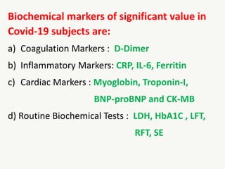
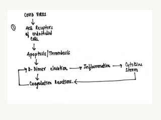
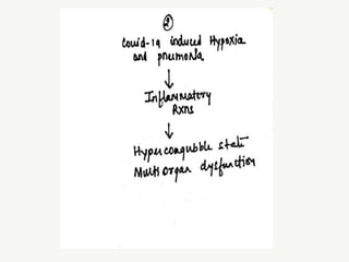
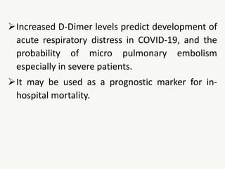
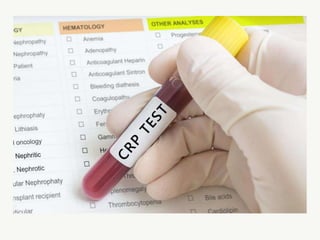
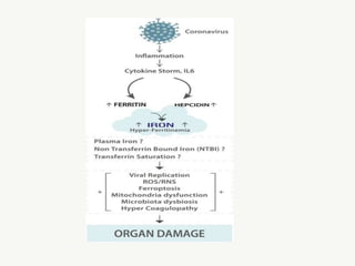
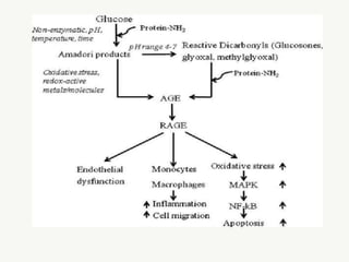
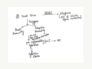
BIOCHEMICAL MARKERS OF COVID-19 AND
CHANGES IN BIOCHEMICAL PROFILE OBSERVED
IN COVID-19 SUBJECTS
AKANKSHA DUBEY
(PhD Scholar)
Under Guidance of
DR PRASHANT J. HISALKAR
Professor & HOD, Dept. Biochemistry
GMCH, Dungarpur (Rajasthan)
Former Dean Academic Affairs &
Controller of Examinations,
People’s University Bhopal (Madhya Pradesh)
India, the current epicenter of the corona virus
pandemic, has reported more than 2 million
new cases and 18,000 Covid-related deaths this
week alone, as a catastrophic second wave
continues throughout the country, pushing
hospitals and crematoriums past their breaking
point.
(Source : Forbes Topline)
 The first case of the COVID-19 pandemic in India was
reported on January 30, 2020.
 Till date 2.95 Cr cases have been reported with 3.71 L
deaths.
 The second wave of Covid-19 infections in India
peaked on May 9, 2021, has come down in June
2021.
 The pace of vaccinations will matter the most in
determining whether or not there is a nation-wide
third wave.
Key Facts :
 India's Health Ministry on Saturday reported
401,993 new infections, the highest number
of new cases reported yet in a single country
in a 24-hour span during second wave
 Hospitals throughout the country continue to
plead for medical supplies, especially oxygen.
 Despite being the world's leading producer of
vaccines, only 2% of India's population has
been vaccinated.
 The clinical characteristics of COVID-19 are
varied and can range from an asymptomatic
infection to a mild to severe pneumonia.
 In this critical situation only group of Laboratory
Tests can categorize patients into mild,
moderate and severe form.
 The hallmark of severe COVID-19 is the hyper
inflammatory host response k/a “Cytokine Storm”,
defined as an uncontrolled systemic inflammatory
response due to the release of large amounts of pro-
inflammatory cytokines, resulting from the SARS-CoV-2
induced activation of both natural and cellular
immunity.
 Hence besides routine biochemical parameters,
special inflammatory markers have been shown to
provide clinical value as they correlate with disease
severity and mortality
Biochemical markers of significant value in
Covid-19 subjects are:
a) Coagulation Markers : D-Dimer
b) Inflammatory Markers: CRP, IL-6, Ferritin
c) Cardiac Markers : Myoglobin, Troponin-I,
BNP-proBNP and CK-MB
d) Routine Biochemical Tests : LDH, HbA1C , LFT,
RFT, SE
 D-Dimer is one of the protein fragments
produced when a blood clot gets dissolved in the
body. It is normally undetectable or detectable at
a very low level, unless the body is forming and
breaking down blood clots.
 D-Dimer tests are used to help rule out the
presence of an inappropriate blood clot
(thrombus)
 D-Dimer is usually measured by CLIA( sandwich
Chemiluminescence immunoassay)
 < 0.5 µgFEU/mL is considered as Reference
range.
Clinical Interpretation:
Any pathologic or non-pathologic process that
increases fibrin production or breakdown
increases plasma D-dimer levels.
The necessities of this test are,
To assess the severity of the disease.
To predict the risks of pulmonary complications
Correlation with COVID-19
D-Dimer is commonly elevated in patients with
COVID-19. The cellular entry of Covid virus is via
angiotensin‐converting enzyme (ACE) 2 receptor
on endothelial cells, resulting in endothelial cell
apoptosis and thrombosis.
Additionally, endothelial cell apoptosis causes
inflammatory cell infiltration and further
increases in the risk of thrombosis.
When large numbers of clots are breaking down,
D-dimer levels are elevated.
The increased inflammatory response in COVID-
19 and hypoxia due to severe pneumonia,
eventually leads to the activation of coagulation
and fibrinolysis, followed by a hypercoagulable
state and multi organ dysfunction.
Increased D-Dimer levels predict development of
acute respiratory distress in COVID-19, and the
probability of micro pulmonary embolism
especially in severe patients.
It may be used as a prognostic marker for in-
hospital mortality.
C-reactive protein (CRP) is a protein synthesized
by the liver.
CRP levels in the blood increase when there is a
condition causing inflammation somewhere in
the body.
A CRP test is done to detect inflammation due
to acute conditions or to monitor the severity of
disease in chronic conditions.
CRP is a non-specific indicator of inflammation
but one of the most sensitive acute phase
reactants. Hence it is released into the blood
within a few hours after an injury, the start of an
infection, or other cause of inflammation.
CRP is assayed by Particle enhanced
Turbidimetric assay.
Reference Range : < 5 mg/L or <3.0 mg/dl
More than 10 mg/dL: Marked elevation (Acute
bacterial infections, viral infections, systemic
vasculitis, major trauma).
More than 50 mg/dL: Severe elevation (Acute
bacterial infections)
CRP in Covid:
Covid-19 is a disease associated with
inflammatory storm.
Elevation of inflammatory markers like IL-6 and
CRP has been well documented.
This elevated IL-6 further induce hepatic
production of CRP an acute phase proteins which
causes alteration in immune response and
systemic inflammation.
 It was also observed that patients with low oxygen saturation
had significantly higher levels of CRP compared with patients
with high oxygen saturation indicating that severe patients
with lung damage have elevated levels of CRP.
 So, higher levels of CRP indicate severity of disease. CRP levels
are correlates very well with the severity of symptoms of
patients with COVID‐19; therefore, it may be a suitable marker
in assessing a patient's conditions together with other clinical
findings.
Myoglobin is a small protein found in heart and
skeletal muscles that binds oxygen.
It traps oxygen within muscle cells, allowing the
cells to produce the energy required for
muscles to contract.
A Myoglobin blood test may be used detect
Muscle Damage.
When heart or skeletal muscle is injured,
myoglobin is released into the blood.
Blood levels of myoglobin can rise very quickly
with severe muscle damage and can be
measured within a few hours following an injury
Measured by ECLIA
Reference Range:
Females: 25-58 ng/ml
Males : 28-72 ng/ml
Myoglobin in Covid:
 Myocarditis is an inflammatory disease of the
myocardium.
 Many viruses are cardiotropic, hence they bind directly
on molecular targets in the myocardium.
 Myocardial damage may be due to different
mechanisms.
Firstly it can be due to direct virus-mediated lysis of
cardiomyocytes. This process is usually followed by a
robust T-cell response, which can lead to further heart
injury and ventricular dysfunction
Secondly, the binding receptor for SARS-CoV-2 cellular
entry ACE-2 is highly expressed in pericytes of adult
human hearts, which indicates an intrinsic susceptibility
of the heart to SARS-CoV-2 infection.
 SARS-CoV-2 seems to not only gain initial entry through
ACE2, but also to subsequently downregulates ACE2
expression, resulting in reduced conversion of angiotensin
II (Ang-II) to angiotensin 1-7 (Ang-1-7). Ang-1-7
physiologically mediates protective cardiovascular effects
in target organs
Due to above mentioned mechanism cardiac
injury leads to elevation of Myoglobin in blood
of covid-19 subjects.
Interleukin-6 is a pleiotropic cytokine produced
in response to tissue damage and infections.
It helps regulate immune responses, which
makes the IL-6 test potentially useful as a marker
of immune system activation.
IL-6 can be elevated with inflammation,
infection, autoimmune disorders, cardiovascular
diseases and some cancers.
Interleukin-6 is one of a large group of molecules
called cytokines.
They have multiple roles to play within the body
but most important is to direct the body's
immune response.
They are a part of the "inflammatory cascade"
that involves the coordinated, sequential
activation of immune response pathways.
IL-6 acts on a variety of cells and tissues. It
promotes differentiation of B-cells (white blood
cells that produce antibodies), promotes cell
growth in some cells, and inhibits growth in
others.
It stimulates the production of acute phase
proteins such as CRP.
IL-6 also plays a role in body temperature
regulation, bone maintenance, and brain
function. It is primarily pro-inflammatory.
 IL-6 is measured by ECL or Electrochemiluminescence
immunoassay, or by ECLIA Sandwich principle or CLIA
sandwich chemiluminescence immunoassay.
 Normally, IL-6 is not detected in the blood or is present in
low levels (< 7.0 pg/ml). This is generally assayed in
inflammatory conditions along with other markers.
Conditions such as Rheumatoid arthritis, lupus and
other autoimmune disorders, Infections, Sepsis, Some
cancers, Diabetes, Cardiovascular disease and Stroke.
IL-6 in Covid
SARS-CoV-2 first binds to alveolar epithelial cells, and
then the virus activates the innate immune system
and the adaptive immune system, resulting in the
release of a large number of cytokines, including IL-6
by activating its receptors.
The SARS-CoV-2 virus replicates rapidly, triggering a
storm characterized by increased levels of cytokines
such as IL-6 which is known as Cytokine storm
Abnormally high levels of these
cytokines/chemokines leads to tissue damage,
accounting for respiratory failure or multiple
organ failure and IL-6 is key factor contributing
to this.
It contributes to COVID-19-associated cytokine
storms, largely enhancing vascular permeability
and impairing the organ function.
IL-6 plays an important role in cytokines storm
and serum levels of IL-6 in covid-19 patients are
evident.
These cytokines storms generate violent attack
on immune system and leads to multiple organ
failure and death.
Troponin are a group of proteins found in skeletal and
heart (cardiac) muscle fibers that regulate muscular
contraction. There are three types of Troponin proteins:
Troponin C, Troponin T, and Troponin I.
 Normally, Troponin is present in very small to undetectable
quantities in the blood. When there is damage to heart
muscle cells, it is released into the blood. Its concentration
is directly proportional to damage . Primarily, Troponin
tests are used to help determine if an individual has
suffered a heart attack.
Troponin I is assayed by ECLIA
Reference Range:
< 0.01 ng/ml
Coronavirus causes cardiac injury via ACE-2
receptors or cytokine storm and elevated
Troponin-I
B-type natriuretic peptide (BNP) and N-terminal
pro b-type natriuretic peptide (NTproBNP) are
peptide (small proteins) that are
either hormones or part of the peptide that
contained the hormone at one time.
They are continually produced in small quantities in
the heart and released in larger quantities when
the heart senses that it needs to work harder.
This inhibits fluid retention and volume
expansion in the arteries and veins.
Subsequently, the heart muscle is stretched and
works hard to pump blood under normal
resting condition.
Tests for BNP and NT-proBNP measure their
levels in the blood in order to detect and
evaluate heart failure.
 Measured by ECLIA
Reference Range :
BNP: <29.4 pg/ml
pro BNP: <172 pg/ml
 Elevated BNP and pro-BNP has been reported in covid-19
subjects. Possible reason that may lead to release of
above two are:
i. Hypoxia induced pulmonary hypertension that may
increase myocardial wall stress and contributing to
increasing level of peptides.
ii. Direct involvement of myocardium tissue by activation of
inflammatory system, oxidative stress, demand-supply
mismatch or by direct virus induced myocardial invasion,
all these leads to release of cardiac peptides.
iii. Occurrence of renal failure may also elevate its
level due to impaired clearance. Elevated level
is associated with mortality
Procalcitonin is a substance produced by many
types of cells in the body, often in response
to bacterial infections but also in response to
tissue injury.
The level of procalcitonin in the blood can
increase significantly in systemic bacterial
infections and sepsis.
Sepsis is the body's serious, overwhelming and
sometimes life-threatening inflammatory
response to a bacterial infection
mildly to moderately elevated PCT is observed
in trauma, surgery, pancreatitis, burns, UTI
sometimes.
Measured by ECLIA
Reference Range: <0.5 ng/ml
 Procalcitonin (PCT) is a widely used biomarker to assess
the risk of bacterial infection and disease progression.
PCT also helps to discriminate between severe bacterial
pneumonia and mild viral pneumonia.
 Most COVID-19 patients have very low PCT levels, but
in some cases PCT level tend to increase. This can be
due to acquired bacterial co-infection or may be
because of inflammatory syndrome due to covid-19
Ferritin
Ferritin is a protein that contains iron and is a
primary form of Iron stored inside the cells.
The small amount of ferritin that is released and
circulates in the blood is reflection of total
amount of iron stored in the body.
Iron is an essential trace element and nutrient
that among other functions is necessary for
production of healthy RBC’s. The body cannot
produce iron and absorb it from the foods we eat
or supplements.
Ferritin is primarily present in liver but also in bone
marrow, spleen and skeletal muscles.
Generally ferritin test is ordered for various iron and
non- iron related disorders like low ferritin is
observed in Iron deficiency, Hemodialysis,
Hypothyroidism and ascorbate deficiency.
Elevated levels of ferritin is seen in Acute and chronic
liver disease, Infection, Inflammation, Alcoholism etc
Serum ferritin assay may be performed by various
methods, including :
(1) immunoradiometric assay,
(2) enzymelinked immunosorbent assay (ELISA)
(3) immunochemiluminescent
(4) immunofluorometric methods
Reference ranges
• Male: 20-250 ng/mL
• Female: 13-150ng/mL
Ferritin in covid-19
 Elevated levels of ferritin (hyperferritinemia) has been well documented
in covid-19 patients. The inflammatory cytokine storm has been
recognized as primary cause of death due to Covid infection. Like other
inflammatory cytokines ferritin is also considered as useful parameter to
predict disease severity and extent of cytokine storm. The SARS-CoV
infection activates macrophages.
 These macrophages along with other cytokines synthesize ferritin. This
active production of ferritin leads to hyperferritinemia, which in turn
might promote the production of several pro-inflammatory cytokines
and associated cytokine storm.
Another possible mechanism of cellular death by
ferritin is due to “Ferroptosis” which is process of
programmed cell damage mediated by iron
dependent peroxidation mechanism.
Iron toxicity due to ferritin leakage from damaged
tissues leads to reaction of iron with molecular
oxygen to generate ROS (Reactive Oxygen Species)
via Haeber weiss and Fenton reactions. Iron
overload may also cause blood coagulation.
LDH is an enzyme that catalyzes the inter
conversion of lactic and pyruvic acid. It is a
hydrogen-transfer enzyme that uses the
coenzyme NAD+.
LDH is widely distributed in the body. High
activities are found in the heart, liver, skeletal
muscle, kidney, and erythrocytes; lesser amounts
are found in the lung, smooth muscle, and brain
The enzyme can be separated into five major
fractions, each comprising four subunits
ISOENZYME TISSUE
LDH-1 (HHHH) Heart
LDH-2 (HHHM) Heart
LDH-3 (HHMM) Lung
LDH-4 (HMMM) Liver
LDH-5 (MMMM) Skeletal muscle
An LDH test is done to find out any kind of
cellular or tissue damage due to its wide
distribution.
LDH is usually assayed by UV assay in
autoanalyzers.
Reference Range:
Males: 135–225 U/L
Females: 135-214 U/L
Clinical Utility
Because LDH is present in so many types of cells,
high levels of LDH may indicate a number of
conditions.
Elevated levels of LDH can include: blood flow
deficiency, cerebrovascular accident also known as
a stroke certain cancers, heart attack, hemolytic
anemia, infectious mononucleosis, liver disease,
such as hepatitis, muscle injury, muscular
dystrophy, pancreatitis, tissue death use of alcohol
or certain drugs sepsis and septic shock.
Correlation with COVID-19
LDH secretion is triggered by necrosis of the cell
membrane, hinting to viral infection or lung
damage, such as the pneumonia induced by
SARS-CoV-2.
Abnormal LDH levels can result from decreased
oxygenation, leading to an upregulation of the
glycolytic pathway and from multiple organ
injury.
The mechanism through which lactate leads to
injury is via the action of metalloproteinases and
enhanced macrophage-mediated angiogenesis.
Creatine kinase-MB (CK-MB) is a form of
an enzyme found primarily in heart muscle cells.
CK-MB is one of three forms (isoenzymes) of the
enzyme creatine kinase (CK). These isoenzymes
include:
i. CK-MM (found in skeletal muscles and the
heart)
ii. CK-MB (found mostly in the heart, but small
amounts found in skeletal muscles)
iii. CK-BB (found mostly in the brain and smooth
muscle, such as the intestines and uterus)
• CK is released from muscle cells and is detectable
in the blood whenever there is muscle damage.
CK-MB will typically only be present in significant
amounts when the heart is damaged.
• CK-MB is usually ordered along with or following
an elevated total CK when a person has chest
pain.
CK-MB assayed by photometric analyzers.
Normal Range is 5-25 IU/L (3-5% of total CK)
Routine Biochemical
Parameters
1. Liver Function Test
Liver Function Tests (LFTs) is a group of tests that
are performed together to detect, evaluate, and
monitor liver disease or damage.
Liver Function Tests in Covid-19
 The Covid-19 outbreak is a significant threat to public health worldwide.
Lung injury is the primary outcome of Covid-19 infection; however,
damage can occur in other organs, including the Liver.
 Liver injury in patients with SARS CoV-2 infection may have happened
directly by the virus itself. It is known that SARS-CoV uses ACE-2
receptors to enter the host cell; these receptors have low activity in the
Liver but are abundantly expressed in cholangiocytes. One of the
possible mechanisms of liver damage in Covid-19 is through
cholangiocytes. SARS-CoV-2 kinds to ACE2 on cholangiocytes and
induce liver damage.
Another reason for liver damage in Covid-19 is the usage of drugs for the
treatment of viral infection. Another possible mechanism for liver
damage is dysregulation of innate immune response due to viral load.
Hence possible ways reading to liver damage maybe
1. Immune-mediated inflammation such as cytokine storm and
pneumonia-related hypoxia
2. Direct cytotoxicity because of active viral replication in liver cells.
3. Drug-induced liver damage antiviral drug-induced
4. Reactivation of pre-hepatic diseases
2. Renal Function Test
 The kidney plays a central role in homeostatic mechanism
of human body. Reduced renal function strongly correlates
with increasing morbidity and mortality. The kidneys are
paired organ located in lumber region. They:
i. Filter the blood
ii. Excrete end products of body metabolism in the form of
urine
iii. Regulated the concentration of hydrogen, sodium,
potassium, phosphate and other ions extra cellular fluid.
The main biological functions of kidneys are
i. Excretion
ii. Homeostatic regulation
iii. Endocrine function
Renal Function Tests in Covid-19
 The corona virus derives its name from its physical form a
spherical virion with spike S protein. This spike protein is
key to its high virality as RNA virus enters cells through
binding between S protein and host receptor.
 The virus efficiently binds to angiotensin converting
enzyme-2 (ACE-2) receptor, which is highly expressed in
many organs including kidney. After binding to ACE-2
receptors the virus enters the cell via clathrin dependent
endocytosis
There are two phases to the immune response
induced by SARS-COV-2
a) An initial specific adaptive immune response
b) Uncontrolled inflammation
The common biochemical change associated
with SARS-COV is altered urea, creatinine values.
The exact mechanism of kidney involvement is
unclear and is multifactorial.
 Kidney impairment caused is due to virus binding to ACE-2 receptors
present on renal cells and allowing virus to enter.
 Research study indicates that this virus can directly damage renal cells
and its functions. Sever tubular necrosis and lymphocyte infiltrations
have been reported in covid-19. Tubular damage could be due to
deposition of MAC complex (final step of complement cascade) on
tubules and infiltration of CD 68+ macrophages in tubular interstitium.
Glomerular capillaries were reported to have erythrocyte aggregation
and fragmentation which can be major reason for disturbance in
filtration capacity of kidney. SARS-COV-2 RNA and viral proteins have
also be reported to be major reason for glomerulus blockage.
These are same dilute mechanism for renal
damage and loss of its function and elevated
urea, creatinine. Other indirect mechanism is
sepsis, cytokine storm syndrome and hypoxia. It is
also suggested that IL-6 and other cytokines have
direct effect on muscle protein breakdown and
muscle wasting.
Serum Electrolytes
 Maintenance of water homeostasis is paramount to life for all
organisms. In human’s maintenance of osmotic pressure and
water distribution in the various body fluid compartments is
primarily a function of four significant electrolytes, sodium
(Na+), potassium (K+) chloride (Cl-), and bicarbonate (HCO3
--).
Apart from water homeostasis, electrolyte plays a vital role in:
 Maintenance of pH
 Proper cardiac and muscle function
 Oxidation and reduction reactions
 Cofactors for enzymatic reactions.
SARS-COV-2 invade human cell through binding
angiotensin converting enzyme-2 (ACE-2) on cell
membrane ACE-2 is widely distributed in many types
of human tissues especially vital organs, such as
heart, liver, kidney, and lungs.
In Covid -19 patients expression of ACE-2 is reduced
and increased concentration of angiotensin –II
causes potassium loss by kidney and ultimately
Hypokalemia.
This Hypokalemia is known to cause
complication of covid-19 such as (ARDS) Acute
Respiratory Distress Syndrome and Acute
cardiac injury which are common in Covid-19.
other possible reason for hypokalemia is
gastrointestinal loss of potassium in covid-19
patients via diarrhoea and vomiting.
Hemoglobin A1c, also called A1c or glycated
hemoglobin, is hemoglobin with glucose
attached. The A1c test evaluates the average
amount of glucose in the blood over the last 2 to
3 months by measuring the percentage of
glycated hemoglobin in the blood.
 Hemoglobin is an oxygen-transporting protein found inside red blood
cells (RBCs). There are several types of normal hemoglobin, but the
predominant form – about 95-96% – is hemoglobin A. As glucose
circulates in the blood, some of it spontaneously binds to hemoglobin A.
 The higher the level of glucose in the blood, the more glycated
hemoglobin is formed. Once the glucose binds to the hemoglobin, it
remains there for the life of the red blood cell – normally about 120
days. The predominant form of glycated hemoglobin is referred to as
A1c. A1c is produced on a daily basis and slowly cleared from the blood
as older RBCs die and younger RBCs (with non-glycated hemoglobin)
take their place.
 There are more than thirty different methods for
determination of HbA1C. These methods distinguish
hemoglobin for GHb using techniques based on charges
difference (Iron - exchange chromatography, HPLC,
electrophoresis and isoelectric focussing), structural
differences (affinity chromatography and immunoassay),
chemical analysis (photometry and spectrophotometery).
Regardless of method used the results is expressed as
percentage of Hb.
As per American Diabetes Association recommendations
 Less than 5.6 % is Normal
 From 5.7% to 6.4 % increased risk of diabetes or Pre – diabetes
 More than 6.5 % is known case of Diabetes.
 The ADA has called for laboratories to express average blood
glucose level along with HbA1C results. ADA believes that eAG
will be easier for patients to understand and will lead to
improve management of Diabetes Mellitus.
eAG (mg/dl) = 28.7 * HbA1c - 46.7
HbA1C in Covid
Initial symptoms reported were from fever, cough,
fatigue, sputum production and myalgia to
development of pneumonia as disease progressed.
Biochemical alterations have been studied in Covid -
19 out of which one is hyperglycemia. In
hyperglycemia, glycation increases which elevates
the values of HbA1C, This may be one reason for
elevated HbA1C in Covid patients.
 The elevated HbA1C is also correlated with other
inflammatory markers due to advance glycation and
products (AGE'S) and receptors for advance glycation and
products (RAGE) play important role in pathogenesis of
covid-19.
 RAGE is a 35 kDa protein and is member of immunoglobulin
superfamily. RAGE exists in two forms membrane bound
RAGE (mRAGE) and Soluble RAGE (sRAGE). AGE'S when
bound to mRAGE, generates signal which stimulates various
inflammatory transcription factors and inflammation.
The other possible mechanism by which HbA1C
causes damage is generation of free radicals with
in blood cells. These free radicals alter the cell
membrane properties, cell aggregation and
increased viscosity of blood finally impaired
blood flow.
Summary
 COVID-19 is a heterogeneous disease spectrum with
manifestations varying with age and presence of co-
morbidities. Biomarkers will play a crucial role in early
suspicion, diagnosis, monitoring, and recognition of
complications, management of patients.
 Needless to say, clinical evaluation will be paramount at
every step and biomarkers will need to be meaningfully
integrated into bedside decision making. Biomarker
panels rather than single biomarkers may provide more
reliable information.
Hence, clinical laboratories has a crucial role for
appropriate Covid-19 management.
Integrating clinical and laboratory data,
monitoring trends rather than a single value,
correlating with the natural course of the
disease and tailoring guidelines to the individual
patient and healthcare setting are essential
Biochemical Markers in Covid-19

Biochemical Markers in Covid-19

  • 1.
    BIOCHEMICAL MARKERS OFCOVID-19 AND CHANGES IN BIOCHEMICAL PROFILE OBSERVED IN COVID-19 SUBJECTS AKANKSHA DUBEY (PhD Scholar) Under Guidance of DR PRASHANT J. HISALKAR Professor & HOD, Dept. Biochemistry GMCH, Dungarpur (Rajasthan) Former Dean Academic Affairs & Controller of Examinations, People’s University Bhopal (Madhya Pradesh)
  • 2.
    India, the currentepicenter of the corona virus pandemic, has reported more than 2 million new cases and 18,000 Covid-related deaths this week alone, as a catastrophic second wave continues throughout the country, pushing hospitals and crematoriums past their breaking point. (Source : Forbes Topline)
  • 6.
     The firstcase of the COVID-19 pandemic in India was reported on January 30, 2020.  Till date 2.95 Cr cases have been reported with 3.71 L deaths.  The second wave of Covid-19 infections in India peaked on May 9, 2021, has come down in June 2021.  The pace of vaccinations will matter the most in determining whether or not there is a nation-wide third wave.
  • 7.
    Key Facts : India's Health Ministry on Saturday reported 401,993 new infections, the highest number of new cases reported yet in a single country in a 24-hour span during second wave  Hospitals throughout the country continue to plead for medical supplies, especially oxygen.  Despite being the world's leading producer of vaccines, only 2% of India's population has been vaccinated.
  • 8.
     The clinicalcharacteristics of COVID-19 are varied and can range from an asymptomatic infection to a mild to severe pneumonia.  In this critical situation only group of Laboratory Tests can categorize patients into mild, moderate and severe form.
  • 9.
     The hallmarkof severe COVID-19 is the hyper inflammatory host response k/a “Cytokine Storm”, defined as an uncontrolled systemic inflammatory response due to the release of large amounts of pro- inflammatory cytokines, resulting from the SARS-CoV-2 induced activation of both natural and cellular immunity.  Hence besides routine biochemical parameters, special inflammatory markers have been shown to provide clinical value as they correlate with disease severity and mortality
  • 10.
    Biochemical markers ofsignificant value in Covid-19 subjects are: a) Coagulation Markers : D-Dimer b) Inflammatory Markers: CRP, IL-6, Ferritin c) Cardiac Markers : Myoglobin, Troponin-I, BNP-proBNP and CK-MB d) Routine Biochemical Tests : LDH, HbA1C , LFT, RFT, SE
  • 12.
     D-Dimer isone of the protein fragments produced when a blood clot gets dissolved in the body. It is normally undetectable or detectable at a very low level, unless the body is forming and breaking down blood clots.  D-Dimer tests are used to help rule out the presence of an inappropriate blood clot (thrombus)
  • 14.
     D-Dimer isusually measured by CLIA( sandwich Chemiluminescence immunoassay)  < 0.5 µgFEU/mL is considered as Reference range.
  • 15.
    Clinical Interpretation: Any pathologicor non-pathologic process that increases fibrin production or breakdown increases plasma D-dimer levels. The necessities of this test are, To assess the severity of the disease. To predict the risks of pulmonary complications
  • 16.
    Correlation with COVID-19 D-Dimeris commonly elevated in patients with COVID-19. The cellular entry of Covid virus is via angiotensin‐converting enzyme (ACE) 2 receptor on endothelial cells, resulting in endothelial cell apoptosis and thrombosis. Additionally, endothelial cell apoptosis causes inflammatory cell infiltration and further increases in the risk of thrombosis.
  • 18.
    When large numbersof clots are breaking down, D-dimer levels are elevated. The increased inflammatory response in COVID- 19 and hypoxia due to severe pneumonia, eventually leads to the activation of coagulation and fibrinolysis, followed by a hypercoagulable state and multi organ dysfunction.
  • 20.
    Increased D-Dimer levelspredict development of acute respiratory distress in COVID-19, and the probability of micro pulmonary embolism especially in severe patients. It may be used as a prognostic marker for in- hospital mortality.
  • 22.
    C-reactive protein (CRP)is a protein synthesized by the liver. CRP levels in the blood increase when there is a condition causing inflammation somewhere in the body. A CRP test is done to detect inflammation due to acute conditions or to monitor the severity of disease in chronic conditions.
  • 23.
    CRP is anon-specific indicator of inflammation but one of the most sensitive acute phase reactants. Hence it is released into the blood within a few hours after an injury, the start of an infection, or other cause of inflammation.
  • 24.
    CRP is assayedby Particle enhanced Turbidimetric assay. Reference Range : < 5 mg/L or <3.0 mg/dl More than 10 mg/dL: Marked elevation (Acute bacterial infections, viral infections, systemic vasculitis, major trauma). More than 50 mg/dL: Severe elevation (Acute bacterial infections)
  • 25.
    CRP in Covid: Covid-19is a disease associated with inflammatory storm. Elevation of inflammatory markers like IL-6 and CRP has been well documented. This elevated IL-6 further induce hepatic production of CRP an acute phase proteins which causes alteration in immune response and systemic inflammation.
  • 27.
     It wasalso observed that patients with low oxygen saturation had significantly higher levels of CRP compared with patients with high oxygen saturation indicating that severe patients with lung damage have elevated levels of CRP.  So, higher levels of CRP indicate severity of disease. CRP levels are correlates very well with the severity of symptoms of patients with COVID‐19; therefore, it may be a suitable marker in assessing a patient's conditions together with other clinical findings.
  • 29.
    Myoglobin is asmall protein found in heart and skeletal muscles that binds oxygen. It traps oxygen within muscle cells, allowing the cells to produce the energy required for muscles to contract.
  • 30.
    A Myoglobin bloodtest may be used detect Muscle Damage. When heart or skeletal muscle is injured, myoglobin is released into the blood. Blood levels of myoglobin can rise very quickly with severe muscle damage and can be measured within a few hours following an injury
  • 31.
    Measured by ECLIA ReferenceRange: Females: 25-58 ng/ml Males : 28-72 ng/ml
  • 32.
    Myoglobin in Covid: Myocarditis is an inflammatory disease of the myocardium.  Many viruses are cardiotropic, hence they bind directly on molecular targets in the myocardium.  Myocardial damage may be due to different mechanisms. Firstly it can be due to direct virus-mediated lysis of cardiomyocytes. This process is usually followed by a robust T-cell response, which can lead to further heart injury and ventricular dysfunction
  • 33.
    Secondly, the bindingreceptor for SARS-CoV-2 cellular entry ACE-2 is highly expressed in pericytes of adult human hearts, which indicates an intrinsic susceptibility of the heart to SARS-CoV-2 infection.  SARS-CoV-2 seems to not only gain initial entry through ACE2, but also to subsequently downregulates ACE2 expression, resulting in reduced conversion of angiotensin II (Ang-II) to angiotensin 1-7 (Ang-1-7). Ang-1-7 physiologically mediates protective cardiovascular effects in target organs
  • 34.
    Due to abovementioned mechanism cardiac injury leads to elevation of Myoglobin in blood of covid-19 subjects.
  • 36.
    Interleukin-6 is apleiotropic cytokine produced in response to tissue damage and infections. It helps regulate immune responses, which makes the IL-6 test potentially useful as a marker of immune system activation. IL-6 can be elevated with inflammation, infection, autoimmune disorders, cardiovascular diseases and some cancers.
  • 37.
    Interleukin-6 is oneof a large group of molecules called cytokines. They have multiple roles to play within the body but most important is to direct the body's immune response. They are a part of the "inflammatory cascade" that involves the coordinated, sequential activation of immune response pathways.
  • 38.
    IL-6 acts ona variety of cells and tissues. It promotes differentiation of B-cells (white blood cells that produce antibodies), promotes cell growth in some cells, and inhibits growth in others. It stimulates the production of acute phase proteins such as CRP. IL-6 also plays a role in body temperature regulation, bone maintenance, and brain function. It is primarily pro-inflammatory.
  • 39.
     IL-6 ismeasured by ECL or Electrochemiluminescence immunoassay, or by ECLIA Sandwich principle or CLIA sandwich chemiluminescence immunoassay.  Normally, IL-6 is not detected in the blood or is present in low levels (< 7.0 pg/ml). This is generally assayed in inflammatory conditions along with other markers. Conditions such as Rheumatoid arthritis, lupus and other autoimmune disorders, Infections, Sepsis, Some cancers, Diabetes, Cardiovascular disease and Stroke.
  • 40.
    IL-6 in Covid SARS-CoV-2first binds to alveolar epithelial cells, and then the virus activates the innate immune system and the adaptive immune system, resulting in the release of a large number of cytokines, including IL-6 by activating its receptors. The SARS-CoV-2 virus replicates rapidly, triggering a storm characterized by increased levels of cytokines such as IL-6 which is known as Cytokine storm
  • 42.
    Abnormally high levelsof these cytokines/chemokines leads to tissue damage, accounting for respiratory failure or multiple organ failure and IL-6 is key factor contributing to this. It contributes to COVID-19-associated cytokine storms, largely enhancing vascular permeability and impairing the organ function.
  • 44.
    IL-6 plays animportant role in cytokines storm and serum levels of IL-6 in covid-19 patients are evident. These cytokines storms generate violent attack on immune system and leads to multiple organ failure and death.
  • 46.
    Troponin are agroup of proteins found in skeletal and heart (cardiac) muscle fibers that regulate muscular contraction. There are three types of Troponin proteins: Troponin C, Troponin T, and Troponin I.  Normally, Troponin is present in very small to undetectable quantities in the blood. When there is damage to heart muscle cells, it is released into the blood. Its concentration is directly proportional to damage . Primarily, Troponin tests are used to help determine if an individual has suffered a heart attack.
  • 47.
    Troponin I isassayed by ECLIA Reference Range: < 0.01 ng/ml
  • 48.
    Coronavirus causes cardiacinjury via ACE-2 receptors or cytokine storm and elevated Troponin-I
  • 51.
    B-type natriuretic peptide(BNP) and N-terminal pro b-type natriuretic peptide (NTproBNP) are peptide (small proteins) that are either hormones or part of the peptide that contained the hormone at one time. They are continually produced in small quantities in the heart and released in larger quantities when the heart senses that it needs to work harder.
  • 52.
    This inhibits fluidretention and volume expansion in the arteries and veins. Subsequently, the heart muscle is stretched and works hard to pump blood under normal resting condition. Tests for BNP and NT-proBNP measure their levels in the blood in order to detect and evaluate heart failure.
  • 53.
     Measured byECLIA Reference Range : BNP: <29.4 pg/ml pro BNP: <172 pg/ml
  • 54.
     Elevated BNPand pro-BNP has been reported in covid-19 subjects. Possible reason that may lead to release of above two are: i. Hypoxia induced pulmonary hypertension that may increase myocardial wall stress and contributing to increasing level of peptides. ii. Direct involvement of myocardium tissue by activation of inflammatory system, oxidative stress, demand-supply mismatch or by direct virus induced myocardial invasion, all these leads to release of cardiac peptides.
  • 55.
    iii. Occurrence ofrenal failure may also elevate its level due to impaired clearance. Elevated level is associated with mortality
  • 58.
    Procalcitonin is asubstance produced by many types of cells in the body, often in response to bacterial infections but also in response to tissue injury. The level of procalcitonin in the blood can increase significantly in systemic bacterial infections and sepsis. Sepsis is the body's serious, overwhelming and sometimes life-threatening inflammatory response to a bacterial infection
  • 59.
    mildly to moderatelyelevated PCT is observed in trauma, surgery, pancreatitis, burns, UTI sometimes. Measured by ECLIA Reference Range: <0.5 ng/ml
  • 60.
     Procalcitonin (PCT)is a widely used biomarker to assess the risk of bacterial infection and disease progression. PCT also helps to discriminate between severe bacterial pneumonia and mild viral pneumonia.  Most COVID-19 patients have very low PCT levels, but in some cases PCT level tend to increase. This can be due to acquired bacterial co-infection or may be because of inflammatory syndrome due to covid-19
  • 61.
  • 62.
    Ferritin is aprotein that contains iron and is a primary form of Iron stored inside the cells. The small amount of ferritin that is released and circulates in the blood is reflection of total amount of iron stored in the body. Iron is an essential trace element and nutrient that among other functions is necessary for production of healthy RBC’s. The body cannot produce iron and absorb it from the foods we eat or supplements.
  • 63.
    Ferritin is primarilypresent in liver but also in bone marrow, spleen and skeletal muscles. Generally ferritin test is ordered for various iron and non- iron related disorders like low ferritin is observed in Iron deficiency, Hemodialysis, Hypothyroidism and ascorbate deficiency. Elevated levels of ferritin is seen in Acute and chronic liver disease, Infection, Inflammation, Alcoholism etc
  • 64.
    Serum ferritin assaymay be performed by various methods, including : (1) immunoradiometric assay, (2) enzymelinked immunosorbent assay (ELISA) (3) immunochemiluminescent (4) immunofluorometric methods
  • 65.
    Reference ranges • Male:20-250 ng/mL • Female: 13-150ng/mL
  • 66.
    Ferritin in covid-19 Elevated levels of ferritin (hyperferritinemia) has been well documented in covid-19 patients. The inflammatory cytokine storm has been recognized as primary cause of death due to Covid infection. Like other inflammatory cytokines ferritin is also considered as useful parameter to predict disease severity and extent of cytokine storm. The SARS-CoV infection activates macrophages.  These macrophages along with other cytokines synthesize ferritin. This active production of ferritin leads to hyperferritinemia, which in turn might promote the production of several pro-inflammatory cytokines and associated cytokine storm.
  • 68.
    Another possible mechanismof cellular death by ferritin is due to “Ferroptosis” which is process of programmed cell damage mediated by iron dependent peroxidation mechanism. Iron toxicity due to ferritin leakage from damaged tissues leads to reaction of iron with molecular oxygen to generate ROS (Reactive Oxygen Species) via Haeber weiss and Fenton reactions. Iron overload may also cause blood coagulation.
  • 71.
    LDH is anenzyme that catalyzes the inter conversion of lactic and pyruvic acid. It is a hydrogen-transfer enzyme that uses the coenzyme NAD+. LDH is widely distributed in the body. High activities are found in the heart, liver, skeletal muscle, kidney, and erythrocytes; lesser amounts are found in the lung, smooth muscle, and brain
  • 72.
    The enzyme canbe separated into five major fractions, each comprising four subunits ISOENZYME TISSUE LDH-1 (HHHH) Heart LDH-2 (HHHM) Heart LDH-3 (HHMM) Lung LDH-4 (HMMM) Liver LDH-5 (MMMM) Skeletal muscle
  • 73.
    An LDH testis done to find out any kind of cellular or tissue damage due to its wide distribution. LDH is usually assayed by UV assay in autoanalyzers. Reference Range: Males: 135–225 U/L Females: 135-214 U/L
  • 74.
    Clinical Utility Because LDHis present in so many types of cells, high levels of LDH may indicate a number of conditions. Elevated levels of LDH can include: blood flow deficiency, cerebrovascular accident also known as a stroke certain cancers, heart attack, hemolytic anemia, infectious mononucleosis, liver disease, such as hepatitis, muscle injury, muscular dystrophy, pancreatitis, tissue death use of alcohol or certain drugs sepsis and septic shock.
  • 75.
    Correlation with COVID-19 LDHsecretion is triggered by necrosis of the cell membrane, hinting to viral infection or lung damage, such as the pneumonia induced by SARS-CoV-2.
  • 76.
    Abnormal LDH levelscan result from decreased oxygenation, leading to an upregulation of the glycolytic pathway and from multiple organ injury. The mechanism through which lactate leads to injury is via the action of metalloproteinases and enhanced macrophage-mediated angiogenesis.
  • 78.
    Creatine kinase-MB (CK-MB)is a form of an enzyme found primarily in heart muscle cells. CK-MB is one of three forms (isoenzymes) of the enzyme creatine kinase (CK). These isoenzymes include: i. CK-MM (found in skeletal muscles and the heart) ii. CK-MB (found mostly in the heart, but small amounts found in skeletal muscles) iii. CK-BB (found mostly in the brain and smooth muscle, such as the intestines and uterus)
  • 79.
    • CK isreleased from muscle cells and is detectable in the blood whenever there is muscle damage. CK-MB will typically only be present in significant amounts when the heart is damaged. • CK-MB is usually ordered along with or following an elevated total CK when a person has chest pain.
  • 80.
    CK-MB assayed byphotometric analyzers. Normal Range is 5-25 IU/L (3-5% of total CK)
  • 81.
  • 82.
    Liver Function Tests(LFTs) is a group of tests that are performed together to detect, evaluate, and monitor liver disease or damage.
  • 85.
    Liver Function Testsin Covid-19  The Covid-19 outbreak is a significant threat to public health worldwide. Lung injury is the primary outcome of Covid-19 infection; however, damage can occur in other organs, including the Liver.  Liver injury in patients with SARS CoV-2 infection may have happened directly by the virus itself. It is known that SARS-CoV uses ACE-2 receptors to enter the host cell; these receptors have low activity in the Liver but are abundantly expressed in cholangiocytes. One of the possible mechanisms of liver damage in Covid-19 is through cholangiocytes. SARS-CoV-2 kinds to ACE2 on cholangiocytes and induce liver damage.
  • 86.
    Another reason forliver damage in Covid-19 is the usage of drugs for the treatment of viral infection. Another possible mechanism for liver damage is dysregulation of innate immune response due to viral load. Hence possible ways reading to liver damage maybe 1. Immune-mediated inflammation such as cytokine storm and pneumonia-related hypoxia 2. Direct cytotoxicity because of active viral replication in liver cells. 3. Drug-induced liver damage antiviral drug-induced 4. Reactivation of pre-hepatic diseases
  • 88.
  • 89.
     The kidneyplays a central role in homeostatic mechanism of human body. Reduced renal function strongly correlates with increasing morbidity and mortality. The kidneys are paired organ located in lumber region. They: i. Filter the blood ii. Excrete end products of body metabolism in the form of urine iii. Regulated the concentration of hydrogen, sodium, potassium, phosphate and other ions extra cellular fluid.
  • 90.
    The main biologicalfunctions of kidneys are i. Excretion ii. Homeostatic regulation iii. Endocrine function
  • 92.
    Renal Function Testsin Covid-19  The corona virus derives its name from its physical form a spherical virion with spike S protein. This spike protein is key to its high virality as RNA virus enters cells through binding between S protein and host receptor.  The virus efficiently binds to angiotensin converting enzyme-2 (ACE-2) receptor, which is highly expressed in many organs including kidney. After binding to ACE-2 receptors the virus enters the cell via clathrin dependent endocytosis
  • 93.
    There are twophases to the immune response induced by SARS-COV-2 a) An initial specific adaptive immune response b) Uncontrolled inflammation The common biochemical change associated with SARS-COV is altered urea, creatinine values. The exact mechanism of kidney involvement is unclear and is multifactorial.
  • 95.
     Kidney impairmentcaused is due to virus binding to ACE-2 receptors present on renal cells and allowing virus to enter.  Research study indicates that this virus can directly damage renal cells and its functions. Sever tubular necrosis and lymphocyte infiltrations have been reported in covid-19. Tubular damage could be due to deposition of MAC complex (final step of complement cascade) on tubules and infiltration of CD 68+ macrophages in tubular interstitium. Glomerular capillaries were reported to have erythrocyte aggregation and fragmentation which can be major reason for disturbance in filtration capacity of kidney. SARS-COV-2 RNA and viral proteins have also be reported to be major reason for glomerulus blockage.
  • 96.
    These are samedilute mechanism for renal damage and loss of its function and elevated urea, creatinine. Other indirect mechanism is sepsis, cytokine storm syndrome and hypoxia. It is also suggested that IL-6 and other cytokines have direct effect on muscle protein breakdown and muscle wasting.
  • 97.
  • 98.
     Maintenance ofwater homeostasis is paramount to life for all organisms. In human’s maintenance of osmotic pressure and water distribution in the various body fluid compartments is primarily a function of four significant electrolytes, sodium (Na+), potassium (K+) chloride (Cl-), and bicarbonate (HCO3 --). Apart from water homeostasis, electrolyte plays a vital role in:  Maintenance of pH  Proper cardiac and muscle function  Oxidation and reduction reactions  Cofactors for enzymatic reactions.
  • 100.
    SARS-COV-2 invade humancell through binding angiotensin converting enzyme-2 (ACE-2) on cell membrane ACE-2 is widely distributed in many types of human tissues especially vital organs, such as heart, liver, kidney, and lungs. In Covid -19 patients expression of ACE-2 is reduced and increased concentration of angiotensin –II causes potassium loss by kidney and ultimately Hypokalemia.
  • 101.
    This Hypokalemia isknown to cause complication of covid-19 such as (ARDS) Acute Respiratory Distress Syndrome and Acute cardiac injury which are common in Covid-19. other possible reason for hypokalemia is gastrointestinal loss of potassium in covid-19 patients via diarrhoea and vomiting.
  • 103.
    Hemoglobin A1c, alsocalled A1c or glycated hemoglobin, is hemoglobin with glucose attached. The A1c test evaluates the average amount of glucose in the blood over the last 2 to 3 months by measuring the percentage of glycated hemoglobin in the blood.
  • 105.
     Hemoglobin isan oxygen-transporting protein found inside red blood cells (RBCs). There are several types of normal hemoglobin, but the predominant form – about 95-96% – is hemoglobin A. As glucose circulates in the blood, some of it spontaneously binds to hemoglobin A.  The higher the level of glucose in the blood, the more glycated hemoglobin is formed. Once the glucose binds to the hemoglobin, it remains there for the life of the red blood cell – normally about 120 days. The predominant form of glycated hemoglobin is referred to as A1c. A1c is produced on a daily basis and slowly cleared from the blood as older RBCs die and younger RBCs (with non-glycated hemoglobin) take their place.
  • 106.
     There aremore than thirty different methods for determination of HbA1C. These methods distinguish hemoglobin for GHb using techniques based on charges difference (Iron - exchange chromatography, HPLC, electrophoresis and isoelectric focussing), structural differences (affinity chromatography and immunoassay), chemical analysis (photometry and spectrophotometery). Regardless of method used the results is expressed as percentage of Hb.
  • 107.
    As per AmericanDiabetes Association recommendations  Less than 5.6 % is Normal  From 5.7% to 6.4 % increased risk of diabetes or Pre – diabetes  More than 6.5 % is known case of Diabetes.  The ADA has called for laboratories to express average blood glucose level along with HbA1C results. ADA believes that eAG will be easier for patients to understand and will lead to improve management of Diabetes Mellitus. eAG (mg/dl) = 28.7 * HbA1c - 46.7
  • 108.
    HbA1C in Covid Initialsymptoms reported were from fever, cough, fatigue, sputum production and myalgia to development of pneumonia as disease progressed. Biochemical alterations have been studied in Covid - 19 out of which one is hyperglycemia. In hyperglycemia, glycation increases which elevates the values of HbA1C, This may be one reason for elevated HbA1C in Covid patients.
  • 109.
     The elevatedHbA1C is also correlated with other inflammatory markers due to advance glycation and products (AGE'S) and receptors for advance glycation and products (RAGE) play important role in pathogenesis of covid-19.  RAGE is a 35 kDa protein and is member of immunoglobulin superfamily. RAGE exists in two forms membrane bound RAGE (mRAGE) and Soluble RAGE (sRAGE). AGE'S when bound to mRAGE, generates signal which stimulates various inflammatory transcription factors and inflammation.
  • 112.
    The other possiblemechanism by which HbA1C causes damage is generation of free radicals with in blood cells. These free radicals alter the cell membrane properties, cell aggregation and increased viscosity of blood finally impaired blood flow.
  • 113.
  • 117.
     COVID-19 isa heterogeneous disease spectrum with manifestations varying with age and presence of co- morbidities. Biomarkers will play a crucial role in early suspicion, diagnosis, monitoring, and recognition of complications, management of patients.  Needless to say, clinical evaluation will be paramount at every step and biomarkers will need to be meaningfully integrated into bedside decision making. Biomarker panels rather than single biomarkers may provide more reliable information.
  • 118.
    Hence, clinical laboratorieshas a crucial role for appropriate Covid-19 management. Integrating clinical and laboratory data, monitoring trends rather than a single value, correlating with the natural course of the disease and tailoring guidelines to the individual patient and healthcare setting are essential