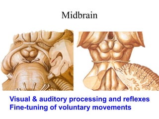
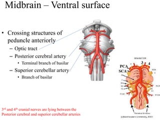
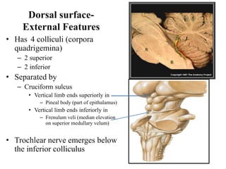
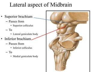
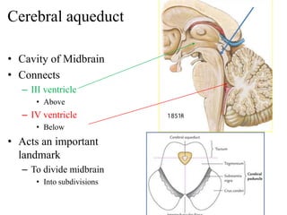
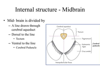
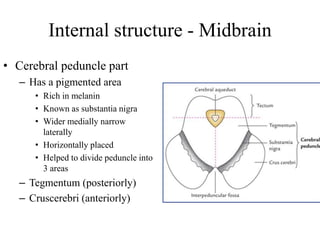
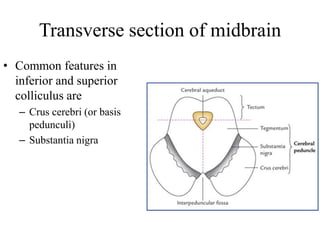
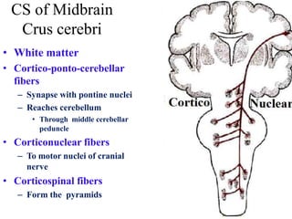
Midbrain
Visual & auditory processing and reflexes
Fine-tuning of voluntary movements
Midbrain
• Shortest part of brain stem
– 2 cm in length
• Connects
– Pons (post. Cranial fossa) & Diencephalon
(mid. cranial fossa)
– Through tentorim cerebelli
External features
• Surfaces
– Ventral
– Dorsal
• Ends
– Superior (rostral)
– Inferior (caudal)
Midbrain – Ventral surface
• Features
– Peduncles
– Interpeduncular fossa
– Emerging structure
• 3rd cranial nerve
– On medial aspect of
peduncle
• 4th cranial nerve
– On lateral aspect of peduncle
– Only nerve emerging from
dorsal aspect of brain stem
Midbrain – Ventral surface
• Crossing structures of
peduncle anteriorly
– Optic tract
– Posterior cerebral artery
• Terminal branch of basilar
– Superior cerebellar artery
• Branch of basilar
3rd and 4th cranial nerves are lying between the
Posterior cerebral and superior cerebellar arteries
Dorsal surface-
External Features
• Has 4 colliculi (corpora
quadrigemina)
– 2 superior
– 2 inferior
• Separated by
– Cruciform sulcus
• Vertical limb ends superiorly in
– Pineal body (part of epithalamus)
• Vertical limb ends inferiorly in
– Frenulum veli (median elevation
on superior medullary velum)
• Trochlear nerve emerges below
the inferior colliculus
Lateral aspect of Midbrain
• Superior brachium
– Passes from
• Superior colliculus
– To
• Lateral geniculate body
• Inferior brachium
– Passes from
• Inferior colliculus
– To
• Medial geniculate body
Cerebral aqueduct
• Cavity of Midbrain
• Connects
– III ventricle
• Above
– IV ventricle
• Below
• Acts an important
landmark
– To divide midbrain
• Into subdivisions
Internal structure - Midbrain
• Mid- brain is divided by
– A line drawn through
cerebral aqueduct
– Dorsal to the line
• Tectum
– Ventral to the line
• Cerebral Peduncle
Internal structure - Midbrain
• Cerebral peduncle part
– Has a pigmented area
• Rich in melanin
• Known as substantia nigra
• Wider medially narrow
laterally
• Horizontally placed
• Helped to divide peduncle into
3 areas
– Tegmentum (posteriorly)
– Cruscerebri (anteriorly)
Transverse section of midbrain
• Common features in
inferior and superior
colliculus are
– Crus cerebri (or basis
pedunculi)
– Substantia nigra
• Pigmented layer of gray matter
– Present in midbrain
– Lies between pons and subthalamus
– Part of extrapyramidal system
– Contains nerve cells with melanin
pigments
– Involves in dopamine synthesis
• 2 parts
– Dorsal compact part (smooth)
– Ventral reticular parts (spiked)
Degeneration of Subtantia nigra
results Parkinson’s Disease
Substantia nigra
Main connections of substantia nigra
• Efferent fibres
– Mainly to striatum
• Which is caudate and
lentiform nucleus
– Constitutes nigrostriatal
pathway
• Rich in dopamine
• Control of muscle tone
• Prevents tremor & other
involuntary movements by
• Inhibiting Globas pallidus
& caudate nucleus
CS of Midbrain
Crus cerebri
– Lies anterior to substantia nigra
– Contains descending tracts
– Medial 1/5
• Fronto pontine fibres
– Middle 3/5
• Corticospinal & cortico-nuclear fibres
– Lateral 1/5
• Parieto-pontine
• Occipito-pontine fibres
• Temporo-pontine
• Above continuous with corona radiata &
below with longitudinal fibres of basilar
part of pons
• White matter
• Cortico-ponto-cerebellar
fibers
– Synapse with pontine nuclei
– Reaches cerebellum
• Through middle cerebellar
peduncle
• Corticonuclear fibers
– To motor nuclei of cranial
nerve
• Corticospinal fibers
– Form the pyramids
CS of Midbrain
Crus cerebri
Internal features – Inferior collicular level
Tectal part - Grey matter
• Nucleus of Inferior
colliculus
• Reflex center for auditory
stimuli
Internal features –Tectal part
Inferior collicular level
• Inferior colliculus
– Act as relay station for auditory pathway
– Reflex centre for response to auditory
stimuli
• Afferent fibres
– Lateral lemniscus
– Cortical fibres
• Efferent fibres
– Medial geniculate body
• By inferior brachium
• Connections for co-ordination
– Efferent to
• Superior colliculi
• Motor nuclei of cranial nerves
– Tecto bulbar
• Spinal motor neurons
– Tecto spinal
» Seen in middle part of tegmentum
Internal features – Tegmental & tectal part
Lower midbrain
• Trapezoid body
– Lies in lower part of pons
– Decussation of the fibres from cochlear
nuclei
– Ascend as lateral lemniscus
• Lateral lemniscus
– Seen
• Upper pons
• Lower midbrain
• End in inferior colliculus
Internal features – Tegmental part
Inferior colliculus level
• Contains
– Grey matter
• Cranial nerve Nuclei
– White matter
• Ascending
• Descending
• Crossing
Internal features – Inferior collicular level
Tegmentum
• Grey matter
– Cranial nerve nuclei
• Trochlear nucleus
– Lies in ventral part of
central grey matter
• Mesencephalic nucleus of
trigeminal
– Lies in lateral part of
central grey matter
– Proprioceptive fibres from
muscles of mastication
Internal features
Inferior collicular level
Tegmentum
• Ascending tracts
• Has 4 lemnisci
– Lateral lemniscus conveys
• Auditory sensation to inferior
colliculus from two ears, mainly
the opposite ear
– Spinal lemniscus conveys
• Pain, temperature, touch and
pressure sensations from
opposite side of body
– Trigeminal lemniscus conveys
• Sensations from opposite side of
face
– Medial lemniscus conveys
• Discriminative touch and
vibration sense from opposite
side of body
Internal features – Tegmental part
Lower midbrain
• Lateral lemniscus
– Seen
• Upper pons
• Lower midbrain
Anterolateral system
• Spinal lemniscus
– Carry contralateral
• Anteior spinothalamic
– Crude touch
• Lateral spinothalamic
– Pain & temperature
– Can be seen
• Medulla
• Pons
• Midbrain
• First order neuron
– Located in
• Trigeminal ganglion
• Second order neuron
– Located in
• Spinal nucleus
– (Pain & temperature)
• Main pontine nucleus
– (touch & pressure)
• Trigeminal lemniscus
– Fibres from second order neuron
– Cross opposite side
– Form trigeminal lemniscus
• Third order neuron
– Lies in thalamus
Trigeminal lemniscus
Dorsal system
• Medial lemniscus
– Carry dorsal column
modalities
• From contralateral side
– Can be seen
• Upper medulla
• Pons
• Midbrain
CS of Midbrain
Inferior collicular level
• Descending tracts
• Present in peripheral
part of tegmentum
– Lateral lemniscus
– Spinal lemniscus
– Trigeminal lemniscus
– Medial lemniscus
Internal features – Tegmental part
Lower midbrain
• Decussating fibres
– Decussation of superior
cerebellar peduncle
• Mainly dentate rubral fibres
– Fibres arising from
• Dentate nucleus
• Passing through superior
cerebellar peduncle
• Main destination is Red
nucleus
• Those fibres decussate here
– In the midline
– At anterior part of
Tegmentum
Internal features – Tegmental part
Inferior collicular level
• White matter
– Fibres interconnecting
cranial nerve nuclei
• Medial longitudinal fasciculus
– Descending tracts
• Tectospinal
• Rubrospinal
– Lies ventral to decussation of
superior cerebellar peduncle
Medial Longitudinal Fasciculus
• Formed of
– Descending & ascending fibers
• Has two components
– Ascending component
• Establishes connections with the
nuclei of
– Occulomotor, Trochlear &
Abducent nerves (motor nuclei
for extraoccular muscles) for
coordination of head & eye
movements
– Descending component
• Extends into the spinal cord as the
medial vestibulospinal tract
Rubrospinal Tract
• Origin
– From red nucleus
– Located in midbrain
• Termination
– Lateral column of spinal
cord
• Seen at
– Lower midbrain
– Pons
– Medulla
Tectospinal Tract
• Origin
– Superior colliculus of
midbrain
• Termination
– Anterior Column
• Function
– Motor function of the
Skeletal muscles of the
head and eyes in response
to visual stimuli
Internal features – Tegmental part
Inferior collicular level
• White matter
– Fibres interconnecting
cranial nerve nuclei
• Medial longitudinal fasciculus
– Descending tracts
• Tectospinal
• Rubrospinal
– Lies ventral to decussation of
superior cerebellar peduncle
Internal features – Tegmental part
Superior colliculus level
• Contains
– Grey matter
• Cranial nerve Nuclei
• Red nucleus
– White matter
• Ascending
• Descending
• Crossing
Internal features – Tegmental part
Superior colliculus level
• Grey matter
– Cranial nerve
• III nerve nuclei
– Nerve emerging through anterior
aspect
– Passing through Red nucleus
– Supplies all extraocular muscles
» Except
• Lateral rectus
• Superior obliquus
• Edinger westphal nucleus (dorsal
part of oculomotor complex)
– Fibres passing along with III N
– Supplies intra ocular muscles
» Sphincter pupillae
» Ciliaris
• Mesencephalic nucleus of V
– Lies in lateral part of central grey
matter
– Proprioceptive fibres from
muscles of mastication
Red Nucleus
• Ovoid mass of grey matter
• Involved in motor control
• Pink in colour – in fresh state
• Has multipolar neurons
• Situation
– In tegmentum
– Dorsal to medial part of
substantia nigra
Red nucleus - connections
• Afferents
– Corticorubral tract:
• from motor and premotor areas of
cerebral cortex
– Dentatorubral tract:
• from opposite dentate nucleus of
cerebellum
• Forms decussation in lower midbrain
Red nucleus - connections
• Efferent
• Cross in ventral tegmental
decussation
• Forms Rubrospinal tract
• Can be seen in lower midbrain &
lower brainstem
– Rubrothalamic tract
• To thalamus (VA & VL nuclei)
• Lesion
– results in signs of cerebellar damage
• contralateral tremor and ataxia
Internal features – Tegmental part
Superior colliculus level
White matter
• In midline
– Medial longitudinal
fasciculus
– Decussating fibres
• Dorsal tegmental decussation
– Fibres arising from superior
colliculus forms
– Continues as tecto spinal &
tectobulbar tracts
• Ventral tegmental decussation
– Fibres arising from red
nucleus forms
– Then continues as
rubrospinal tract
Internal features – superior collicular level
Tectal part - Grey matter
• Superior colliculus
– Part of visual system
– Optic reflex center
• Changing the position of eyes &
head in response to moving objects
• Afferents are
– From retina
• Through superior brachium (visual)
– From spinal cord
• Through Spinotectal (touch sense)
– From inferior colliculus
• Auditory impulses
– Corticotectal fibres
• From
– Visual cortex of occipital lobe
– Frontal eye field of frontal lobe
Internal features – superior collicular level
Tectal part - Grey matter
Superior colliculus
• Efferent fibres
– Tectospinal
– Tecto bulbar
• Forms dorsal tegmental
decussation
Internal features – superior collicular level
Tectal part - Grey matter
• Pretectal nucleus
– Response for pupillary
light reflex
• Situation
– Close to superior
colliculus
– At junction of midbrain
& diencephalon
• Afferents
– Retina, superior
colliculus, visual cortex
• Efferent
– Edingerwestphal
Arterial supply
• Basilar arteries
• Branches of basilar
– Posterior cerebral
– Superior cerebellar
• Posterior communicating
• Anterior choroidal
Weber’s syndrome
• Lesion in ventral part of
midbrain
– Mainly due to occlusion of
posterior cerebral artery
• Involves oculomotor nerve &
basis pedunculi
– Upper motor neuron paralaysis
of opposite side
– Paralysis of all muscles
supplied by oculomotor
• Lateral squint
Benedikt Syndrome
• Necrosis involves
medial lemniscus, spinal
lemniscus and red
nucleus
– Ipsilateral lateral squint
• Due to oculomotor
involvement
– Contralateral pain &
temperature loss
• Due to spinal lemniscus
involvement

Midbrain

  • 1.
    Midbrain Visual & auditoryprocessing and reflexes Fine-tuning of voluntary movements
  • 2.
    Midbrain • Shortest partof brain stem – 2 cm in length • Connects – Pons (post. Cranial fossa) & Diencephalon (mid. cranial fossa) – Through tentorim cerebelli
  • 3.
    External features • Surfaces –Ventral – Dorsal • Ends – Superior (rostral) – Inferior (caudal)
  • 4.
    Midbrain – Ventralsurface • Features – Peduncles – Interpeduncular fossa – Emerging structure • 3rd cranial nerve – On medial aspect of peduncle • 4th cranial nerve – On lateral aspect of peduncle – Only nerve emerging from dorsal aspect of brain stem
  • 5.
    Midbrain – Ventralsurface • Crossing structures of peduncle anteriorly – Optic tract – Posterior cerebral artery • Terminal branch of basilar – Superior cerebellar artery • Branch of basilar 3rd and 4th cranial nerves are lying between the Posterior cerebral and superior cerebellar arteries
  • 6.
    Dorsal surface- External Features •Has 4 colliculi (corpora quadrigemina) – 2 superior – 2 inferior • Separated by – Cruciform sulcus • Vertical limb ends superiorly in – Pineal body (part of epithalamus) • Vertical limb ends inferiorly in – Frenulum veli (median elevation on superior medullary velum) • Trochlear nerve emerges below the inferior colliculus
  • 7.
    Lateral aspect ofMidbrain • Superior brachium – Passes from • Superior colliculus – To • Lateral geniculate body • Inferior brachium – Passes from • Inferior colliculus – To • Medial geniculate body
  • 8.
    Cerebral aqueduct • Cavityof Midbrain • Connects – III ventricle • Above – IV ventricle • Below • Acts an important landmark – To divide midbrain • Into subdivisions
  • 9.
    Internal structure -Midbrain • Mid- brain is divided by – A line drawn through cerebral aqueduct – Dorsal to the line • Tectum – Ventral to the line • Cerebral Peduncle
  • 10.
    Internal structure -Midbrain • Cerebral peduncle part – Has a pigmented area • Rich in melanin • Known as substantia nigra • Wider medially narrow laterally • Horizontally placed • Helped to divide peduncle into 3 areas – Tegmentum (posteriorly) – Cruscerebri (anteriorly)
  • 11.
    Transverse section ofmidbrain • Common features in inferior and superior colliculus are – Crus cerebri (or basis pedunculi) – Substantia nigra
  • 12.
    • Pigmented layerof gray matter – Present in midbrain – Lies between pons and subthalamus – Part of extrapyramidal system – Contains nerve cells with melanin pigments – Involves in dopamine synthesis • 2 parts – Dorsal compact part (smooth) – Ventral reticular parts (spiked) Degeneration of Subtantia nigra results Parkinson’s Disease Substantia nigra
  • 13.
    Main connections ofsubstantia nigra • Efferent fibres – Mainly to striatum • Which is caudate and lentiform nucleus – Constitutes nigrostriatal pathway • Rich in dopamine • Control of muscle tone • Prevents tremor & other involuntary movements by • Inhibiting Globas pallidus & caudate nucleus
  • 14.
    CS of Midbrain Cruscerebri – Lies anterior to substantia nigra – Contains descending tracts – Medial 1/5 • Fronto pontine fibres – Middle 3/5 • Corticospinal & cortico-nuclear fibres – Lateral 1/5 • Parieto-pontine • Occipito-pontine fibres • Temporo-pontine • Above continuous with corona radiata & below with longitudinal fibres of basilar part of pons
  • 15.
    • White matter •Cortico-ponto-cerebellar fibers – Synapse with pontine nuclei – Reaches cerebellum • Through middle cerebellar peduncle • Corticonuclear fibers – To motor nuclei of cranial nerve • Corticospinal fibers – Form the pyramids CS of Midbrain Crus cerebri
  • 16.
    Internal features –Inferior collicular level Tectal part - Grey matter • Nucleus of Inferior colliculus • Reflex center for auditory stimuli
  • 17.
    Internal features –Tectalpart Inferior collicular level • Inferior colliculus – Act as relay station for auditory pathway – Reflex centre for response to auditory stimuli • Afferent fibres – Lateral lemniscus – Cortical fibres • Efferent fibres – Medial geniculate body • By inferior brachium • Connections for co-ordination – Efferent to • Superior colliculi • Motor nuclei of cranial nerves – Tecto bulbar • Spinal motor neurons – Tecto spinal » Seen in middle part of tegmentum
  • 18.
    Internal features –Tegmental & tectal part Lower midbrain • Trapezoid body – Lies in lower part of pons – Decussation of the fibres from cochlear nuclei – Ascend as lateral lemniscus • Lateral lemniscus – Seen • Upper pons • Lower midbrain • End in inferior colliculus
  • 19.
    Internal features –Tegmental part Inferior colliculus level • Contains – Grey matter • Cranial nerve Nuclei – White matter • Ascending • Descending • Crossing
  • 20.
    Internal features –Inferior collicular level Tegmentum • Grey matter – Cranial nerve nuclei • Trochlear nucleus – Lies in ventral part of central grey matter • Mesencephalic nucleus of trigeminal – Lies in lateral part of central grey matter – Proprioceptive fibres from muscles of mastication
  • 21.
    Internal features Inferior collicularlevel Tegmentum • Ascending tracts • Has 4 lemnisci – Lateral lemniscus conveys • Auditory sensation to inferior colliculus from two ears, mainly the opposite ear – Spinal lemniscus conveys • Pain, temperature, touch and pressure sensations from opposite side of body – Trigeminal lemniscus conveys • Sensations from opposite side of face – Medial lemniscus conveys • Discriminative touch and vibration sense from opposite side of body
  • 22.
    Internal features –Tegmental part Lower midbrain • Lateral lemniscus – Seen • Upper pons • Lower midbrain
  • 23.
    Anterolateral system • Spinallemniscus – Carry contralateral • Anteior spinothalamic – Crude touch • Lateral spinothalamic – Pain & temperature – Can be seen • Medulla • Pons • Midbrain
  • 24.
    • First orderneuron – Located in • Trigeminal ganglion • Second order neuron – Located in • Spinal nucleus – (Pain & temperature) • Main pontine nucleus – (touch & pressure) • Trigeminal lemniscus – Fibres from second order neuron – Cross opposite side – Form trigeminal lemniscus • Third order neuron – Lies in thalamus Trigeminal lemniscus
  • 25.
    Dorsal system • Mediallemniscus – Carry dorsal column modalities • From contralateral side – Can be seen • Upper medulla • Pons • Midbrain
  • 26.
    CS of Midbrain Inferiorcollicular level • Descending tracts • Present in peripheral part of tegmentum – Lateral lemniscus – Spinal lemniscus – Trigeminal lemniscus – Medial lemniscus
  • 27.
    Internal features –Tegmental part Lower midbrain • Decussating fibres – Decussation of superior cerebellar peduncle • Mainly dentate rubral fibres – Fibres arising from • Dentate nucleus • Passing through superior cerebellar peduncle • Main destination is Red nucleus • Those fibres decussate here – In the midline – At anterior part of Tegmentum
  • 28.
    Internal features –Tegmental part Inferior collicular level • White matter – Fibres interconnecting cranial nerve nuclei • Medial longitudinal fasciculus – Descending tracts • Tectospinal • Rubrospinal – Lies ventral to decussation of superior cerebellar peduncle
  • 29.
    Medial Longitudinal Fasciculus •Formed of – Descending & ascending fibers • Has two components – Ascending component • Establishes connections with the nuclei of – Occulomotor, Trochlear & Abducent nerves (motor nuclei for extraoccular muscles) for coordination of head & eye movements – Descending component • Extends into the spinal cord as the medial vestibulospinal tract
  • 30.
    Rubrospinal Tract • Origin –From red nucleus – Located in midbrain • Termination – Lateral column of spinal cord • Seen at – Lower midbrain – Pons – Medulla
  • 31.
    Tectospinal Tract • Origin –Superior colliculus of midbrain • Termination – Anterior Column • Function – Motor function of the Skeletal muscles of the head and eyes in response to visual stimuli
  • 32.
    Internal features –Tegmental part Inferior collicular level • White matter – Fibres interconnecting cranial nerve nuclei • Medial longitudinal fasciculus – Descending tracts • Tectospinal • Rubrospinal – Lies ventral to decussation of superior cerebellar peduncle
  • 33.
    Internal features –Tegmental part Superior colliculus level • Contains – Grey matter • Cranial nerve Nuclei • Red nucleus – White matter • Ascending • Descending • Crossing
  • 34.
    Internal features –Tegmental part Superior colliculus level • Grey matter – Cranial nerve • III nerve nuclei – Nerve emerging through anterior aspect – Passing through Red nucleus – Supplies all extraocular muscles » Except • Lateral rectus • Superior obliquus • Edinger westphal nucleus (dorsal part of oculomotor complex) – Fibres passing along with III N – Supplies intra ocular muscles » Sphincter pupillae » Ciliaris • Mesencephalic nucleus of V – Lies in lateral part of central grey matter – Proprioceptive fibres from muscles of mastication
  • 35.
    Red Nucleus • Ovoidmass of grey matter • Involved in motor control • Pink in colour – in fresh state • Has multipolar neurons • Situation – In tegmentum – Dorsal to medial part of substantia nigra
  • 36.
    Red nucleus -connections • Afferents – Corticorubral tract: • from motor and premotor areas of cerebral cortex – Dentatorubral tract: • from opposite dentate nucleus of cerebellum • Forms decussation in lower midbrain
  • 37.
    Red nucleus -connections • Efferent • Cross in ventral tegmental decussation • Forms Rubrospinal tract • Can be seen in lower midbrain & lower brainstem – Rubrothalamic tract • To thalamus (VA & VL nuclei) • Lesion – results in signs of cerebellar damage • contralateral tremor and ataxia
  • 38.
    Internal features –Tegmental part Superior colliculus level White matter • In midline – Medial longitudinal fasciculus – Decussating fibres • Dorsal tegmental decussation – Fibres arising from superior colliculus forms – Continues as tecto spinal & tectobulbar tracts • Ventral tegmental decussation – Fibres arising from red nucleus forms – Then continues as rubrospinal tract
  • 39.
    Internal features –superior collicular level Tectal part - Grey matter • Superior colliculus – Part of visual system – Optic reflex center • Changing the position of eyes & head in response to moving objects • Afferents are – From retina • Through superior brachium (visual) – From spinal cord • Through Spinotectal (touch sense) – From inferior colliculus • Auditory impulses – Corticotectal fibres • From – Visual cortex of occipital lobe – Frontal eye field of frontal lobe
  • 40.
    Internal features –superior collicular level Tectal part - Grey matter Superior colliculus • Efferent fibres – Tectospinal – Tecto bulbar • Forms dorsal tegmental decussation
  • 41.
    Internal features –superior collicular level Tectal part - Grey matter • Pretectal nucleus – Response for pupillary light reflex • Situation – Close to superior colliculus – At junction of midbrain & diencephalon • Afferents – Retina, superior colliculus, visual cortex • Efferent – Edingerwestphal
  • 42.
    Arterial supply • Basilararteries • Branches of basilar – Posterior cerebral – Superior cerebellar • Posterior communicating • Anterior choroidal
  • 43.
    Weber’s syndrome • Lesionin ventral part of midbrain – Mainly due to occlusion of posterior cerebral artery • Involves oculomotor nerve & basis pedunculi – Upper motor neuron paralaysis of opposite side – Paralysis of all muscles supplied by oculomotor • Lateral squint
  • 44.
    Benedikt Syndrome • Necrosisinvolves medial lemniscus, spinal lemniscus and red nucleus – Ipsilateral lateral squint • Due to oculomotor involvement – Contralateral pain & temperature loss • Due to spinal lemniscus involvement