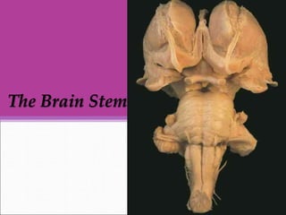
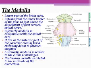
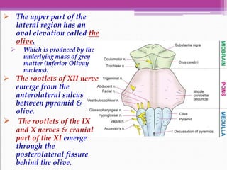
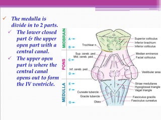
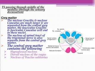
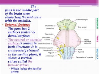
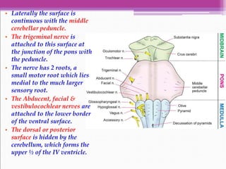
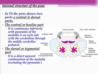
The Brain Stem
• The brain stem
consists of
▫ The medulla
oblongata
▫ The pons
▫ The midbrain
• It is situated in the
posterior cranial
fossa.
The Medulla
• Lower part of the brain stem.
• Extents from the lower border
of the pons to just above the
attachment of first cervical
spinal nerve.
• Inferiorly medulla is
continuous with the spinal
cord
• It lies in the anterior part of
the posterior cranial fossa
extending down to foramen
magnum
• Anteriorly, medulla is related
to the clivus & meninges.
• Posteriorly medulla is related
to the vallecula of the
cerebellum.
External Features
The medulla is divided in to right
& left halves by the anterior &
posterior median fissure.
• Each half is further divided
in to anterior, lateral &
posterior regions by the
anterolateral &
posterolateral sulci.
• The anterior region is in the
form of longitudinal elevation
called as the pyramid.
▫ It is made of corticospinal
fibers
• In the lower part of the
medulla many fibers of the
right & left pyramids cross in
the midline forming
pyramidal decussation.
 The upper part of the
lateral region has an
oval elevation called the
olive.
 Which is produced by the
underlying mass of grey
matter (inferior Olivay
nucleus).
 The rootlets of XII nerve
emerge from the
anterolateral sulcus
between pyramid &
olive.
 The rootlets of the IX
and X nerves & cranial
part of the XI emerge
through the
posterolateral fissure
behind the olive.
 The posterolateral region lies
between the posterolateral sulcus
& the posterior median fissure,
 The upper part of this region has
a “V” shaped depression which
is the lower part of the floor of
the IV ventricle.
 Below the floor there is
longitudinal elevations,
 From medial to lateral side they
are
 The fasciculus Gracilis,
 The fasciculus Cuneatus &
 The inferior cerebellar
peduncle.
 The upper ends of the fasciculus
gracilis & cuneatus expand to form
the gracile & cuneate tubercles.
 These tubercles are formed by the
underlying masses of grey matter
called the nucleus Gracilis, &
nucleus Cuneatus.
 The medulla is
divide in to 2 parts.
 The lower closed
part & the upper
open part with a
central canal.
 The upper open
part is where the
central canal
opens out to form
the IV ventricle.
Internal Structure Of Medulla
• The internal structure of
medulla is studied by
examining TS through it at 3
places
TS through the Lower part of the
medulla passing through the
Pyramidal decussation
• It resembles the TS of the
spinal cord
White matter
• There is Pyramids anteriorly.
• The decussation of the
pyramidal tract forms the
important feature of the
medulla at this level, the
fibers run laterally & form
the lateral cortcospinal
tracts.
• The fasciculus gracilis &
the fasciculus cuneatus
occupy the broad posterior
white column
• The other features are
similar to spinal cord
Grey matter
• The decussating
Pyramidal fibers separate
the anterior horn from the
grey matter & forms the
spinal nucleus of the
spinal accessory nerve &
supra Nucleus for the 1st
cervical nerve.
• The nucleus gracilis, &
nucleus cuneatus are
continuous with the
central grey matter.
TS passing through middle of the
medulla (through the sensory
decussation)
Grey matter
 The nucleus Gracilis & nucleus
Cuneatus are much larger & are
separated from the central grey
matter, the fasciculus Gracilis
& fasciculus Cuneatus will end
in these nuclei.
 The nucleus of spinal tract of
the trigeminal nerve is also
separate from the central grey
matter.
 The central grey matter
contains the following
 Hypoglossal nucleus
 Dorsal nucleus of the vagus
 Nucleus of Tractus solitarius
White matter
• The nucleus
Gracilis &
Cuneatus give
rise to internal
arcuate fibers,
these fibers
cross to
oppossite side
where they
form Para
median band
of fibers called
the medial
meniscus.
• The pyramidal
tracts lie
anteriorly
TS through the upper part of the medulla
passing through the IV ventricle
Grey matter
• The nuclei of several
cranial nerves are
seen in the floor of
the IV ventricle
▫ Hypoglossal nucleus
▫ The dorsal nucleus
of the vagus
▫ The nucleus of
tractus solitarius
▫ The inferior&
medial vestibular
nuclei
• The nucleus of the
spinal tract of the
trigeminal nerve lies in
the dorsolateral part.
• The inferior olivary
nucleus is the largest
mass of grey matter
seen at this level.
White matter
• The inferior
cerebellar peduncle
occupies the
posterolateral part,
lateral to IV
ventricle
• Other tracts seen in
the spinal cord can
also be seen.
Applied Anatomy
• The vital centers ( respiratory & vaso- motor ) are
situated in the lower part of the floor of the IV
ventricle formed by the medulla.
▫ An injury to the medulla is therefore usually fatal
• Common vascular lesions involving the medulla
are
▫ Thrombosis of the posteior inferior cerebellar artery &
Thrombosis of the vertebral artery. These two lesions
cause lateral & medial medullary syndromes,
respectively.
• Bulbar paralysis may be acute.
▫ It is characterized by the paralysis of the muscles
supplied by the last 4 cranial nerves which arise
from the medulla.
▫ Paralysis of respiratory & vasomotor would be
fatal
The Pons
The
pons is the middle part
of the brain stem
connecting the mid brain
with the medulla.
• External features
▫ The pons has 2
surfaces ventral &
dorsal surfaces
▫ The ventral or anterior
surface is convex in
both directions & is
transversely striated.
▫ In the median plane, it
shows a vertical
sulcus called the
basilar sulcus
 Which lodges the basilar
artery.
• Laterally the surface is
continuous with the middle
cerebellar peduncle.
• The trigeminal nerve is
attached to this surface at
the junction of the pons with
the peduncle.
• The nerve has 2 roots, a
small motor root which lies
medial to the much larger
sensory root.
• The Abducent, facial &
vestibulocochlear nerves are
attached to the lower border
of the ventral surface.
• The dorsal or posterior
surface is hidden by the
cerebellum, which forms the
upper ½ of the IV ventricle.
Internal structure of the pons
• In TS the pons shows two
parts a ventral & dorsal
parts.
• The ventral or basilar part
▫ It is continuous inferiorly
with pyramids of the
medulla & on each side
with the cerebellum through
the middle cerebellar
peduncle
• The dorsal or tegmental
part
▫ It is a direct upward
continuation of the medulla
(excluding the pyramids )
• The basillar part has same structure
through out the pons & has the
following features.
Grey matter
• It is represented by the pontine nuclei
which are scattered among the
longitudinal & transverse fibers.
• The pontine nuclei form an important
part of the corticoponto-cerebellar
pathway.
• Fibers from all these nuclei will go to
the opposite ½ of the cerebellum.
White matter
• It consists of the longitudinal &
transverse fibers
• The longitudinal fibers include
▫ The corticospinal &
corticonuclear(pyramidal)
▫ The corticopontine fibres ending in the
pontine nuclei
• The transverse fibers are
pontocerebellar fibers beginning from
the pontine nuclei & going to the
opposite ½ of the cerebellum through
the inferior cerebellar peduncle
Tegmentum in the upper part of the pons
Grey matter
▫ The special features are the
motor & superior nuclei of
the trigeminal nerve.
▫ The motor nucleus is
medial to the superior
sensory nucleus
White matter
▫ Immediately behind the
ventral part of the pons
there is transverse band of
fibers that is made up (
from medial to lateral side )
of
 The medial leminiscus
 The trigeminal leminiscus
 The spinal leminiscus
 The lateral leminiscus.
▫ The trigeminal leminiscus
contains fibers arising in
the spinal nucleus of the
trigeminal nerve &
traveling to the thalamus.
▫ The lateral leminiscus is the
part of auditory pathway.
• Applied Anatomy
▫ Unilateral lesion in the lower part of the pons
results in paralysis of the facial nerve on the side
of the lesion, & paralysis of the limbs (
hemiplegia ) on the opposite side (crossed
hemiplegia or Millard Gubler Syndrome)
The Mid
Brain
• The mid brain is also called the
mesencephalon.
• It connects the hind brain with the
fore brain.
• Its cavity is known as the cerebral
aqueduct,
▫ It connects the 3rd ventricle with 4th
ventricle
• The mid brain passes through the
tentorial notch
• The medial & lateral geniculate
bodies ( meta thalamus ) are
situated on the posterolateral aspect
of the mid brain.
• The superior colliculus is connected
to the lateral geniculate body by the
superior brachium.
• The inferior colliculus is connected
to the medial geniculate body by the
inferior brachium.
In TS the following are
the major
subdivisions
• The Tectum
▫ It is the part posterior
to the aqueduct.
▫ It is made up of right &
left, superior & inferior
colliculi
• The cerebral peduncle.
▫ Each ½ of the midbrain
anterior to the aqueduct
is called the cerebral
peduncle.
▫ Each cerebral peduncle
is subdivided in to
 The crus cerebri
anteriorly
 The substasia nigra
in the middle
 The tegmentum
posteriorly
Internal structure of the mid brain
This can be studied at the level of inferior &
superior colliculi
T S of mid brain at the level of inferior
colliculus
Grey matter
• The inferior colliculus receives
the afferents of the lateral
meniscus, & gives efferents to
the medial geniculate body,
which helps in localizing the
source of sounds.
• The substantia nigra is a lamina
of grey matter made up of
deeply pigmented nerve cells.
• The central ( peri aqueductal )grey
matter contains
▫ The nucleus of the trochlear nerve in
the ventromedial part.
▫ The mesencephalic nucleus of the
trigeminal nerve in the
lateral part.
White matter
• The crus cerebri contains
▫ The pyramidal tract in the middle
▫ Frontopontine fibres,
parietopontine, & occipitopontine
fibres in the lateral 1/6th .
The tegmentum contains
ascending tracts as follows
• The leminisci ( medial,
spinal, trigeminal, & lateral
) are arranged in the form of
a band in which they lie in
order mentioned ( from med
to lateral )
• The decussating of the
superior cerebellar
peduncles is seen in the
median plane.
• The tectospinal &
rubrospinal tracts are
present.
TS of mid brain at the level of superior colliculus
Grey matter
• The central grey matter
contains
▫ The nucleus of the occulomotor
nerve in the ventromedial part
▫ The mesencephalic nucleus of
the trigeminal nerve in the
lateral part
• The superior colliculus receives
afferents from the retina (
visual ) & various other
centres.
▫ It gives afferents to the spinal
cord (tectospinal ).
▫ It controls the reflex of the
movements of the eyes, & of the
head & neck in response to the
visual stimuli.
• The pretectal nucleus lies deep
to the superolateral part of the
superior colliculus.
▫ It receives the afferents from
lateral root of the optic nerve.
▫ It gives efferents to the Edinger
Westphal nuclei of both sides.
• The red nucleus is about
0.5 cm in diameter
▫ It receives the afferent
fibres from the superior
cerebellar peduncle, the
globus pallidus, the
subtahalamic nucleus &
the cerebral cortex.
▫ It gives efferents to the
spinal cord (rubrospinal
tract), reticular
formation, the thalamus,
the olivary nucleus, the
subthalamic nucleus.
▫ It has an inhibitory
influence on muscle tone.
White matter
• The crus cerebri has same
tracts as described above
Applied Anatomy
• Lesion in the upper part of the midbrain can
produce a paralysis of muscles supplied by the
oculomotor nerve on the side of lesion, along
with a hemiplegia on the opposite side (Weber’s
syndrome).
• A similar lesion in the pons, results in a
paralysis of the lateral rectus muscle (abducent
nerve) on the side of lesion with hemiplegia on
the opposite side (Raymond’s syndrome).
• Alternatively, facial paralysis of one side can be
combined with contralateral hemiplegia
(Millard Gubler syndrome).

Brain stem New.ppsx

  • 1.
  • 2.
    • The brainstem consists of ▫ The medulla oblongata ▫ The pons ▫ The midbrain • It is situated in the posterior cranial fossa.
  • 3.
    The Medulla • Lowerpart of the brain stem. • Extents from the lower border of the pons to just above the attachment of first cervical spinal nerve. • Inferiorly medulla is continuous with the spinal cord • It lies in the anterior part of the posterior cranial fossa extending down to foramen magnum • Anteriorly, medulla is related to the clivus & meninges. • Posteriorly medulla is related to the vallecula of the cerebellum.
  • 4.
    External Features The medullais divided in to right & left halves by the anterior & posterior median fissure. • Each half is further divided in to anterior, lateral & posterior regions by the anterolateral & posterolateral sulci. • The anterior region is in the form of longitudinal elevation called as the pyramid. ▫ It is made of corticospinal fibers • In the lower part of the medulla many fibers of the right & left pyramids cross in the midline forming pyramidal decussation.
  • 5.
     The upperpart of the lateral region has an oval elevation called the olive.  Which is produced by the underlying mass of grey matter (inferior Olivay nucleus).  The rootlets of XII nerve emerge from the anterolateral sulcus between pyramid & olive.  The rootlets of the IX and X nerves & cranial part of the XI emerge through the posterolateral fissure behind the olive.
  • 6.
     The posterolateralregion lies between the posterolateral sulcus & the posterior median fissure,  The upper part of this region has a “V” shaped depression which is the lower part of the floor of the IV ventricle.  Below the floor there is longitudinal elevations,  From medial to lateral side they are  The fasciculus Gracilis,  The fasciculus Cuneatus &  The inferior cerebellar peduncle.  The upper ends of the fasciculus gracilis & cuneatus expand to form the gracile & cuneate tubercles.  These tubercles are formed by the underlying masses of grey matter called the nucleus Gracilis, & nucleus Cuneatus.
  • 7.
     The medullais divide in to 2 parts.  The lower closed part & the upper open part with a central canal.  The upper open part is where the central canal opens out to form the IV ventricle.
  • 8.
    Internal Structure OfMedulla • The internal structure of medulla is studied by examining TS through it at 3 places TS through the Lower part of the medulla passing through the Pyramidal decussation • It resembles the TS of the spinal cord White matter • There is Pyramids anteriorly. • The decussation of the pyramidal tract forms the important feature of the medulla at this level, the fibers run laterally & form the lateral cortcospinal tracts.
  • 9.
    • The fasciculusgracilis & the fasciculus cuneatus occupy the broad posterior white column • The other features are similar to spinal cord Grey matter • The decussating Pyramidal fibers separate the anterior horn from the grey matter & forms the spinal nucleus of the spinal accessory nerve & supra Nucleus for the 1st cervical nerve. • The nucleus gracilis, & nucleus cuneatus are continuous with the central grey matter.
  • 10.
    TS passing throughmiddle of the medulla (through the sensory decussation) Grey matter  The nucleus Gracilis & nucleus Cuneatus are much larger & are separated from the central grey matter, the fasciculus Gracilis & fasciculus Cuneatus will end in these nuclei.  The nucleus of spinal tract of the trigeminal nerve is also separate from the central grey matter.  The central grey matter contains the following  Hypoglossal nucleus  Dorsal nucleus of the vagus  Nucleus of Tractus solitarius
  • 11.
    White matter • Thenucleus Gracilis & Cuneatus give rise to internal arcuate fibers, these fibers cross to oppossite side where they form Para median band of fibers called the medial meniscus. • The pyramidal tracts lie anteriorly
  • 12.
    TS through theupper part of the medulla passing through the IV ventricle Grey matter • The nuclei of several cranial nerves are seen in the floor of the IV ventricle ▫ Hypoglossal nucleus ▫ The dorsal nucleus of the vagus ▫ The nucleus of tractus solitarius ▫ The inferior& medial vestibular nuclei • The nucleus of the spinal tract of the trigeminal nerve lies in the dorsolateral part. • The inferior olivary nucleus is the largest mass of grey matter seen at this level.
  • 13.
    White matter • Theinferior cerebellar peduncle occupies the posterolateral part, lateral to IV ventricle • Other tracts seen in the spinal cord can also be seen.
  • 14.
    Applied Anatomy • Thevital centers ( respiratory & vaso- motor ) are situated in the lower part of the floor of the IV ventricle formed by the medulla. ▫ An injury to the medulla is therefore usually fatal • Common vascular lesions involving the medulla are ▫ Thrombosis of the posteior inferior cerebellar artery & Thrombosis of the vertebral artery. These two lesions cause lateral & medial medullary syndromes, respectively. • Bulbar paralysis may be acute. ▫ It is characterized by the paralysis of the muscles supplied by the last 4 cranial nerves which arise from the medulla. ▫ Paralysis of respiratory & vasomotor would be fatal
  • 15.
  • 16.
    The pons is themiddle part of the brain stem connecting the mid brain with the medulla. • External features ▫ The pons has 2 surfaces ventral & dorsal surfaces ▫ The ventral or anterior surface is convex in both directions & is transversely striated. ▫ In the median plane, it shows a vertical sulcus called the basilar sulcus  Which lodges the basilar artery.
  • 17.
    • Laterally thesurface is continuous with the middle cerebellar peduncle. • The trigeminal nerve is attached to this surface at the junction of the pons with the peduncle. • The nerve has 2 roots, a small motor root which lies medial to the much larger sensory root. • The Abducent, facial & vestibulocochlear nerves are attached to the lower border of the ventral surface. • The dorsal or posterior surface is hidden by the cerebellum, which forms the upper ½ of the IV ventricle.
  • 18.
    Internal structure ofthe pons • In TS the pons shows two parts a ventral & dorsal parts. • The ventral or basilar part ▫ It is continuous inferiorly with pyramids of the medulla & on each side with the cerebellum through the middle cerebellar peduncle • The dorsal or tegmental part ▫ It is a direct upward continuation of the medulla (excluding the pyramids )
  • 19.
    • The basillarpart has same structure through out the pons & has the following features. Grey matter • It is represented by the pontine nuclei which are scattered among the longitudinal & transverse fibers. • The pontine nuclei form an important part of the corticoponto-cerebellar pathway. • Fibers from all these nuclei will go to the opposite ½ of the cerebellum. White matter • It consists of the longitudinal & transverse fibers • The longitudinal fibers include ▫ The corticospinal & corticonuclear(pyramidal) ▫ The corticopontine fibres ending in the pontine nuclei • The transverse fibers are pontocerebellar fibers beginning from the pontine nuclei & going to the opposite ½ of the cerebellum through the inferior cerebellar peduncle
  • 20.
    Tegmentum in theupper part of the pons Grey matter ▫ The special features are the motor & superior nuclei of the trigeminal nerve. ▫ The motor nucleus is medial to the superior sensory nucleus White matter ▫ Immediately behind the ventral part of the pons there is transverse band of fibers that is made up ( from medial to lateral side ) of  The medial leminiscus  The trigeminal leminiscus  The spinal leminiscus  The lateral leminiscus. ▫ The trigeminal leminiscus contains fibers arising in the spinal nucleus of the trigeminal nerve & traveling to the thalamus. ▫ The lateral leminiscus is the part of auditory pathway.
  • 21.
    • Applied Anatomy ▫Unilateral lesion in the lower part of the pons results in paralysis of the facial nerve on the side of the lesion, & paralysis of the limbs ( hemiplegia ) on the opposite side (crossed hemiplegia or Millard Gubler Syndrome)
  • 22.
  • 23.
    • The midbrain is also called the mesencephalon. • It connects the hind brain with the fore brain. • Its cavity is known as the cerebral aqueduct, ▫ It connects the 3rd ventricle with 4th ventricle • The mid brain passes through the tentorial notch • The medial & lateral geniculate bodies ( meta thalamus ) are situated on the posterolateral aspect of the mid brain. • The superior colliculus is connected to the lateral geniculate body by the superior brachium. • The inferior colliculus is connected to the medial geniculate body by the inferior brachium.
  • 25.
    In TS thefollowing are the major subdivisions • The Tectum ▫ It is the part posterior to the aqueduct. ▫ It is made up of right & left, superior & inferior colliculi • The cerebral peduncle. ▫ Each ½ of the midbrain anterior to the aqueduct is called the cerebral peduncle. ▫ Each cerebral peduncle is subdivided in to  The crus cerebri anteriorly  The substasia nigra in the middle  The tegmentum posteriorly
  • 26.
    Internal structure ofthe mid brain This can be studied at the level of inferior & superior colliculi T S of mid brain at the level of inferior colliculus Grey matter • The inferior colliculus receives the afferents of the lateral meniscus, & gives efferents to the medial geniculate body, which helps in localizing the source of sounds. • The substantia nigra is a lamina of grey matter made up of deeply pigmented nerve cells. • The central ( peri aqueductal )grey matter contains ▫ The nucleus of the trochlear nerve in the ventromedial part. ▫ The mesencephalic nucleus of the trigeminal nerve in the lateral part.
  • 27.
    White matter • Thecrus cerebri contains ▫ The pyramidal tract in the middle ▫ Frontopontine fibres, parietopontine, & occipitopontine fibres in the lateral 1/6th . The tegmentum contains ascending tracts as follows • The leminisci ( medial, spinal, trigeminal, & lateral ) are arranged in the form of a band in which they lie in order mentioned ( from med to lateral ) • The decussating of the superior cerebellar peduncles is seen in the median plane. • The tectospinal & rubrospinal tracts are present.
  • 28.
    TS of midbrain at the level of superior colliculus Grey matter • The central grey matter contains ▫ The nucleus of the occulomotor nerve in the ventromedial part ▫ The mesencephalic nucleus of the trigeminal nerve in the lateral part • The superior colliculus receives afferents from the retina ( visual ) & various other centres. ▫ It gives afferents to the spinal cord (tectospinal ). ▫ It controls the reflex of the movements of the eyes, & of the head & neck in response to the visual stimuli. • The pretectal nucleus lies deep to the superolateral part of the superior colliculus. ▫ It receives the afferents from lateral root of the optic nerve. ▫ It gives efferents to the Edinger Westphal nuclei of both sides.
  • 29.
    • The rednucleus is about 0.5 cm in diameter ▫ It receives the afferent fibres from the superior cerebellar peduncle, the globus pallidus, the subtahalamic nucleus & the cerebral cortex. ▫ It gives efferents to the spinal cord (rubrospinal tract), reticular formation, the thalamus, the olivary nucleus, the subthalamic nucleus. ▫ It has an inhibitory influence on muscle tone. White matter • The crus cerebri has same tracts as described above
  • 30.
    Applied Anatomy • Lesionin the upper part of the midbrain can produce a paralysis of muscles supplied by the oculomotor nerve on the side of lesion, along with a hemiplegia on the opposite side (Weber’s syndrome). • A similar lesion in the pons, results in a paralysis of the lateral rectus muscle (abducent nerve) on the side of lesion with hemiplegia on the opposite side (Raymond’s syndrome). • Alternatively, facial paralysis of one side can be combined with contralateral hemiplegia (Millard Gubler syndrome).