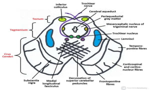
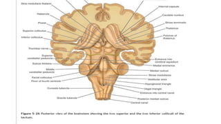
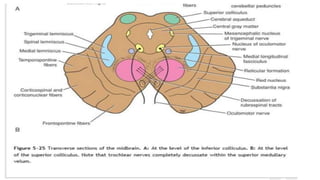
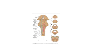
SlideShare a Scribd company logo
Lecture of midbrain
Murad Ali
DPT
Gross Appearance of the Midbrain
• The midbrain measures about 0.8 inch (2 cm) in length
• and connects the pons and cerebellum with the forebrain (Fig. 5-23).
• Its long axis inclines anteriorly as it ascends through the opening in
the tentorium cerebelli.
• The midbrain is traversed by a narrow channel, the cerebral
aqueduct, which is filled with cerebrospinal fluid (Figs. 5-24, 5-25, 5-
26, 5-27 and 5-28).
entorium or tentorium cerebelli(Latin: "tent
m") is an extension of the dura mater that
erebellum from the inferior portion of the
Anterior aspect :
On the anterior aspect of the
midbrain (Fig. 5-23), there is a
deep depression in the midline,
the interpeduncular fossa, which
is bounded on either side by the
crus cerebri. Many small blood
vessels perforate ( Hole) the
floor of the interpeduncular
fossa, and this region is termed
the posterior perforated
substance (Fig. 5-23).
The oculomotor nerve III
emerges from a groove on the
medial side of the crus cerebri
and passes forward in the lateral
wall of the cavernous sinus.
Posterior surface
• On the posterior surface are four colliculi (corpora quadrigemina).
• These are rounded eminences that are divided into superior and
inferior pairs by a vertical and a transverse groove (Fig. 5-26).
• The superior colliculi are centers for visual reflexes (see p. 338),
• and the inferior colliculi are lower auditory centers.
• Origin of Trochlear nerves:
• In the midline below the inferior colliculi, the trochlear nerves
emerge. These are small-diameter nerves that wind around the lateral
aspect of the midbrain to enter the lateral wall of the cavernous
sinus.
0
Lateral aspect:
• On the lateral aspect of the midbrain, the superior and inferior
brachia ascend in an anterolateral direction (Fig. 5-23).
• The superior brachium passes from the superior colliculus to the
lateral geniculate body and the optic tract.
• The inferior brachium connects the inferior colliculus to the medial
geniculate body.
Internal Structure of the Midbrain
• The midbrain comprises two lateral halves, called the cerebral
peduncles; each of these is divided into an anterior part, the crus
cerebri, and a posterior part, the tegmentum, by a pigmented band
of gray matter, the substantia nigra (Figs. 5-24 and 5-25)
• The narrow cavity of the midbrain is the cerebral aqueduct, which
connects the third and fourth ventricles. The tectum is the part of
the midbrain posterior to the cerebral aqueduct; it has four small
surface swellings referred to previously; these are the two superior
and two inferior colliculi (Figs. 5-24 and 5-25). The cerebral aqueduct
is lined by ependyma and is surrounded by the central gray matter.
On transverse sections of the midbrain, the interpeduncular fossa can
be seen to separate the crura cerebri, whereas the tegmentum is
continuous across the median plane (Fig. 5-24).
Transverse Section of the Midbrain at the
Level of the Inferior Colliculi
• The inferior colliculus, consisting of a large nucleus of gray matter, lies
beneath the corresponding surface elevation and forms part of the
auditory pathway (Figs. 5-25A and 5-27). It receives many of the
terminal fibers of the lateral lemniscus. The pathway then continues
through the inferior brachium to the medial geniculate body
• The trochlear nucleus is situated in the central gray matter close to the
median plane just. posterior to the medial longitudinal fasciculus. The
emerging fibers of the trochlear nucleus pass laterally and posteriorly
around the central gray matter and leave the midbrain just below the
inferior colliculi.
• The fibers of the trochlear nerve now decussate completely in the superior
medullary velum.
• The mesencephalic nuclei of the trigeminal nerve are lateral to the
cerebral aqueduct (Figs. 5-25A and 5-27).
• The decussation of the superior cerebellar peduncles occupies the central
part of the tegmentum anterior to the cerebral aqueduct.
• The reticular formation is smaller than that of the pons and is situated
lateral to the decussation.
• The medial lemniscus ascends posterior to the substantia nigra;
• the spinal and trigeminal lemnisci are situated lateral to the medial
lemniscus (Figs. 5-25 and 5-27).
• The lateral lemniscus is located posterior to the trigeminal lemniscus.
• The substantia nigra (Figs. 5-25 and 5-27) is a large motor nucleus situated
between the tegmentum, and the crus cerebri and is found throughout the
midbrain. The nucleus is composed of medium-size multipolar neurons that
possess inclusion granules of melanin pigment within their cytoplasm.
• The substantia nigra is concerned with muscle tone and is connected to the
cerebral cortex, spinal cord, hypothalamus, and basal nuclei.
Descending Tracts
• The crus cerebri contains important descending tracts and is separated
from the tegmentum by the substantia nigra (Figs. 5-25 and 5-27).
• The corticospinal and corticonuclear fibers occupy the middle two-thirds of
the crus.
• The frontopontine fibers occupy the medial part of the crus, and
• the temporopontine fibers occupy the lateral part of the crus (Figs. 5-25
and 5-27).
• These descending tracts connect the cerebral cortex to the anterior gray
column cells of the spinal cord, the cranial nerve nuclei, the pons, and the
cerebellum (Table 5-4).
Transverse Section of the Midbrain at the
Level of the Superior Colliculi
• The superior colliculus (Figs. 5-25B and 5-28), a large nucleus of gray
matter that lies beneath the corresponding surface elevation, forms
part of the visual reflexes. It is connected to the lateral geniculate
body by the superior brachium. It receives afferent fibers from the
optic nerve, the visual cortex, and the spinotectal tract.
• The efferent fibers form the tectospinal and tectobulbar tracts, which
are probably responsible for the reflex movements of the eyes, head,
and neck in response to visual stimuli
• The afferent pathway for the light reflex ends in the pretectal
nucleus. This is a small group of neurons situated close to the lateral
part of the superior colliculus. After relaying in the pretectal nucleus,
the fibers pass to the parasympathetic nucleus of the oculomotor
nerve (Edinger-Westphal nucleus). The emerging fibers then pass to
the oculomotor nerve. The oculomotor nucleus is situated in the
central gray matter close to the median plane, just posterior to the
medial longitudinal fasciculus (Figs. 5-25B and 528). The fibers of the
oculomotor nucleus pass anteriorly through the red nucleus to emerge
on the medial side of the crus cerebri in the interpeduncular fossa. The
nucleus of the oculomotor nerve is divisible into a number of cell
groups.
• The medial, spinal, and trigeminal lemnisci form a curved band posterior to the substantia nigra, but the
lateral lemniscus does not extend superiorly to this level (Figs. 5-25B and 5-28).
• The red nucleus (Figs. 5-25B and 5-28) is a rounded mass of gray matter situated between the cerebral
aqueduct and the substantia nigra. Its reddish hue, seen in fresh specimens, is due to its vascularity and the
presence of an iron-containing pigment in the cytoplasm of many of its neurons.
• Afferent fibers reach the red nucleus from
• (1) the cerebral cortex through the corticospinal fibers,
• (2) the cerebellum through the superior cerebellar peduncle, and
• (3) the lentiform nucleus, subthalamic and hypothalamic nuclei, substantia nigra, and spinal cord. Efferent
fibers leave the red nucleus and pass to
• (1) the spinal cord through the rubrospinal tract (as this tract descends, it decussates)
• , (2) the reticular formation through the rubroreticular tract,
• (3) the thalamus, and
• (4) the substantia nigra.
• The reticular formation is situated in the tegmentum lateral and
posterior to the red nucleus (Figs. 5-25B and 5-28).
• The crus cerebri contains the identical important descending
tractsâ!”the corticospinal, corticonuclear, and corticopontine
fibersâ!”that are present at the level of the inferior colliculus (see
Table 5-4).
• The continuity of the various cranial nerve nuclei through the
different regions of the brainstem is shown diagrammatically in Figure
5-29.

More Related Content

What's hot

Midbrain
MidbrainMidbrain
Midbrain
DrTanujaSawant
 
Epithalamus & metathalamus
Epithalamus & metathalamusEpithalamus & metathalamus
Epithalamus & metathalamus
DINESH KUMAR D
 
Anatomy of cerebellum and 4th ventricle
Anatomy of cerebellum and 4th ventricleAnatomy of cerebellum and 4th ventricle
Anatomy of cerebellum and 4th ventricle
Dr. Mohammad Mahmoud
 
Cranial nerve nuclei
Cranial nerve nucleiCranial nerve nuclei
Cranial nerve nuclei
Lucidante1
 
Cervical Plexus
Cervical PlexusCervical Plexus
Cervical Plexus
Rashmi Priyem Saravanan
 
Commissural fibres
Commissural fibresCommissural fibres
Commissural fibres
Chaitra Chinmayee M K
 
Thalamus, anatomy of thalamus, Thalamus PPT
Thalamus, anatomy of thalamus, Thalamus PPTThalamus, anatomy of thalamus, Thalamus PPT
Thalamus, anatomy of thalamus, Thalamus PPT
drasarma1947
 
Anatomy of midbrain
Anatomy of midbrainAnatomy of midbrain
Anatomy of midbrain
Dr. Mohammad Mahmoud
 
Cerebellum
CerebellumCerebellum
Cerebellum
Prakhar Jain
 
Thalamus
ThalamusThalamus
Thalamus
Siva Pesala
 
Development of nervous system
Development of nervous systemDevelopment of nervous system
Development of nervous system
Dr. Mohammad Mahmoud
 
Third ventricle
Third ventricleThird ventricle
Third ventricle
PGINeurosurgery
 
BRAINSTEM-DETAILED.pptx
BRAINSTEM-DETAILED.pptxBRAINSTEM-DETAILED.pptx
BRAINSTEM-DETAILED.pptx
DrRashidKaziMahbubur
 
Functional anatomy of brainstem and clinical correlates
Functional anatomy of brainstem and clinical correlatesFunctional anatomy of brainstem and clinical correlates
Functional anatomy of brainstem and clinical correlates
Dr. Arun Mathai Mani
 
Cranial fossa-SCALP
Cranial fossa-SCALPCranial fossa-SCALP
Cranial fossa-SCALP
Mosese HULKSTAH Tuapati JNR
 
BACK OF NECK & SUB OCCIPITAL TRIANGLE
BACK OF NECK & SUB OCCIPITAL TRIANGLEBACK OF NECK & SUB OCCIPITAL TRIANGLE
BACK OF NECK & SUB OCCIPITAL TRIANGLE
Kanav Bhanot
 
Midbrain-Neuroradiology
Midbrain-NeuroradiologyMidbrain-Neuroradiology
Midbrain-Neuroradiology
Parvathy Nair
 
5. Brainstem (2): Pons & Midbrain
5. Brainstem (2): Pons & Midbrain5. Brainstem (2): Pons & Midbrain
5. Brainstem (2): Pons & Midbrain
Ahmed Eljack
 
Anterior cranial-fossa
Anterior cranial-fossaAnterior cranial-fossa
Anterior cranial-fossa
Dr.Maliha Nawar
 
Anatomy of pons
Anatomy of ponsAnatomy of pons
Anatomy of pons
Dr. Mohammad Mahmoud
 

What's hot (20)

Midbrain
MidbrainMidbrain
Midbrain
 
Epithalamus & metathalamus
Epithalamus & metathalamusEpithalamus & metathalamus
Epithalamus & metathalamus
 
Anatomy of cerebellum and 4th ventricle
Anatomy of cerebellum and 4th ventricleAnatomy of cerebellum and 4th ventricle
Anatomy of cerebellum and 4th ventricle
 
Cranial nerve nuclei
Cranial nerve nucleiCranial nerve nuclei
Cranial nerve nuclei
 
Cervical Plexus
Cervical PlexusCervical Plexus
Cervical Plexus
 
Commissural fibres
Commissural fibresCommissural fibres
Commissural fibres
 
Thalamus, anatomy of thalamus, Thalamus PPT
Thalamus, anatomy of thalamus, Thalamus PPTThalamus, anatomy of thalamus, Thalamus PPT
Thalamus, anatomy of thalamus, Thalamus PPT
 
Anatomy of midbrain
Anatomy of midbrainAnatomy of midbrain
Anatomy of midbrain
 
Cerebellum
CerebellumCerebellum
Cerebellum
 
Thalamus
ThalamusThalamus
Thalamus
 
Development of nervous system
Development of nervous systemDevelopment of nervous system
Development of nervous system
 
Third ventricle
Third ventricleThird ventricle
Third ventricle
 
BRAINSTEM-DETAILED.pptx
BRAINSTEM-DETAILED.pptxBRAINSTEM-DETAILED.pptx
BRAINSTEM-DETAILED.pptx
 
Functional anatomy of brainstem and clinical correlates
Functional anatomy of brainstem and clinical correlatesFunctional anatomy of brainstem and clinical correlates
Functional anatomy of brainstem and clinical correlates
 
Cranial fossa-SCALP
Cranial fossa-SCALPCranial fossa-SCALP
Cranial fossa-SCALP
 
BACK OF NECK & SUB OCCIPITAL TRIANGLE
BACK OF NECK & SUB OCCIPITAL TRIANGLEBACK OF NECK & SUB OCCIPITAL TRIANGLE
BACK OF NECK & SUB OCCIPITAL TRIANGLE
 
Midbrain-Neuroradiology
Midbrain-NeuroradiologyMidbrain-Neuroradiology
Midbrain-Neuroradiology
 
5. Brainstem (2): Pons & Midbrain
5. Brainstem (2): Pons & Midbrain5. Brainstem (2): Pons & Midbrain
5. Brainstem (2): Pons & Midbrain
 
Anterior cranial-fossa
Anterior cranial-fossaAnterior cranial-fossa
Anterior cranial-fossa
 
Anatomy of pons
Anatomy of ponsAnatomy of pons
Anatomy of pons
 

Similar to Lecture of midbrain

MIDBRAIN.DK.pptx
MIDBRAIN.DK.pptxMIDBRAIN.DK.pptx
MIDBRAIN.DK.pptx
Deepak Khedekar
 
Mid brain
Mid brainMid brain
Mid brain
DrSamadhanPatil1
 
thebrainstem-170219034215.pdf
thebrainstem-170219034215.pdfthebrainstem-170219034215.pdf
thebrainstem-170219034215.pdf
DrRashidKaziMahbubur
 
The brainstem
The brainstemThe brainstem
The brainstem
Vyjayanthi Kadambi
 
Brain stem.pptx
Brain stem.pptxBrain stem.pptx
Brain stem.pptx
Sundip Charmode
 
Mid brain part 1
Mid brain part 1Mid brain part 1
Mid brain part 1
DHABHAI
 
Brachial plexus
Brachial plexusBrachial plexus
Brachial plexus
Idris Siddiqui
 
embryology_developmentofcentralnervoussystem_100708132545_phpapp02.pdf
embryology_developmentofcentralnervoussystem_100708132545_phpapp02.pdfembryology_developmentofcentralnervoussystem_100708132545_phpapp02.pdf
embryology_developmentofcentralnervoussystem_100708132545_phpapp02.pdf
Joharseid1
 
Embryology developmentofcentralnervoussystem-100708132545-phpapp02
Embryology developmentofcentralnervoussystem-100708132545-phpapp02Embryology developmentofcentralnervoussystem-100708132545-phpapp02
Embryology developmentofcentralnervoussystem-100708132545-phpapp02
PTCHANDAN221
 
Medulla oblongata
Medulla oblongata Medulla oblongata
Medulla oblongata
Prabhakar Yadav
 
SPINAL CORD AND APPLIED ANATOMY NEW.pptx
SPINAL CORD AND APPLIED ANATOMY NEW.pptxSPINAL CORD AND APPLIED ANATOMY NEW.pptx
SPINAL CORD AND APPLIED ANATOMY NEW.pptx
maniteja16jan1998
 
ANATOMY OF VERTEBRAL COLUMN AND SPINAL CORD.pptx
ANATOMY OF VERTEBRAL COLUMN AND SPINAL CORD.pptxANATOMY OF VERTEBRAL COLUMN AND SPINAL CORD.pptx
ANATOMY OF VERTEBRAL COLUMN AND SPINAL CORD.pptx
ranjitharadhakrishna3
 
Embryological development of the nervous system and special
Embryological development of the nervous system and specialEmbryological development of the nervous system and special
Embryological development of the nervous system and special
Vernon Pashi
 
4th ventricle 21.12.2015.dk.
4th  ventricle 21.12.2015.dk.4th  ventricle 21.12.2015.dk.
4th ventricle 21.12.2015.dk.
Deepak Khedekar
 
Detailed Anatomy of Medulla Oblongata ppt
Detailed Anatomy of Medulla Oblongata pptDetailed Anatomy of Medulla Oblongata ppt
Detailed Anatomy of Medulla Oblongata ppt
Dr Geeta Dolli
 
Posterior triangle (1) Dental student / general anatomy .pdf
Posterior triangle (1) Dental student / general anatomy .pdfPosterior triangle (1) Dental student / general anatomy .pdf
Posterior triangle (1) Dental student / general anatomy .pdf
sanjeevani34
 
FINAL--PARTS OF BRAIN.pptx
FINAL--PARTS OF BRAIN.pptxFINAL--PARTS OF BRAIN.pptx
FINAL--PARTS OF BRAIN.pptx
AsifBahadurAli
 
Midbrain And Cerebellum
Midbrain And CerebellumMidbrain And Cerebellum
Midbrain And Cerebellum
Dr.Faris Muhammed
 
Neuroanatomy Cerebellum-5 (Muhadharaty).pdf
Neuroanatomy Cerebellum-5 (Muhadharaty).pdfNeuroanatomy Cerebellum-5 (Muhadharaty).pdf
Neuroanatomy Cerebellum-5 (Muhadharaty).pdf
maktabaalarbaeen
 
Occulo motor nerve dnm
Occulo motor nerve dnmOcculo motor nerve dnm
Occulo motor nerve dnm
Dharma Niranjan Mishra
 

Similar to Lecture of midbrain (20)

MIDBRAIN.DK.pptx
MIDBRAIN.DK.pptxMIDBRAIN.DK.pptx
MIDBRAIN.DK.pptx
 
Mid brain
Mid brainMid brain
Mid brain
 
thebrainstem-170219034215.pdf
thebrainstem-170219034215.pdfthebrainstem-170219034215.pdf
thebrainstem-170219034215.pdf
 
The brainstem
The brainstemThe brainstem
The brainstem
 
Brain stem.pptx
Brain stem.pptxBrain stem.pptx
Brain stem.pptx
 
Mid brain part 1
Mid brain part 1Mid brain part 1
Mid brain part 1
 
Brachial plexus
Brachial plexusBrachial plexus
Brachial plexus
 
embryology_developmentofcentralnervoussystem_100708132545_phpapp02.pdf
embryology_developmentofcentralnervoussystem_100708132545_phpapp02.pdfembryology_developmentofcentralnervoussystem_100708132545_phpapp02.pdf
embryology_developmentofcentralnervoussystem_100708132545_phpapp02.pdf
 
Embryology developmentofcentralnervoussystem-100708132545-phpapp02
Embryology developmentofcentralnervoussystem-100708132545-phpapp02Embryology developmentofcentralnervoussystem-100708132545-phpapp02
Embryology developmentofcentralnervoussystem-100708132545-phpapp02
 
Medulla oblongata
Medulla oblongata Medulla oblongata
Medulla oblongata
 
SPINAL CORD AND APPLIED ANATOMY NEW.pptx
SPINAL CORD AND APPLIED ANATOMY NEW.pptxSPINAL CORD AND APPLIED ANATOMY NEW.pptx
SPINAL CORD AND APPLIED ANATOMY NEW.pptx
 
ANATOMY OF VERTEBRAL COLUMN AND SPINAL CORD.pptx
ANATOMY OF VERTEBRAL COLUMN AND SPINAL CORD.pptxANATOMY OF VERTEBRAL COLUMN AND SPINAL CORD.pptx
ANATOMY OF VERTEBRAL COLUMN AND SPINAL CORD.pptx
 
Embryological development of the nervous system and special
Embryological development of the nervous system and specialEmbryological development of the nervous system and special
Embryological development of the nervous system and special
 
4th ventricle 21.12.2015.dk.
4th  ventricle 21.12.2015.dk.4th  ventricle 21.12.2015.dk.
4th ventricle 21.12.2015.dk.
 
Detailed Anatomy of Medulla Oblongata ppt
Detailed Anatomy of Medulla Oblongata pptDetailed Anatomy of Medulla Oblongata ppt
Detailed Anatomy of Medulla Oblongata ppt
 
Posterior triangle (1) Dental student / general anatomy .pdf
Posterior triangle (1) Dental student / general anatomy .pdfPosterior triangle (1) Dental student / general anatomy .pdf
Posterior triangle (1) Dental student / general anatomy .pdf
 
FINAL--PARTS OF BRAIN.pptx
FINAL--PARTS OF BRAIN.pptxFINAL--PARTS OF BRAIN.pptx
FINAL--PARTS OF BRAIN.pptx
 
Midbrain And Cerebellum
Midbrain And CerebellumMidbrain And Cerebellum
Midbrain And Cerebellum
 
Neuroanatomy Cerebellum-5 (Muhadharaty).pdf
Neuroanatomy Cerebellum-5 (Muhadharaty).pdfNeuroanatomy Cerebellum-5 (Muhadharaty).pdf
Neuroanatomy Cerebellum-5 (Muhadharaty).pdf
 
Occulo motor nerve dnm
Occulo motor nerve dnmOcculo motor nerve dnm
Occulo motor nerve dnm
 

Recently uploaded

RESULTS OF THE EVALUATION QUESTIONNAIRE.pptx
RESULTS OF THE EVALUATION QUESTIONNAIRE.pptxRESULTS OF THE EVALUATION QUESTIONNAIRE.pptx
RESULTS OF THE EVALUATION QUESTIONNAIRE.pptx
zuzanka
 
Beyond Degrees - Empowering the Workforce in the Context of Skills-First.pptx
Beyond Degrees - Empowering the Workforce in the Context of Skills-First.pptxBeyond Degrees - Empowering the Workforce in the Context of Skills-First.pptx
Beyond Degrees - Empowering the Workforce in the Context of Skills-First.pptx
EduSkills OECD
 
RHEOLOGY Physical pharmaceutics-II notes for B.pharm 4th sem students
RHEOLOGY Physical pharmaceutics-II notes for B.pharm 4th sem studentsRHEOLOGY Physical pharmaceutics-II notes for B.pharm 4th sem students
RHEOLOGY Physical pharmaceutics-II notes for B.pharm 4th sem students
Himanshu Rai
 
THE SACRIFICE HOW PRO-PALESTINE PROTESTS STUDENTS ARE SACRIFICING TO CHANGE T...
THE SACRIFICE HOW PRO-PALESTINE PROTESTS STUDENTS ARE SACRIFICING TO CHANGE T...THE SACRIFICE HOW PRO-PALESTINE PROTESTS STUDENTS ARE SACRIFICING TO CHANGE T...
THE SACRIFICE HOW PRO-PALESTINE PROTESTS STUDENTS ARE SACRIFICING TO CHANGE T...
indexPub
 
NEWSPAPERS - QUESTION 1 - REVISION POWERPOINT.pptx
NEWSPAPERS - QUESTION 1 - REVISION POWERPOINT.pptxNEWSPAPERS - QUESTION 1 - REVISION POWERPOINT.pptx
NEWSPAPERS - QUESTION 1 - REVISION POWERPOINT.pptx
iammrhaywood
 
How to Predict Vendor Bill Product in Odoo 17
How to Predict Vendor Bill Product in Odoo 17How to Predict Vendor Bill Product in Odoo 17
How to Predict Vendor Bill Product in Odoo 17
Celine George
 
CIS 4200-02 Group 1 Final Project Report (1).pdf
CIS 4200-02 Group 1 Final Project Report (1).pdfCIS 4200-02 Group 1 Final Project Report (1).pdf
CIS 4200-02 Group 1 Final Project Report (1).pdf
blueshagoo1
 
Temple of Asclepius in Thrace. Excavation results
Temple of Asclepius in Thrace. Excavation resultsTemple of Asclepius in Thrace. Excavation results
Temple of Asclepius in Thrace. Excavation results
Krassimira Luka
 
How Barcodes Can Be Leveraged Within Odoo 17
How Barcodes Can Be Leveraged Within Odoo 17How Barcodes Can Be Leveraged Within Odoo 17
How Barcodes Can Be Leveraged Within Odoo 17
Celine George
 
Pharmaceutics Pharmaceuticals best of brub
Pharmaceutics Pharmaceuticals best of brubPharmaceutics Pharmaceuticals best of brub
Pharmaceutics Pharmaceuticals best of brub
danielkiash986
 
spot a liar (Haiqa 146).pptx Technical writhing and presentation skills
spot a liar (Haiqa 146).pptx Technical writhing and presentation skillsspot a liar (Haiqa 146).pptx Technical writhing and presentation skills
spot a liar (Haiqa 146).pptx Technical writhing and presentation skills
haiqairshad
 
SWOT analysis in the project Keeping the Memory @live.pptx
SWOT analysis in the project Keeping the Memory @live.pptxSWOT analysis in the project Keeping the Memory @live.pptx
SWOT analysis in the project Keeping the Memory @live.pptx
zuzanka
 
HYPERTENSION - SLIDE SHARE PRESENTATION.
HYPERTENSION - SLIDE SHARE PRESENTATION.HYPERTENSION - SLIDE SHARE PRESENTATION.
HYPERTENSION - SLIDE SHARE PRESENTATION.
deepaannamalai16
 
Oliver Asks for More by Charles Dickens (9)
Oliver Asks for More by Charles Dickens (9)Oliver Asks for More by Charles Dickens (9)
Oliver Asks for More by Charles Dickens (9)
nitinpv4ai
 
Mule event processing models | MuleSoft Mysore Meetup #47
Mule event processing models | MuleSoft Mysore Meetup #47Mule event processing models | MuleSoft Mysore Meetup #47
Mule event processing models | MuleSoft Mysore Meetup #47
MysoreMuleSoftMeetup
 
Bossa N’ Roll Records by Ismael Vazquez.
Bossa N’ Roll Records by Ismael Vazquez.Bossa N’ Roll Records by Ismael Vazquez.
Bossa N’ Roll Records by Ismael Vazquez.
IsmaelVazquez38
 
Chapter wise All Notes of First year Basic Civil Engineering.pptx
Chapter wise All Notes of First year Basic Civil Engineering.pptxChapter wise All Notes of First year Basic Civil Engineering.pptx
Chapter wise All Notes of First year Basic Civil Engineering.pptx
Denish Jangid
 
Standardized tool for Intelligence test.
Standardized tool for Intelligence test.Standardized tool for Intelligence test.
Standardized tool for Intelligence test.
deepaannamalai16
 
Skimbleshanks-The-Railway-Cat by T S Eliot
Skimbleshanks-The-Railway-Cat by T S EliotSkimbleshanks-The-Railway-Cat by T S Eliot
Skimbleshanks-The-Railway-Cat by T S Eliot
nitinpv4ai
 
Juneteenth Freedom Day 2024 David Douglas School District
Juneteenth Freedom Day 2024 David Douglas School DistrictJuneteenth Freedom Day 2024 David Douglas School District
Juneteenth Freedom Day 2024 David Douglas School District
David Douglas School District
 

Recently uploaded (20)

RESULTS OF THE EVALUATION QUESTIONNAIRE.pptx
RESULTS OF THE EVALUATION QUESTIONNAIRE.pptxRESULTS OF THE EVALUATION QUESTIONNAIRE.pptx
RESULTS OF THE EVALUATION QUESTIONNAIRE.pptx
 
Beyond Degrees - Empowering the Workforce in the Context of Skills-First.pptx
Beyond Degrees - Empowering the Workforce in the Context of Skills-First.pptxBeyond Degrees - Empowering the Workforce in the Context of Skills-First.pptx
Beyond Degrees - Empowering the Workforce in the Context of Skills-First.pptx
 
RHEOLOGY Physical pharmaceutics-II notes for B.pharm 4th sem students
RHEOLOGY Physical pharmaceutics-II notes for B.pharm 4th sem studentsRHEOLOGY Physical pharmaceutics-II notes for B.pharm 4th sem students
RHEOLOGY Physical pharmaceutics-II notes for B.pharm 4th sem students
 
THE SACRIFICE HOW PRO-PALESTINE PROTESTS STUDENTS ARE SACRIFICING TO CHANGE T...
THE SACRIFICE HOW PRO-PALESTINE PROTESTS STUDENTS ARE SACRIFICING TO CHANGE T...THE SACRIFICE HOW PRO-PALESTINE PROTESTS STUDENTS ARE SACRIFICING TO CHANGE T...
THE SACRIFICE HOW PRO-PALESTINE PROTESTS STUDENTS ARE SACRIFICING TO CHANGE T...
 
NEWSPAPERS - QUESTION 1 - REVISION POWERPOINT.pptx
NEWSPAPERS - QUESTION 1 - REVISION POWERPOINT.pptxNEWSPAPERS - QUESTION 1 - REVISION POWERPOINT.pptx
NEWSPAPERS - QUESTION 1 - REVISION POWERPOINT.pptx
 
How to Predict Vendor Bill Product in Odoo 17
How to Predict Vendor Bill Product in Odoo 17How to Predict Vendor Bill Product in Odoo 17
How to Predict Vendor Bill Product in Odoo 17
 
CIS 4200-02 Group 1 Final Project Report (1).pdf
CIS 4200-02 Group 1 Final Project Report (1).pdfCIS 4200-02 Group 1 Final Project Report (1).pdf
CIS 4200-02 Group 1 Final Project Report (1).pdf
 
Temple of Asclepius in Thrace. Excavation results
Temple of Asclepius in Thrace. Excavation resultsTemple of Asclepius in Thrace. Excavation results
Temple of Asclepius in Thrace. Excavation results
 
How Barcodes Can Be Leveraged Within Odoo 17
How Barcodes Can Be Leveraged Within Odoo 17How Barcodes Can Be Leveraged Within Odoo 17
How Barcodes Can Be Leveraged Within Odoo 17
 
Pharmaceutics Pharmaceuticals best of brub
Pharmaceutics Pharmaceuticals best of brubPharmaceutics Pharmaceuticals best of brub
Pharmaceutics Pharmaceuticals best of brub
 
spot a liar (Haiqa 146).pptx Technical writhing and presentation skills
spot a liar (Haiqa 146).pptx Technical writhing and presentation skillsspot a liar (Haiqa 146).pptx Technical writhing and presentation skills
spot a liar (Haiqa 146).pptx Technical writhing and presentation skills
 
SWOT analysis in the project Keeping the Memory @live.pptx
SWOT analysis in the project Keeping the Memory @live.pptxSWOT analysis in the project Keeping the Memory @live.pptx
SWOT analysis in the project Keeping the Memory @live.pptx
 
HYPERTENSION - SLIDE SHARE PRESENTATION.
HYPERTENSION - SLIDE SHARE PRESENTATION.HYPERTENSION - SLIDE SHARE PRESENTATION.
HYPERTENSION - SLIDE SHARE PRESENTATION.
 
Oliver Asks for More by Charles Dickens (9)
Oliver Asks for More by Charles Dickens (9)Oliver Asks for More by Charles Dickens (9)
Oliver Asks for More by Charles Dickens (9)
 
Mule event processing models | MuleSoft Mysore Meetup #47
Mule event processing models | MuleSoft Mysore Meetup #47Mule event processing models | MuleSoft Mysore Meetup #47
Mule event processing models | MuleSoft Mysore Meetup #47
 
Bossa N’ Roll Records by Ismael Vazquez.
Bossa N’ Roll Records by Ismael Vazquez.Bossa N’ Roll Records by Ismael Vazquez.
Bossa N’ Roll Records by Ismael Vazquez.
 
Chapter wise All Notes of First year Basic Civil Engineering.pptx
Chapter wise All Notes of First year Basic Civil Engineering.pptxChapter wise All Notes of First year Basic Civil Engineering.pptx
Chapter wise All Notes of First year Basic Civil Engineering.pptx
 
Standardized tool for Intelligence test.
Standardized tool for Intelligence test.Standardized tool for Intelligence test.
Standardized tool for Intelligence test.
 
Skimbleshanks-The-Railway-Cat by T S Eliot
Skimbleshanks-The-Railway-Cat by T S EliotSkimbleshanks-The-Railway-Cat by T S Eliot
Skimbleshanks-The-Railway-Cat by T S Eliot
 
Juneteenth Freedom Day 2024 David Douglas School District
Juneteenth Freedom Day 2024 David Douglas School DistrictJuneteenth Freedom Day 2024 David Douglas School District
Juneteenth Freedom Day 2024 David Douglas School District
 

Lecture of midbrain

  • 2. Gross Appearance of the Midbrain • The midbrain measures about 0.8 inch (2 cm) in length • and connects the pons and cerebellum with the forebrain (Fig. 5-23). • Its long axis inclines anteriorly as it ascends through the opening in the tentorium cerebelli. • The midbrain is traversed by a narrow channel, the cerebral aqueduct, which is filled with cerebrospinal fluid (Figs. 5-24, 5-25, 5- 26, 5-27 and 5-28). entorium or tentorium cerebelli(Latin: "tent m") is an extension of the dura mater that erebellum from the inferior portion of the
  • 3.
  • 4. Anterior aspect : On the anterior aspect of the midbrain (Fig. 5-23), there is a deep depression in the midline, the interpeduncular fossa, which is bounded on either side by the crus cerebri. Many small blood vessels perforate ( Hole) the floor of the interpeduncular fossa, and this region is termed the posterior perforated substance (Fig. 5-23). The oculomotor nerve III emerges from a groove on the medial side of the crus cerebri and passes forward in the lateral wall of the cavernous sinus.
  • 5.
  • 6. Posterior surface • On the posterior surface are four colliculi (corpora quadrigemina). • These are rounded eminences that are divided into superior and inferior pairs by a vertical and a transverse groove (Fig. 5-26). • The superior colliculi are centers for visual reflexes (see p. 338), • and the inferior colliculi are lower auditory centers. • Origin of Trochlear nerves: • In the midline below the inferior colliculi, the trochlear nerves emerge. These are small-diameter nerves that wind around the lateral aspect of the midbrain to enter the lateral wall of the cavernous sinus.
  • 7. 0
  • 8.
  • 9. Lateral aspect: • On the lateral aspect of the midbrain, the superior and inferior brachia ascend in an anterolateral direction (Fig. 5-23). • The superior brachium passes from the superior colliculus to the lateral geniculate body and the optic tract. • The inferior brachium connects the inferior colliculus to the medial geniculate body.
  • 10. Internal Structure of the Midbrain • The midbrain comprises two lateral halves, called the cerebral peduncles; each of these is divided into an anterior part, the crus cerebri, and a posterior part, the tegmentum, by a pigmented band of gray matter, the substantia nigra (Figs. 5-24 and 5-25)
  • 11. • The narrow cavity of the midbrain is the cerebral aqueduct, which connects the third and fourth ventricles. The tectum is the part of the midbrain posterior to the cerebral aqueduct; it has four small surface swellings referred to previously; these are the two superior and two inferior colliculi (Figs. 5-24 and 5-25). The cerebral aqueduct is lined by ependyma and is surrounded by the central gray matter. On transverse sections of the midbrain, the interpeduncular fossa can be seen to separate the crura cerebri, whereas the tegmentum is continuous across the median plane (Fig. 5-24).
  • 12. Transverse Section of the Midbrain at the Level of the Inferior Colliculi • The inferior colliculus, consisting of a large nucleus of gray matter, lies beneath the corresponding surface elevation and forms part of the auditory pathway (Figs. 5-25A and 5-27). It receives many of the terminal fibers of the lateral lemniscus. The pathway then continues through the inferior brachium to the medial geniculate body
  • 13.
  • 14.
  • 15. • The trochlear nucleus is situated in the central gray matter close to the median plane just. posterior to the medial longitudinal fasciculus. The emerging fibers of the trochlear nucleus pass laterally and posteriorly around the central gray matter and leave the midbrain just below the inferior colliculi. • The fibers of the trochlear nerve now decussate completely in the superior medullary velum. • The mesencephalic nuclei of the trigeminal nerve are lateral to the cerebral aqueduct (Figs. 5-25A and 5-27). • The decussation of the superior cerebellar peduncles occupies the central part of the tegmentum anterior to the cerebral aqueduct. • The reticular formation is smaller than that of the pons and is situated lateral to the decussation.
  • 16. • The medial lemniscus ascends posterior to the substantia nigra; • the spinal and trigeminal lemnisci are situated lateral to the medial lemniscus (Figs. 5-25 and 5-27). • The lateral lemniscus is located posterior to the trigeminal lemniscus. • The substantia nigra (Figs. 5-25 and 5-27) is a large motor nucleus situated between the tegmentum, and the crus cerebri and is found throughout the midbrain. The nucleus is composed of medium-size multipolar neurons that possess inclusion granules of melanin pigment within their cytoplasm. • The substantia nigra is concerned with muscle tone and is connected to the cerebral cortex, spinal cord, hypothalamus, and basal nuclei.
  • 17. Descending Tracts • The crus cerebri contains important descending tracts and is separated from the tegmentum by the substantia nigra (Figs. 5-25 and 5-27). • The corticospinal and corticonuclear fibers occupy the middle two-thirds of the crus. • The frontopontine fibers occupy the medial part of the crus, and • the temporopontine fibers occupy the lateral part of the crus (Figs. 5-25 and 5-27). • These descending tracts connect the cerebral cortex to the anterior gray column cells of the spinal cord, the cranial nerve nuclei, the pons, and the cerebellum (Table 5-4).
  • 18.
  • 19. Transverse Section of the Midbrain at the Level of the Superior Colliculi • The superior colliculus (Figs. 5-25B and 5-28), a large nucleus of gray matter that lies beneath the corresponding surface elevation, forms part of the visual reflexes. It is connected to the lateral geniculate body by the superior brachium. It receives afferent fibers from the optic nerve, the visual cortex, and the spinotectal tract. • The efferent fibers form the tectospinal and tectobulbar tracts, which are probably responsible for the reflex movements of the eyes, head, and neck in response to visual stimuli
  • 20. • The afferent pathway for the light reflex ends in the pretectal nucleus. This is a small group of neurons situated close to the lateral part of the superior colliculus. After relaying in the pretectal nucleus, the fibers pass to the parasympathetic nucleus of the oculomotor nerve (Edinger-Westphal nucleus). The emerging fibers then pass to the oculomotor nerve. The oculomotor nucleus is situated in the central gray matter close to the median plane, just posterior to the medial longitudinal fasciculus (Figs. 5-25B and 528). The fibers of the oculomotor nucleus pass anteriorly through the red nucleus to emerge on the medial side of the crus cerebri in the interpeduncular fossa. The nucleus of the oculomotor nerve is divisible into a number of cell groups.
  • 21.
  • 22.
  • 23. • The medial, spinal, and trigeminal lemnisci form a curved band posterior to the substantia nigra, but the lateral lemniscus does not extend superiorly to this level (Figs. 5-25B and 5-28). • The red nucleus (Figs. 5-25B and 5-28) is a rounded mass of gray matter situated between the cerebral aqueduct and the substantia nigra. Its reddish hue, seen in fresh specimens, is due to its vascularity and the presence of an iron-containing pigment in the cytoplasm of many of its neurons. • Afferent fibers reach the red nucleus from • (1) the cerebral cortex through the corticospinal fibers, • (2) the cerebellum through the superior cerebellar peduncle, and • (3) the lentiform nucleus, subthalamic and hypothalamic nuclei, substantia nigra, and spinal cord. Efferent fibers leave the red nucleus and pass to • (1) the spinal cord through the rubrospinal tract (as this tract descends, it decussates) • , (2) the reticular formation through the rubroreticular tract, • (3) the thalamus, and • (4) the substantia nigra.
  • 24.
  • 25. • The reticular formation is situated in the tegmentum lateral and posterior to the red nucleus (Figs. 5-25B and 5-28). • The crus cerebri contains the identical important descending tractsâ!”the corticospinal, corticonuclear, and corticopontine fibersâ!”that are present at the level of the inferior colliculus (see Table 5-4). • The continuity of the various cranial nerve nuclei through the different regions of the brainstem is shown diagrammatically in Figure 5-29.

Editor's Notes

  1. Gross anatomy: In medicine, the study of human structures that can be seen with the naked eye
  2. Murad Ali
  3. Murad Ali