maxillary nerve blocks
•Download as PPT, PDF•
221 likes•76,863 views

This document discusses various techniques for maxillary nerve blocks and anesthesia. It begins by outlining the maxillary nerve and its branches, then describes 10 different injection techniques in detail. These include supraperiosteal, posterior superior alveolar, anterior superior alveolar, middle superior alveolar, greater palatine, nasopalatine, and maxillary nerve blocks. Each technique section explains the nerves anesthetized, areas anesthetized, anatomical landmarks, advantages and disadvantages, and procedural steps. Images are provided to illustrate the injection sites and anatomical relationships.

Recommended
Recommended
More Related Content
What's hot
What's hot (20)
Similar to maxillary nerve blocks
Similar to maxillary nerve blocks (20)
More from Government Dental College and Hospital, Shimla
More from Government Dental College and Hospital, Shimla (20)
Recently uploaded
Recently uploaded (20)
maxillary nerve blocks
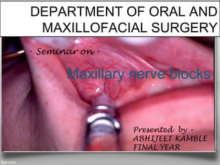
- 1. DEPARTMENT OF ORAL AND MAXILLOFACIAL SURGERY Presented by – ABHIJEET KAMBLE FINAL YEAR Maxillary nerve blocks • Seminar on -
- 5. www.themegallery.com Techniques of Maxillary Anesthesia
- 6. www.themegallery.com Types of Injections I. Supraperiosteal (infiltration) II. Periodontal ligament (PDL, intraligamentary) III. Intraseptal injection IV. Posterior superior alveolar nerve block V. Middle superior alveolar nerve block VI. Anterior superior alveolar nerve block VII. Greater (anterior) palatine nerve block VIII. Nasopalatine nerve block IX. Maxillary (second division) nerve block X. Anterior meddle superior alveolar nerve block XI. Palatal approach-anterior superior alveolar n block
- 8. www.themegallery.com Nerves anesthetized– terminal branch of dental plexus Areas anesthetized Entire region innervated by the large terminal branches of this plexus Indications 1. Pulpal anesthesia of maxillary teeth when treatment is limited to 1 or 2 teeth 2. Soft tissue anesthesia when indicated for surgical procedure Contraindications 1. Infection or acute inflammation 2. Dense bone covering the apices of teeth
- 9. www.themegallery.com Advantages 1. High success rate (>95%) 2. Easy & usually entirely atraumatic Disadvantages Not recommended for larger areas because of multiple injection Alternatives– PDL, IO, regional block Anatomical landmark: Mucobuccal fold Crown of the tooth Root contour of the tooth
- 10. www.themegallery.com Technique 1. Lift the lip, pulling the tissue taut 2. Hold the syringe parallel to the long axis of the tooth 3. Insert the needle at the height of the mucobuccal fold over the target tooth 4. Advance the needle until its bevel is at or above the apical region of the tooth 5. Aspirate, if –ve , deposit 0.6 ml slowly over 20 seconds Sighs & symptoms 1. Subjective: feeling of numbness in the area of administration 2. Objective: no pain during therapy
- 11. www.themegallery.com Safety features 1. Minimal risk of intravascular administration 2. Slowness of injection, aspiration Precautions should not be used for larger areas Complications pain on needle insertion with the tip against periosteum
- 12. www.themegallery.com Posterior superior alveolar nerve blockPosterior superior alveolar nerve block
- 13. www.themegallery.com Nerves Anesthetized- Posterior superior alveolar and its branches Areas Anesthetized- 1) Pulps of the maxillary 3rd , 2nd and 1st molars 2) Buccal periodontium and bone overlying these teeth Anatomical Landmarks- 1. Mucobuccal fold and its concavity 2. Zygomatic process of the maxilla 3. Infratemporal surface of the maxilla 4. Anterior border and coronoid process of the ramus of the mandible 5. Maxillary tuberosity
- 20. www.themegallery.com Anterior superior alveolar(ASA) nerve block
- 22. www.themegallery.com 3. Buccal(labial) periodontium and bone of these teeth 4. Lower eyelid, lateral aspect of the nose, upper lip Anatomical landmarks 1. Infraorbotal notch 2. Infraorbital depression 3. Infraorbital ridge 4. Supraorbital notch 5. Anterior teeth 6. Pupils of eye
- 25. www.themegallery.com Middle Superior Alveolar Nerve Block
- 26. www.themegallery.com Nerves anaesthetized MSA & terminal branch Areas anaesthetized 1. Pulps of maxillary 1st & 2nd premolar & mesiobuccal root of 1st molar(28%) 2. Buccal periodontal tissues & bone of these teeth Anatomical landmarks Mucobuccal fold above the maxillary 2nd premolar Advantages– minimizes no. of injection & volume of solution
- 34. www.themegallery.com Technique Two types of technique– 1.single penetration 2. multiple penetration Technique-1 (single) 1. Area of insertion– palatal mucosa just lateral to the incisive papilla 2. Target area– incisive foramen beneath the papilla 3. Path– approach the injection site at 45 degree angle toward the papilla 4. Chair position– 9 or 10 o’clock position facing in the same direction as the patient 5. Slowly advance the needle towards the foramen until bone is gently contacted (depth approx. 5 mm) 6. Slowly deposit 0.45 ml in 15-30 second minimum
- 37. www.themegallery.com 3. Procedure 4. a) 1st injection: retract the upper lip to stretch tissues & improve visibility. Gently insert in the frenum & deposit 0.3 ml in approx. 15 seconds b) 2nd injection: at 11 or 12 o’clock position, tilting the patients head in the right, & needle at right angle to interdental papilla needle is inserted into the papilla just above the level of crestal bone. Aspirate when ischemia is noted in the incisive papilla or needle tip become visible just beneath the tissue surface Signs & symptoms 1. Subjective: numbness in the upper lip & anterior portion of the hard palate 2. Objective: no pain therapy Safety features 1. Aspiration 2. Contact with bone
- 39. www.themegallery.com Alternatives 1. PSA nerve block 2. ASA nerve block 3. GP nerve block 4. Nasopalatine nerve block Technique– 2-type: high tuberosity approach & GP canal approach High-tuberosity approach 1. Area of insertion– height of mucobuccal fold above the distal aspect of 2nd molar 2. Target area– maxillary n. as it passes through the pterygopalatine fossa • superior and medial to the target area of PSA n. block
- 41. www.themegallery.com Anterior middle superior alveolar nerve block
- 44. www.themegallery.com Advantages 1. Provides anesthesia of multiple teeth with single injection 2. Minimizes volume of anesthesia & no. of puncture 3. Allows effective soft tissue & pulpal anesthesia for periodontal scaling 7 root planing 4. Allows accurate smile line assessment 5. Eliminates postoperative inconvenience of numbness to the upper lip & muscle of facial expression 6. Can be perform comfortably with a CCLAD Disadvantages 1. Requires a slow administration time ( 0.5 ml/min) 2. Can cause operator fatigue with a manual syringe 3. May need supplemental anesthesia for C.I. & L.I.
Editor's Notes
- It arises from medial part of convex anterior border of trigeminal ganglion.Then it pierces the duramater of trigeminal cave and enters into lateral wall of cavernous sinus.Finally,it entera the orbit through the superior orbital fissure and divides into three branches namely lacrimal,frontal and nasocilliary.
- It arises