SlideShare a Scribd company logo
1 of 60
FRONTAL BONE FRACTURE AND
ITS MANAGEMENT
 A presentation by Dr. Abhijeet Kamble
Junior resident II
GOOD MORNING
CONTENTS
1 Introduction
2 embryology
3 SURGICAL ANATOMY
4 DIAGNOSIS
5 TREATMENT PLANNING
19th Century skull showing sword-blade
trauma on frontal bone.
INTRODUCTION
Fontal sinus is most frequently damaged as a result of high
velocity motor vehicle accidents. 70% of frontal sinus
fractures were due to automobile accidents and 20% due to
assaults..
Fractures involving frontal bone is rather rare because of its
protected location
1 . It is basically protected from trauma by the prominence
formed by the nasal pyramid.
2 . Incidence of fractures involving this area ranges between
5-15%
3 . Fractures involving this bone is considered to be rather
dangerous because of its proximity to brain as well as due to
the cosmetic defects it can produce. The proximity of this
bone to the orbit and naso frontal duct doesn’t help matters
either.
Fractures involving this area if not treated promptly can lead
to :
1. Meningitis 2. Mucopyocele 3. Encephalitis 4. Cerebral
abscess
4 F r o n t a l b o n e f a c t u r e a n d i t s m a n a g e m e n t 2 0 X X
EMBRYOLOGY
FRONTAL BONE
FRACTURE
EMBRYOLOGY OF FRONTAL BONE
6 p r e s e n t a t i o n t i t l e 2 0 X X
Structure of the frontal bone
The frontal bone is one of the skull bones enclosing the brain
(neurocranium) and it consists of the following parts:
•The squamous part;
•The nasal part;
•Two orbital plates;
•Two zygomatic processes.
Begin as an outgrowth from nasal chamber in utero
Absent at birth
Do not develop until 2" year of life
Develop from ethmoidal infundibular air cells by invagination of
frontal bone through frontal recess or from superior meatus
Cannot be identified radiographically until about age of 8
years
Development
Orbital surface of frontal
bone
EMBRYOLOGY OF FRONTAL BONE
7 p r e s e n t a t i o n t i t l e 2 0 X X
EMBRYOLOGY OF FRONTAL BONE
8 p r e s e n t a t i o n t i t l e 2 0 X X
Structure of the frontal bone
The frontal bone is made up of two main parts. These are the squamous part, and the orbital part.
The squamous part marks the vertical, flat, and also the biggest part, and the main region of
the forehead. The orbital part is the horizontal and second biggest region of the frontal bone. It
enters into the formation of the roofs of the orbital and nasal cavities. Sometimes a third part is
included as the nasal part of the frontal bone, and sometimes this is included with the squamous
part. The nasal part is between the brow ridges, and ends in a serrated nasal notch that articulates
with the nasal bones inferiorly, and with the lacrimal and maxilla bones laterally
The frontal bone is presumed to be derived from neural crest cells.[3]
The frontal bone is ossified in membrane from two primary centers, one for each half, which appear
toward the end of the second month of fetal life, one above each supraorbital margin. From each of these
centers, ossification extends upward to form the corresponding half of the squama, and backwards to
form the orbital plate. The spine is ossified from a pair of secondary centers, on either side of the middle
line; similar centers appear in the nasal part and zygomatic processes.
At birth the bone consists of two pieces, separated by the frontal suture, which is usually obliterated
by Intramembranous ossification, except at its lower part, by the eighth year, but occasionally persists
throughout life. It is generally maintained that the development of the frontal sinuses begins at the end of
the first or beginning of the second year, but may begin at birth. The sinuses are of considerable size by
the seventh or eighth year, but do not attain their full proportions until after puberty.
Development
9 p r e s e n t a t i o n t i t l e 2 0 X X
SURGICAL ANATOMY
From the Nasion the bone extends approximately 12.5cm
superiorly, 8.0 cm laterally and 5.5 cm posteriorly.
Two frontal tuberosities are noted lateral to the midline and
superior to the supraorbial rim.
The thickest area of the bone is the supraorbial rim from the
frontozygomatic process to the nasal bones.
The supraorbital foramen are located at the most superior
portion of orbital rim.
supratrochlear foramen is located medial to the
supraorbital foramen or notch and lateral to the nasal
bones.
A spine or concavity exists on the frontal bone along the
medial orbital roof; the trocha of the superior oblique
muscle is attached to this spine.
1 0 p r e s e n t a t i o n t i t l e 2 0 X X
FRONTAL SINUS
Anatomy
Evolution over age
The frontal sinus is generally absent at birth. At one year
the anterior ethmoid air cells begin to invade the frontal
bone. Frontal sinus growth is then complete at
approximately 15 years of age.
The frontal sinus is irregularly shaped and scalloped at its
margins. Asymmetry of the sinus is the rule rather the
exception (10% of individuals have a unilateral sinus, 9%
of individuals have a rudimentary or absent sinus).
Applied Anatomy of Frontal Sinus
1 1 p r e s e n t a t i o n t i t l e 2 0 X X
FRONTAL SINUS
Functions of Frontal Sinus
Following are the various functions of
the frontal sinus:
1. Production and storage of mucus
2. Resonator for voice
3. Humidification and warming of
inhaled air
4. Accessory area of olfaction
5. Conservation of heat from the nasal
fossae
6. Definition of facial contours
7.“ Surge Tank” to dampen the
pressure differential that develops
during inspiration
1 2 p r e s e n t a t i o n t i t l e 2 0 X X
FRONTAL SINUS
Anatomic relationships
The frontal sinus has several critical anatomic
relationships. These include:
Sinus floor -> orbital roof/anterior ethmoid air cells
Posterior table -> anterior cranial fossa
Anterior table -> frontal contour
Drainage
The frontal sinus drains via a small outflow tract into the
ethmoid sinus/nasal cavity. The outflow tract is hour-glass
shaped with the true ostium (3-4 mm) at the narrowest
portion.
Epidemiolog
y
1 3 p r e s e n t a t i o n t i t l e 2 0 X X
Frontal sinus fractures are relatively uncommon and account for only 5-15% of maxillofacial fractures with
a preponderance of male patients.
The most common frontal sinus fractures involve a combination of the anterior and posterior tables with or
without frontal recess involvement (about 2/3).
Isolated posterior table fractures are extremely uncommon
(< 1%).
This 3D CT reconstruction shows an isolated anterior
table fracture.
CLASSIFICATION
1 4 2 0 X X
Frontal sinus fractures are usually classified
based on:
•Location
•Extent of injury
•Involvement of frontonasal duct
•Current injury to the dura
Stanley’s Classification of Frontal Sinus
Fracture
•Type I: Anterior Table Fracture
• Isolated to anterior table
• Accompanied by supraorbital rim fracture
• Accompanied by naso-ethmoid complex
fracture
•Type II: Anterior and Posterior Table Fractures
• It is a linear fracture either on transverse
direction or in vertical direction
•Type III: Comminuted Fractures
• Isolated to both tables
• Accompanied by naso-ethmoid complex
Gonty Et al. Classification of Frontal Sinus
Fracture
•Type I: Anterior Table Fracture
• Isolated to anterior table
• Accompanied by supraorbital rim fracture
• Accompanied by naso-ethmoid complex
fracture
•Type II: Anterior and Posterior Table Fractures
• A linear fracture either on transverse
direction or in vertical direction
• Comminuted fracture either isolated to both
tables or accompanied by naso-ethmoid
complex fracture
•Type III: Posterior Table Fracture
•Type IV: Through and Through Frontal Sinus
Fracture
CLASSIFICATION
1 5
Type I : Anterior table fracture with minimum
comminution
Type II : Anterior Wall comminuted fracture with -
Possible NOE or Orbital rim fracture
Type III : Anterior & Posterior Wall fracture
(Posterior wall fracture without significant
displacement or ductal injury)
Type IV : Anterior & Posterior Wall fracture with
Dural injury & CSF leak
Type V :Anterior & Posterior Wall fracture with
Dural injury, CSF leak, soft tissue or bone loss
and/or severe disruption of anterior cranial fossa
Gerbino G, Roccia F, Benoch A, ot al. Analyss of 158 fontalsinus fractures: Current surgical management
and complications. J Craniomaxilofac Surg 2000; 28133
CLASSIFICATION
1 Frontal recess
involvement
A frontal recess injury involves the floor of the
frontal sinus and the outflow tract. It may also
involve the anterior skull base.
Anterior table
fractures
Fractures of the anterior table of the
frontal sinus vary from minimally
displaced to severely
displaced/comminuted depending
upon the severity of the trauma and
size of the sinus.
Minimal displacement
Less severe trauma may result in
fractures of the anterior table which
are minimally (as illustrated) or
nondisplaced. 2
3
4
Posterior table
fracture
Posterior table fractures carry a higher
risk of intracranial injury because they
create a communication with the
intracranial space. The degree of
fracture displacement gives some
insight into the severity of the injury.
While there is no commonly agreed
upon classification for displacement,
many authors have used the width of
the posterior table (0.1 mm – 4 mm) as
a “measuring stick” to describe
displacement because it is readily
visible on axial CT-scans.
3
1 6 p r e s e n t a t i o n t i t l e 2 0 X X
DIAGNOSIS
 The diagnosis of frontal bone fracture is based upon
proper history and physical examination of the patient
 This includes Inspection and palpation of the affected
area
Detailed History includes:
 Information about events
 Visual difficulties
 Numbness
 Pain
 Rhinorrhea
 Sense of smell
 Previous history of nasal or sinus disease
surgery
1 7 p r e s e n t a t i o n t i t l e 2 0 X X
CLINICAL FEATURES
Forehead laceration
(58%)
Forehead pain (82%)
swelling
1 8 p r e s e n t a t i o n t i t l e 2 0 X X
Frontal bone depression (25%)
Csf rhinorrhea (1/3rd patients
Periorbital ecchymosis
1 9 p r e s e n t a t i o n t i t l e 2 0 X X
Clinical evaluation
General
Facial pain
Forehead paraesthesia or anaesthesia
Forehead laceration
Visible and/or palpable frontal bone depression
CSF Rhinorrhoea
Neurological injuries
Cerebral contusion
Subdural & epidural haematoma.
Ophthalmic injuries
Pupillary defect
Optic neuropathy
Hyphaema
Disc oedema
Corneal defect
Loss of globe integrity
Associated maxillofacial Injuries
NOE fracture
Orbital fracture
Zygomatic fracture
RADIOGRAPHIC
EVALUATION
2 0 p r e s e n t a t i o n t i t l e 2 0 X X
PLAIN RADIOGRAPHS are of little use in the diagnosis of frontal sinus
fractures
Caldwell's view
EVIDENCE OF AIR FLUID LEVEL, CLOUDING
OF FRONTAL SINUS, PNEUMOCEPHALUS
Lateral view High Resolution CT Scan
CT Scans are the GOLD standard for imaging
of these fracture
CT SCANS
2 1 p r e s e n t a t i o n t i t l e 2 0 X X
AXIAL CT VIEW
Axial images reveals location, severity and degree of
comminution of anterior and posterior table fractures.
A thin cut (1.0 to 1.5 mm), axial computed tomography (CT) scan is
the gold standard.
It is recommended to obtain coronal, sagittal, and
three-dimensional (3-D) reconstructions for
diagnostic accuracy
Coronal images for the Frontal sinus floor and orbital Roof fracture
Coronal CT VIEW
CT SCANS
2 2 p r e s e n t a t i o n t i t l e 2 0 X X
Sagittal images can be useful in assessing
the patency of the frontal recess
while 3-D reconstructions help define the shape,
location, and orientation of individual bone
fragments that are seen less clearly on 2-D views
sagittal CT VIEW
The 3-D information can
reduce the need for surgical
dissection, because the
surgeon knows the number,
location, and orientation of the
larger bone fragments. It can
also help the patient and/or
family to understand the bony
anatomy and severity of the
injury.
MANAGEMENT.
FRONTAL BONE FRACTURE
2 3 p r e s e n t a t i o n t i t l e 2 0 X X
PRINCIPLES OF FRONTAL BONE FRACTURE MANAGEMENT
2 4 p r e s e n t a t i o n t i t l e 2 0 X X
SAFE
SINUS
RESTORE
FACIAL
CONTOUR
AVOID
SHORT &
LONG
TERM
COMPLICATIONS
Objectives of management
• To avoid immediate and short-term complications such as CSF
leak, meningitis, spreading infection.
• To avoid long-term complications such as frontal bone
osteomyelitis, chronic frontal sinusitis, mucocele,
mycopyocele, and brain abscess.
• To provide adequate exposure for anatomic reduction of naso-
orbito-ethmoidal (NEO) fractures.
• To restore proper aesthetic contour of the forehead.
2 5 p r e s e n t a t i o n t i t l e 2 0 X X
TREATMENT OPTIONS
Conservative
Fracture reduction & fixation
Sinus obliteration
Sinus cranialization
Management generally based on
Three clinical factors:
2 6 p r e s e n t a t i o n t i t l e 2 0 X X
• Fracture location and displacement
• Dural and cerebral involvement
• Damage to the frontal sinus drainage systems
Guiding Principles for Frontal Sinus Management
2 7 p r e s e n t a t i o n t i t l e 2 0 X X
•To separate nasal cavity from sinus
•To eliminate dead space
•To separate the frontonasal duct from frontal
sinus by obstructing the duct
•To eliminate a functional sinus, sinus mucosa
is removed, and sinus is obliterated
MANAGEMENT
Surgical Access
2 8 p r e s e n t a t i o n t i t l e 2 0 X X
1. Through existing laceration
2. Butterfly incision
3. Gullwing or eyeglass incision
4. Bicoronal approach suggests sterile surgical glove tourniquet intraoperatively to get the hemorrhage
control and blood less surgical field in elevation of bicoronal flap for the surgical management of frontal
sinus fracture.
Gullwing or eyeglass incision
Surgical Access
2 9 p r e s e n t a t i o n t i t l e 2 0 X X
Use of existing laceration to access left-sided
frontal sinus fracture.
Extensive transverse laceration of the forehead with easy
access to the frontal sinus. The fractured segments have
been removed to gain access to the nasofrontal duct.
Through existing laceration
Surgical Access
3 0 p r e s e n t a t i o n t i t l e 2 0 X X
Irregular outline of a coronal incision in two different patients
Surgical Access
3 1 p r e s e n t a t i o n t i t l e 2 0 X X
Bicoronal approach
Access areas
The following areas can be
exposed:
•Entire calvarial vault
•Anterior and lateral skull base
•Frontal sinus/Ethmoid
•Zygoma
•Zygomatic arch
•Orbit (lateral/cranial/medial)
•Nasal dorsum
•Temporomandibular joint (TMJ)
and condylar/subcondylar region
3 2 p r e s e n t a t i o n t i t l e 2 0 X X
Indications of Surgery in Frontal Sinus
Fractures
1. To avoid immediate complications such as CSF leak, meningitis
2. To avoid long-term complications such as frontal sinusitis, meningitis and brain abscess formation
3. To provide aesthetic contour to the forehead
4. To provide exposure for anatomic reduction of NOE fractures
3 3 p r e s e n t a t i o n t i t l e 2 0 X X
History of frontal sinus surgery
 Surgical approaches to frontal sinus
• Frontal sinus surgery was first described in the
18th century. It is noted that as early as 1750
Runge performed an obliteration procedure of
the frontal sinus.
• The first published report in 1870 by Wells
described an external and intracranial drainage
procedure for a frontal sinus mucocele.
• In 1884 Alexander Ogston described a
trephination procedure through the anterior
table to evacuate the frontal sinus
• Kuhnt in 1895 described removing the
anterior wall of the frontal sinus in an attempt
to clear disease
In 1898 Riedel/Schenke described the first
procedure for obliteration of the frontal sinus
the Lynch and Howarth
operation
Lynch incision (A) with resulting access
to frontal sinus and ethmoid sinuses (B)
3 4 p r e s e n t a t i o n t i t l e 2 0 X X
History of frontal sinus surgery
 Surgical approaches to frontal sinus
MANAGEMENT.
3 5 p r e s e n t a t i o n t i t l e 2 0 X X
3 6 p r e s e n t a t i o n t i t l e 2 0 X X
Management of Anterior Table Fracture
Decision-Making
•Simple greenstick or undisplaced fracture does not
require surgical intervention.
•In depressed anterior wall fracture, frontal sinus explored,
careful irrigation carried out, fragments reduced and
stabilize by internal fixation. In posterior wall fracture
without CSF leak or pneumocephalus, reconstruction of
only anterior wall is done.
• Other than surgical intervention, antibiotics,
sinus decongestants and analgesics are
prescribed to keep the frontonasal duct patent
and to prevent infection.
Rai et al. suggested bone mapping/sketching in management of
anterior table frontal sinus fracture with great success
Bone mapping/sketching in management of
anterior table frontal sinus fracture
Yoo MH et al. suggested endoscopic trans nasal reduction of
anterior table fracture To support the reduced fragments, they
advocated use of custom made latex glove balloon to be inserted
into the frontal sinus then expanded and maintained for 3 weeks
ANTERIOR table fracture
FRONTAL BONE FRACTURE
3 7 p r e s e n t a t i o n t i t l e 2 0 X X
(Nondisplaced, linear/isolated)
Conservative treatment
• Local wound care
• Antibiotics, nasal decongestant, analgesics
• Follow up evaluation
Operative Indication of FS injury
FRONTAL BONE FRACTURE
3 8 p r e s e n t a t i o n t i t l e 2 0 X X
• Nasofrontal duct involvement/obstruction
• Displacement of posterior table with underlying
neurological injury
• Aesthetic forehead deformity
ANTERIOR table fracture
3 9 p r e s e n t a t i o n t i t l e 2 0 X X
Modified treatment algorithm for highly selected patients
ANTERIOR table fracture
4 0 p r e s e n t a t i o n t i t l e 2 0 X X
(Displaced)
Approaches
Intraoperative assessment
• Any kind of fluid
• Nasofrontal duct patency
Nasofrontal duct injury
• Nasofrontal duct filling with frontal sinus obliteration
Grafting
Reduction & fixation
• Existing laceration
• Butterfly/seagull/open sky approaches, coronal
flap
Isolated Anterior Table Fracture
FRONTAL BONE FRACTURE
4 1 p r e s e n t a t i o n t i t l e 2 0 X X
• Open Reduction Internal Fixation
If the degree of displacement, as
visualized on axial or sagittal sections of
CT scans, is >1–2 mm, that is, more than a
table’s width, it warrants an open reduction
and fixation within 7–10 days*
Open reduction internal fixation
4 2
Principle
s
The goal of open reduction is to reapproximate the bone fragments into their premorbid position
and use internal fixation hardware to maintain them in their position.
Approac
h
coronal
approach
Reduction
General considerations
The anterior table of the frontal sinus
is normally convex. Compressive
forces on the frontal bone deform the
convexity into a concavity. This may
or may not result in fracture
comminution.
Mobilization of the depressed bone
fragment(s) may require significant
effort to overcome compressive
forces between bone fragments.
Reduction techniques
4 3
Mobilizing the bone fragments
using an elevator
There are several techniques to reduce the fragments:
Inserting a screw into a bony
fragment and reducing it by
pulling it outwards
Fixation
Simple reduction
Postop care following management of frontal sinus
fractures
4 4
Evaluation of the patient’s vision
performed as soon as they are awakened from anesthesia and then at
regular intervals until they are discharged from the hospital.
Postoperative
positioning Keeping the patient’s head in a raised position both
preoperatively and postoperatively may significantly
improve edema and pain.
Nose-
blowing
Nose-blowing should be avoided for at least 3 weeks
following frontal sinus and skull base repair.
Frontal sinus Obliteration
FRONTAL BONE FRACTURE
4 5 p r e s e n t a t i o n t i t l e 2 0 X X
• Steps
• Sinus Exploration
• Mucosal Exenteration
• Nasofrontal Duct obturation
• Frontal sinus Obliteration
• Fracture Reduction
Frontal sinus
FRONTAL BONE FRACTURE
4 6 p r e s e n t a t i o n t i t l e 2 0 X X
• Filling materials for obturation and obliteration
• Autogenous Material Synthetic materials
- Autologous Fat Polyfluorotetraethylene, ePTFE
- Cancellous bone’ Methylmethacrylate, MMA
- Muscle
- Pericranial flap
- Banked cadaveric tissue
Indication for Bone Grafting
FRONTAL BONE FRACTURE
4 7 p r e s e n t a t i o n t i t l e 2 0 X X
1. Extensive loss of support at the skull base over the fovea
ethmoidalis and cribriform plate, in combination with a
peri cranial flap.
2. Superior orbital roof fractures, to avoid pulsatile
exophthalmos and orbital deformity.
3. Extensive bone loss of the anterior table, which cannot be
replaced with elements of the posterior table.
4. In combination with NOE and orbital reconstruction
as layered bone grafts to obliterate the ethmoids and reconstruct
the medial orbital wall.
posterior table fracture
FRONTAL BONE FRACTURE
4 8 p r e s e n t a t i o n t i t l e 2 0 X X
CSF leak Absent and No displacement
Frontal Sinus Obliteration
CSF leak Absent and/or Displacement
Frontal Sinus Cranialization
FRONTAL BONE FRACTURE
4 9 p r e s e n t a t i o n t i t l e 2 0 X X
• It is another technique to minimize the dead
space in sinus. It involves removing
posterior table thus permitting brain to
expand into frontal sinus resulting in
confuence between sinus cavity and
anterior cranial fossa.
• It is done in cases with CSF leak and
neurological injury due to displaced
posterior table fracture. To isolate the
splanchnocranium from the frontal sinus, a
pericranial fap is used.
Frontal Sinus Cranialization
FRONTAL BONE FRACTURE
5 0 p r e s e n t a t i o n t i t l e 2 0 X X
Although obliteration has been touted as the gold standard
and safest method to treat the injured frontal sinus, there
are many disadvantages including :
Complications
FRONTAL BONE FRACTURE
5 1 p r e s e n t a t i o n t i t l e 2 0 X X
Recent Advancements
FRONTAL BONE FRACTURE
5 2 p r e s e n t a t i o n t i t l e 2 0 X X
Virtual Surgery
Augmented Reality
Stereolithography or 3D printing
Virtual Endoscopy
Preoperative Photographs and radiographs
5 3 A 48-year-old female patient who sustained a frontal bone fracture right side having a H/O fall from a hill
Intraoperative & postoperative Photographs
5 4 A 48-year-old female patient who sustained a frontal bone fracture right side having a H/O fall from a hill
Preoperative Photographs and radiographs
5 5 A 22-year-old male patient who sustained a frontal bone having a H/O RSA, having fallen form his bike
Intraoperative & postoperative Photographs
5 6 A 22-year-old male patient who sustained a frontal bone having a H/O RSA, having fallen form his bike
Follow up Photographs
5 7 A 22-year-old male patient who sustained a frontal bone having a H/O RSA, having fallen form his bike
Conclusion
The management of NOE and frontal sinus fracture is
always a challenging task .The proper handling of MCL
and nasofrontal duct is mandatory to get good post-
operative results. Proper treatment planning in the form of
incision selection, method of fixation and use of bone graft
should be done to avoid unaesthetic results.
5 8 p r e s e n t a t i o n t i t l e 2 0 X X
references
5 9 N O E F R A C T U R E S 2 0 X X
• 1. Milad Etemadi SH, Shahnaseri S, Soltani P, Motamedi MRK. Management of naso-orbito-ethmoid fractures: a 10-year review. Trauma
Mon. 2017;22:292–30.
• 2. Brasileiro BF, Passeri LA. Epidemiological analysis of maxillofacial fractures in Brazil: a 5-year prospective study. Oral Surg Oral Med
Oral Pathol Oral Radiol Endod. 2006;102(1):28–34.
• 3. Rosenberger E, Kriet JD, Humphrey C. Management of nasoethmoid fractures. Curr Opin Otolaryngol Head Neck Surg. 2013;21:410–6.
(Anatomy of MCL, physical examination of the complex and displacement)
• 4. Poh E, Kakizaki H, Selva D, Leibovitch I. Anatomy of medial canthal tendon in Caucasians. Clin Exp Ophthalmol. 2012;40:170–3.
• 5. Merkx MA, Freihofer HP, Borstlap WA, van’t Hoff MA. Effectiveness of primary correction of traumatic telecanthus. Int J Oral Maxillofac
Surg. 1995;24:344–7.
• 6. Heine D, Catone GA, Bavitz B, Grenadier MR. Naso-orbitalethmoid injury: report of a case and review of the literature. Oral Surg Oral
Med Oral Path. 2005;69(5):542–9.
• 7. Rowe NL. Rowe and Williams’ maxillofacial injuries, vol. 2. 2nd ed. Edinburgh: Churchill Livinstone; 1994.
• 8. Markowitz BL, Manson PN, Sargent L, et al. Management of the medial canthal tendon in nasoethmoid orbital fractures: the importance of
the central fragment in classifcation and treatment. Plast Reconstr Surg. 1991;87:843–853.
• 9. Burstein F, Cohen S, Hudgins R, Boydston W. Frontal basilar trauma: classifcation and treatment. Plast Reconstr Surg.
1997;99(5):1314–21.
• 10. Balaraman K, Ramani V, Bharathi R, Venkataramani H, Sabapathy SR. Outcome assessment of Nasoethmoid fractures. Int J Oral
Maxillofac Surg. 2013;42(10):1220.
• 11. Ellis E III. Sequencing treatment for naso-orbito-ethmoid fractures. J Oral Maxillofac Surg. 1993;51:543–558.
• 12. Nguyen M, Koshy JC, Hollier LH Jr. Pearls of nasoorbitoethmoid trauma management. Semin Plast Surg. 2010;24:383–8.
• 13. Dingman RO, Grabb WC, Oneal RM. Management of injuries of the naso-orbital complex. Arch Surg. 1969;98:566–71.
• 14. Mustarde JC. Epicanthus and telecanthus. Int Opthalmol Clin. 1964;4:359.
• 15. Cruse C, Blevins PK, Luce EA. Naso-ethmoid-orbital fractures. J Trauma. 1980;20:551.
THANK YOU

More Related Content

What's hot

Submandibular and retromandibular approach
Submandibular and retromandibular approachSubmandibular and retromandibular approach
Submandibular and retromandibular approachJamil Kifayatullah
 
Zygomatic Complex Fracture- ZMC
Zygomatic Complex Fracture- ZMCZygomatic Complex Fracture- ZMC
Zygomatic Complex Fracture- ZMCHimanshu Soni
 
3 approaches to the tmj
3 approaches to the tmj3 approaches to the tmj
3 approaches to the tmjDrKamini Dadsena
 
Weber ferguson incison (poster)
Weber ferguson incison (poster)Weber ferguson incison (poster)
Weber ferguson incison (poster)Sk Aziz Ikbal
 
04 frontal sinus FRACTURE
04 frontal sinus FRACTURE04 frontal sinus FRACTURE
04 frontal sinus FRACTUREJamil Kifayatullah
 
Naso orbito ethmoid (noe) complex fracture
Naso orbito ethmoid (noe) complex fractureNaso orbito ethmoid (noe) complex fracture
Naso orbito ethmoid (noe) complex fracturesailesh kumar
 
NOE fractures
NOE fractures NOE fractures
NOE fractures anchalag8
 
mandibular molar Impactions
mandibular molar Impactionsmandibular molar Impactions
mandibular molar ImpactionsNishant Tewari
 
Condylar fractures
Condylar fracturesCondylar fractures
Condylar fracturesZeeshan Arif
 
Fluids and electrolytes in Maxillofacial Surgery
Fluids and electrolytes in Maxillofacial SurgeryFluids and electrolytes in Maxillofacial Surgery
Fluids and electrolytes in Maxillofacial SurgeryVarun Mittal
 
Naso-orbito-ethmoidal fracture
Naso-orbito-ethmoidal fractureNaso-orbito-ethmoidal fracture
Naso-orbito-ethmoidal fractureDr Bhavik Miyani
 
Pedicle flap in Maxillofacial Surgery
 Pedicle flap in Maxillofacial Surgery Pedicle flap in Maxillofacial Surgery
Pedicle flap in Maxillofacial SurgeryDrKamini Dadsena
 
Mid facial fractures and their management
Mid facial fractures and their managementMid facial fractures and their management
Mid facial fractures and their managementRuhi Kashmiri
 
Apertognathia and its surgical management
Apertognathia and its surgical managementApertognathia and its surgical management
Apertognathia and its surgical managementHimanshu Soni
 
Symphysis & Angle MANDIBULAR FRACTURES
Symphysis & Angle MANDIBULAR FRACTURES Symphysis & Angle MANDIBULAR FRACTURES
Symphysis & Angle MANDIBULAR FRACTURES Dr-Faisal Al-Qahtani
 
Ligation of arteries in maxillofacial region
Ligation of arteries in maxillofacial regionLigation of arteries in maxillofacial region
Ligation of arteries in maxillofacial regionIndian dental academy
 
Classification of Mandible, Midface, ZMC and NOE Fractures
Classification of Mandible, Midface, ZMC and NOE FracturesClassification of Mandible, Midface, ZMC and NOE Fractures
Classification of Mandible, Midface, ZMC and NOE FracturesArjun Shenoy
 
Mandibular fractures
Mandibular fracturesMandibular fractures
Mandibular fracturesArjun Shenoy
 

What's hot (20)

Zygomatic fractures
Zygomatic fracturesZygomatic fractures
Zygomatic fractures
 
Submandibular and retromandibular approach
Submandibular and retromandibular approachSubmandibular and retromandibular approach
Submandibular and retromandibular approach
 
Zygomatic Complex Fracture- ZMC
Zygomatic Complex Fracture- ZMCZygomatic Complex Fracture- ZMC
Zygomatic Complex Fracture- ZMC
 
3 approaches to the tmj
3 approaches to the tmj3 approaches to the tmj
3 approaches to the tmj
 
Weber ferguson incison (poster)
Weber ferguson incison (poster)Weber ferguson incison (poster)
Weber ferguson incison (poster)
 
04 frontal sinus FRACTURE
04 frontal sinus FRACTURE04 frontal sinus FRACTURE
04 frontal sinus FRACTURE
 
Naso orbito ethmoid (noe) complex fracture
Naso orbito ethmoid (noe) complex fractureNaso orbito ethmoid (noe) complex fracture
Naso orbito ethmoid (noe) complex fracture
 
NOE fractures
NOE fractures NOE fractures
NOE fractures
 
mandibular molar Impactions
mandibular molar Impactionsmandibular molar Impactions
mandibular molar Impactions
 
Condylar fractures
Condylar fracturesCondylar fractures
Condylar fractures
 
Lefort 1 osteotomy
Lefort 1 osteotomyLefort 1 osteotomy
Lefort 1 osteotomy
 
Fluids and electrolytes in Maxillofacial Surgery
Fluids and electrolytes in Maxillofacial SurgeryFluids and electrolytes in Maxillofacial Surgery
Fluids and electrolytes in Maxillofacial Surgery
 
Naso-orbito-ethmoidal fracture
Naso-orbito-ethmoidal fractureNaso-orbito-ethmoidal fracture
Naso-orbito-ethmoidal fracture
 
Pedicle flap in Maxillofacial Surgery
 Pedicle flap in Maxillofacial Surgery Pedicle flap in Maxillofacial Surgery
Pedicle flap in Maxillofacial Surgery
 
Mid facial fractures and their management
Mid facial fractures and their managementMid facial fractures and their management
Mid facial fractures and their management
 
Apertognathia and its surgical management
Apertognathia and its surgical managementApertognathia and its surgical management
Apertognathia and its surgical management
 
Symphysis & Angle MANDIBULAR FRACTURES
Symphysis & Angle MANDIBULAR FRACTURES Symphysis & Angle MANDIBULAR FRACTURES
Symphysis & Angle MANDIBULAR FRACTURES
 
Ligation of arteries in maxillofacial region
Ligation of arteries in maxillofacial regionLigation of arteries in maxillofacial region
Ligation of arteries in maxillofacial region
 
Classification of Mandible, Midface, ZMC and NOE Fractures
Classification of Mandible, Midface, ZMC and NOE FracturesClassification of Mandible, Midface, ZMC and NOE Fractures
Classification of Mandible, Midface, ZMC and NOE Fractures
 
Mandibular fractures
Mandibular fracturesMandibular fractures
Mandibular fractures
 

Similar to FRONTAL BONE FRACTURE AND ITS MANAGEMENT.pptx

FRONTAL BONE FRACTURE
FRONTAL BONE FRACTUREFRONTAL BONE FRACTURE
FRONTAL BONE FRACTUREankitaraj63
 
Fractures of the Middle-third of the Facial Skeleton (1).pptx
Fractures of the Middle-third of the Facial Skeleton (1).pptxFractures of the Middle-third of the Facial Skeleton (1).pptx
Fractures of the Middle-third of the Facial Skeleton (1).pptxPalPal12
 
Naso orbito ethmoidal fracture
Naso orbito ethmoidal fractureNaso orbito ethmoidal fracture
Naso orbito ethmoidal fractureDr.Preethi Gopinath
 
Nasal and nasoethmoidal fractures
Nasal and nasoethmoidal fracturesNasal and nasoethmoidal fractures
Nasal and nasoethmoidal fracturesAhmed Adawy
 
Surgical Anatomy For Orbital Procedures .pptx
Surgical Anatomy For Orbital Procedures .pptxSurgical Anatomy For Orbital Procedures .pptx
Surgical Anatomy For Orbital Procedures .pptxAnwar Almahmode
 
Management of Facial Fractures in ED
Management of Facial Fractures in EDManagement of Facial Fractures in ED
Management of Facial Fractures in EDtmit2
 
Nasal fracture ent ppt
Nasal fracture ent ppt Nasal fracture ent ppt
Nasal fracture ent ppt Ayushree Sharma
 
Imaging in facial trauma
Imaging in facial traumaImaging in facial trauma
Imaging in facial traumaSumiya Arshad
 
Orbit and adenexa
Orbit  and adenexaOrbit  and adenexa
Orbit and adenexaRakesh Sharma
 
Maxillofacial Trauma.pptx
Maxillofacial       Trauma.pptxMaxillofacial       Trauma.pptx
Maxillofacial Trauma.pptxfaheem411362
 
Naso orbito-ethmoid fractures
Naso orbito-ethmoid fracturesNaso orbito-ethmoid fractures
Naso orbito-ethmoid fracturesSaarang Hansraj
 
Proptosis by Dr.Ashwin Menon
Proptosis by Dr.Ashwin MenonProptosis by Dr.Ashwin Menon
Proptosis by Dr.Ashwin MenonDr.Ashwin Menon
 
Nasal septum and its diseases
Nasal septum and its diseasesNasal septum and its diseases
Nasal septum and its diseasesVinay Bhat
 
Imaging of paranasal sinuses
Imaging of paranasal sinusesImaging of paranasal sinuses
Imaging of paranasal sinusesArchana Koshy
 
mandibular fracture
mandibular fracturemandibular fracture
mandibular fractureIbraheem AlRawi
 

Similar to FRONTAL BONE FRACTURE AND ITS MANAGEMENT.pptx (20)

FRONTAL BONE FRACTURE
FRONTAL BONE FRACTUREFRONTAL BONE FRACTURE
FRONTAL BONE FRACTURE
 
Fractures of the Middle-third of the Facial Skeleton (1).pptx
Fractures of the Middle-third of the Facial Skeleton (1).pptxFractures of the Middle-third of the Facial Skeleton (1).pptx
Fractures of the Middle-third of the Facial Skeleton (1).pptx
 
Naso orbito ethmoidal fracture
Naso orbito ethmoidal fractureNaso orbito ethmoidal fracture
Naso orbito ethmoidal fracture
 
Mid facial fracture
Mid facial fractureMid facial fracture
Mid facial fracture
 
Mandibular fractures
Mandibular fracturesMandibular fractures
Mandibular fractures
 
Nasal and nasoethmoidal fractures
Nasal and nasoethmoidal fracturesNasal and nasoethmoidal fractures
Nasal and nasoethmoidal fractures
 
Surgical Anatomy For Orbital Procedures .pptx
Surgical Anatomy For Orbital Procedures .pptxSurgical Anatomy For Orbital Procedures .pptx
Surgical Anatomy For Orbital Procedures .pptx
 
Medical
MedicalMedical
Medical
 
Management of Facial Fractures in ED
Management of Facial Fractures in EDManagement of Facial Fractures in ED
Management of Facial Fractures in ED
 
The septum
The septumThe septum
The septum
 
Nasal fracture ent ppt
Nasal fracture ent ppt Nasal fracture ent ppt
Nasal fracture ent ppt
 
Nasal fracture
Nasal fractureNasal fracture
Nasal fracture
 
Imaging in facial trauma
Imaging in facial traumaImaging in facial trauma
Imaging in facial trauma
 
Orbit and adenexa
Orbit  and adenexaOrbit  and adenexa
Orbit and adenexa
 
Maxillofacial Trauma.pptx
Maxillofacial       Trauma.pptxMaxillofacial       Trauma.pptx
Maxillofacial Trauma.pptx
 
Naso orbito-ethmoid fractures
Naso orbito-ethmoid fracturesNaso orbito-ethmoid fractures
Naso orbito-ethmoid fractures
 
Proptosis by Dr.Ashwin Menon
Proptosis by Dr.Ashwin MenonProptosis by Dr.Ashwin Menon
Proptosis by Dr.Ashwin Menon
 
Nasal septum and its diseases
Nasal septum and its diseasesNasal septum and its diseases
Nasal septum and its diseases
 
Imaging of paranasal sinuses
Imaging of paranasal sinusesImaging of paranasal sinuses
Imaging of paranasal sinuses
 
mandibular fracture
mandibular fracturemandibular fracture
mandibular fracture
 

More from Government Dental College and Hospital, Shimla

More from Government Dental College and Hospital, Shimla (20)

MENINGIOMA CASE REPORT.pptx
MENINGIOMA CASE REPORT.pptxMENINGIOMA CASE REPORT.pptx
MENINGIOMA CASE REPORT.pptx
 
case presentation.pptx
case presentation.pptxcase presentation.pptx
case presentation.pptx
 
case presentaion.pptx
case presentaion.pptxcase presentaion.pptx
case presentaion.pptx
 
MENINGIOMA.pptx
MENINGIOMA.pptxMENINGIOMA.pptx
MENINGIOMA.pptx
 
oral cancer case presentation.pptx
oral cancer case presentation.pptxoral cancer case presentation.pptx
oral cancer case presentation.pptx
 
case presentation in maxillofacial surgery.pptx
case presentation in maxillofacial surgery.pptxcase presentation in maxillofacial surgery.pptx
case presentation in maxillofacial surgery.pptx
 
Gorlin-Goltz syndrome case report maxillofacial surgery.pptx
Gorlin-Goltz syndrome case report maxillofacial surgery.pptxGorlin-Goltz syndrome case report maxillofacial surgery.pptx
Gorlin-Goltz syndrome case report maxillofacial surgery.pptx
 
Journal club presentation in maxillofacial surgery.pptx
Journal club presentation in maxillofacial surgery.pptxJournal club presentation in maxillofacial surgery.pptx
Journal club presentation in maxillofacial surgery.pptx
 
Journal Club Presentation in maxillofacial surgery.pptx
Journal Club Presentation in maxillofacial surgery.pptxJournal Club Presentation in maxillofacial surgery.pptx
Journal Club Presentation in maxillofacial surgery.pptx
 
CLINICAL CASE PRESENTATION in maxillofacial surgery.pptx
CLINICAL CASE PRESENTATION in maxillofacial surgery.pptxCLINICAL CASE PRESENTATION in maxillofacial surgery.pptx
CLINICAL CASE PRESENTATION in maxillofacial surgery.pptx
 
CLINICAL CASE PRESENTATION lateral wall orbit.pptx
CLINICAL CASE PRESENTATION lateral wall orbit.pptxCLINICAL CASE PRESENTATION lateral wall orbit.pptx
CLINICAL CASE PRESENTATION lateral wall orbit.pptx
 
CLINICAL CASE PRESENTATION TMJ ANKYLOSIS.pptx
CLINICAL CASE PRESENTATION TMJ ANKYLOSIS.pptxCLINICAL CASE PRESENTATION TMJ ANKYLOSIS.pptx
CLINICAL CASE PRESENTATION TMJ ANKYLOSIS.pptx
 
DIAGNOSIS AND MANAGEMENT OF MAXILLOFACIAL INJURIES.pptx
DIAGNOSIS AND MANAGEMENT OF MAXILLOFACIAL INJURIES.pptxDIAGNOSIS AND MANAGEMENT OF MAXILLOFACIAL INJURIES.pptx
DIAGNOSIS AND MANAGEMENT OF MAXILLOFACIAL INJURIES.pptx
 
PHYSICAL ASSESSMENT OF A TRAUMA PATIENT PRESSENTING WITH acute injury particu...
PHYSICAL ASSESSMENT OF A TRAUMA PATIENT PRESSENTING WITH acute injury particu...PHYSICAL ASSESSMENT OF A TRAUMA PATIENT PRESSENTING WITH acute injury particu...
PHYSICAL ASSESSMENT OF A TRAUMA PATIENT PRESSENTING WITH acute injury particu...
 
POST OPERATIVE CARE OF MAXILLOFACIAL SURGERY PATIENTS.pptx
POST OPERATIVE CARE OF MAXILLOFACIAL SURGERY PATIENTS.pptxPOST OPERATIVE CARE OF MAXILLOFACIAL SURGERY PATIENTS.pptx
POST OPERATIVE CARE OF MAXILLOFACIAL SURGERY PATIENTS.pptx
 
Fascial SPACE INFECTION
Fascial SPACE INFECTION Fascial SPACE INFECTION
Fascial SPACE INFECTION
 
Imagine modalities in Maxillofacial trauma patients.pptx
Imagine modalities in Maxillofacial trauma patients.pptxImagine modalities in Maxillofacial trauma patients.pptx
Imagine modalities in Maxillofacial trauma patients.pptx
 
Journal club presentation Surgery-first orthognathic approach for the correct...
Journal club presentation Surgery-first orthognathic approach for the correct...Journal club presentation Surgery-first orthognathic approach for the correct...
Journal club presentation Surgery-first orthognathic approach for the correct...
 
Periodontal ligaments
Periodontal ligamentsPeriodontal ligaments
Periodontal ligaments
 
Osteomyelitis of jaw--department of oral medicine and radiology
Osteomyelitis of jaw--department of oral medicine and radiologyOsteomyelitis of jaw--department of oral medicine and radiology
Osteomyelitis of jaw--department of oral medicine and radiology
 

Recently uploaded

Bangalore Call Girls Majestic đź“ž 9907093804 High Profile Service 100% Safe
Bangalore Call Girls Majestic đź“ž 9907093804 High Profile Service 100% SafeBangalore Call Girls Majestic đź“ž 9907093804 High Profile Service 100% Safe
Bangalore Call Girls Majestic đź“ž 9907093804 High Profile Service 100% Safenarwatsonia7
 
Call Girls In Andheri East Call 9920874524 Book Hot And Sexy Girls
Call Girls In Andheri East Call 9920874524 Book Hot And Sexy GirlsCall Girls In Andheri East Call 9920874524 Book Hot And Sexy Girls
Call Girls In Andheri East Call 9920874524 Book Hot And Sexy Girlsnehamumbai
 
Call Girls Hebbal Just Call 7001305949 Top Class Call Girl Service Available
Call Girls Hebbal Just Call 7001305949 Top Class Call Girl Service AvailableCall Girls Hebbal Just Call 7001305949 Top Class Call Girl Service Available
Call Girls Hebbal Just Call 7001305949 Top Class Call Girl Service Availablenarwatsonia7
 
Call Girls ITPL Just Call 7001305949 Top Class Call Girl Service Available
Call Girls ITPL Just Call 7001305949 Top Class Call Girl Service AvailableCall Girls ITPL Just Call 7001305949 Top Class Call Girl Service Available
Call Girls ITPL Just Call 7001305949 Top Class Call Girl Service Availablenarwatsonia7
 
Call Girls Kanakapura Road Just Call 7001305949 Top Class Call Girl Service A...
Call Girls Kanakapura Road Just Call 7001305949 Top Class Call Girl Service A...Call Girls Kanakapura Road Just Call 7001305949 Top Class Call Girl Service A...
Call Girls Kanakapura Road Just Call 7001305949 Top Class Call Girl Service A...narwatsonia7
 
Aspirin presentation slides by Dr. Rewas Ali
Aspirin presentation slides by Dr. Rewas AliAspirin presentation slides by Dr. Rewas Ali
Aspirin presentation slides by Dr. Rewas AliRewAs ALI
 
Call Girl Indore Vrinda 9907093804 Independent Escort Service Indore
Call Girl Indore Vrinda 9907093804 Independent Escort Service IndoreCall Girl Indore Vrinda 9907093804 Independent Escort Service Indore
Call Girl Indore Vrinda 9907093804 Independent Escort Service IndoreRiya Pathan
 
High Profile Call Girls Jaipur Vani 8445551418 Independent Escort Service Jaipur
High Profile Call Girls Jaipur Vani 8445551418 Independent Escort Service JaipurHigh Profile Call Girls Jaipur Vani 8445551418 Independent Escort Service Jaipur
High Profile Call Girls Jaipur Vani 8445551418 Independent Escort Service Jaipurparulsinha
 
Bangalore Call Girls Marathahalli đź“ž 9907093804 High Profile Service 100% Safe
Bangalore Call Girls Marathahalli đź“ž 9907093804 High Profile Service 100% SafeBangalore Call Girls Marathahalli đź“ž 9907093804 High Profile Service 100% Safe
Bangalore Call Girls Marathahalli đź“ž 9907093804 High Profile Service 100% Safenarwatsonia7
 
Low Rate Call Girls Mumbai Suman 9910780858 Independent Escort Service Mumbai
Low Rate Call Girls Mumbai Suman 9910780858 Independent Escort Service MumbaiLow Rate Call Girls Mumbai Suman 9910780858 Independent Escort Service Mumbai
Low Rate Call Girls Mumbai Suman 9910780858 Independent Escort Service Mumbaisonalikaur4
 
Call Girls Service Surat Samaira ❤️🍑 8250192130 👄 Independent Escort Service ...
Call Girls Service Surat Samaira ❤️🍑 8250192130 👄 Independent Escort Service ...Call Girls Service Surat Samaira ❤️🍑 8250192130 👄 Independent Escort Service ...
Call Girls Service Surat Samaira ❤️🍑 8250192130 👄 Independent Escort Service ...CALL GIRLS
 
Russian Call Girls in Pune Riya 9907093804 Short 1500 Night 6000 Best call gi...
Russian Call Girls in Pune Riya 9907093804 Short 1500 Night 6000 Best call gi...Russian Call Girls in Pune Riya 9907093804 Short 1500 Night 6000 Best call gi...
Russian Call Girls in Pune Riya 9907093804 Short 1500 Night 6000 Best call gi...Miss joya
 
VIP Call Girls Pune Vrinda 9907093804 Short 1500 Night 6000 Best call girls S...
VIP Call Girls Pune Vrinda 9907093804 Short 1500 Night 6000 Best call girls S...VIP Call Girls Pune Vrinda 9907093804 Short 1500 Night 6000 Best call girls S...
VIP Call Girls Pune Vrinda 9907093804 Short 1500 Night 6000 Best call girls S...Miss joya
 
Low Rate Call Girls Pune Esha 9907093804 Short 1500 Night 6000 Best call girl...
Low Rate Call Girls Pune Esha 9907093804 Short 1500 Night 6000 Best call girl...Low Rate Call Girls Pune Esha 9907093804 Short 1500 Night 6000 Best call girl...
Low Rate Call Girls Pune Esha 9907093804 Short 1500 Night 6000 Best call girl...Miss joya
 
Kolkata Call Girls Services 9907093804 @24x7 High Class Babes Here Call Now
Kolkata Call Girls Services 9907093804 @24x7 High Class Babes Here Call NowKolkata Call Girls Services 9907093804 @24x7 High Class Babes Here Call Now
Kolkata Call Girls Services 9907093804 @24x7 High Class Babes Here Call NowNehru place Escorts
 
Housewife Call Girls Hoskote | 7001305949 At Low Cost Cash Payment Booking
Housewife Call Girls Hoskote | 7001305949 At Low Cost Cash Payment BookingHousewife Call Girls Hoskote | 7001305949 At Low Cost Cash Payment Booking
Housewife Call Girls Hoskote | 7001305949 At Low Cost Cash Payment Bookingnarwatsonia7
 
Call Girl Koramangala | 7001305949 At Low Cost Cash Payment Booking
Call Girl Koramangala | 7001305949 At Low Cost Cash Payment BookingCall Girl Koramangala | 7001305949 At Low Cost Cash Payment Booking
Call Girl Koramangala | 7001305949 At Low Cost Cash Payment Bookingnarwatsonia7
 
Call Girls Electronic City Just Call 7001305949 Top Class Call Girl Service A...
Call Girls Electronic City Just Call 7001305949 Top Class Call Girl Service A...Call Girls Electronic City Just Call 7001305949 Top Class Call Girl Service A...
Call Girls Electronic City Just Call 7001305949 Top Class Call Girl Service A...narwatsonia7
 
Vip Call Girls Anna Salai Chennai 👉 8250192130 ❣️💯 Top Class Girls Available
Vip Call Girls Anna Salai Chennai 👉 8250192130 ❣️💯 Top Class Girls AvailableVip Call Girls Anna Salai Chennai 👉 8250192130 ❣️💯 Top Class Girls Available
Vip Call Girls Anna Salai Chennai 👉 8250192130 ❣️💯 Top Class Girls AvailableNehru place Escorts
 

Recently uploaded (20)

Bangalore Call Girls Majestic đź“ž 9907093804 High Profile Service 100% Safe
Bangalore Call Girls Majestic đź“ž 9907093804 High Profile Service 100% SafeBangalore Call Girls Majestic đź“ž 9907093804 High Profile Service 100% Safe
Bangalore Call Girls Majestic đź“ž 9907093804 High Profile Service 100% Safe
 
Call Girls In Andheri East Call 9920874524 Book Hot And Sexy Girls
Call Girls In Andheri East Call 9920874524 Book Hot And Sexy GirlsCall Girls In Andheri East Call 9920874524 Book Hot And Sexy Girls
Call Girls In Andheri East Call 9920874524 Book Hot And Sexy Girls
 
Call Girls Hebbal Just Call 7001305949 Top Class Call Girl Service Available
Call Girls Hebbal Just Call 7001305949 Top Class Call Girl Service AvailableCall Girls Hebbal Just Call 7001305949 Top Class Call Girl Service Available
Call Girls Hebbal Just Call 7001305949 Top Class Call Girl Service Available
 
sauth delhi call girls in Bhajanpura 🔝 9953056974 🔝 escort Service
sauth delhi call girls in Bhajanpura 🔝 9953056974 🔝 escort Servicesauth delhi call girls in Bhajanpura 🔝 9953056974 🔝 escort Service
sauth delhi call girls in Bhajanpura 🔝 9953056974 🔝 escort Service
 
Call Girls ITPL Just Call 7001305949 Top Class Call Girl Service Available
Call Girls ITPL Just Call 7001305949 Top Class Call Girl Service AvailableCall Girls ITPL Just Call 7001305949 Top Class Call Girl Service Available
Call Girls ITPL Just Call 7001305949 Top Class Call Girl Service Available
 
Call Girls Kanakapura Road Just Call 7001305949 Top Class Call Girl Service A...
Call Girls Kanakapura Road Just Call 7001305949 Top Class Call Girl Service A...Call Girls Kanakapura Road Just Call 7001305949 Top Class Call Girl Service A...
Call Girls Kanakapura Road Just Call 7001305949 Top Class Call Girl Service A...
 
Aspirin presentation slides by Dr. Rewas Ali
Aspirin presentation slides by Dr. Rewas AliAspirin presentation slides by Dr. Rewas Ali
Aspirin presentation slides by Dr. Rewas Ali
 
Call Girl Indore Vrinda 9907093804 Independent Escort Service Indore
Call Girl Indore Vrinda 9907093804 Independent Escort Service IndoreCall Girl Indore Vrinda 9907093804 Independent Escort Service Indore
Call Girl Indore Vrinda 9907093804 Independent Escort Service Indore
 
High Profile Call Girls Jaipur Vani 8445551418 Independent Escort Service Jaipur
High Profile Call Girls Jaipur Vani 8445551418 Independent Escort Service JaipurHigh Profile Call Girls Jaipur Vani 8445551418 Independent Escort Service Jaipur
High Profile Call Girls Jaipur Vani 8445551418 Independent Escort Service Jaipur
 
Bangalore Call Girls Marathahalli đź“ž 9907093804 High Profile Service 100% Safe
Bangalore Call Girls Marathahalli đź“ž 9907093804 High Profile Service 100% SafeBangalore Call Girls Marathahalli đź“ž 9907093804 High Profile Service 100% Safe
Bangalore Call Girls Marathahalli đź“ž 9907093804 High Profile Service 100% Safe
 
Low Rate Call Girls Mumbai Suman 9910780858 Independent Escort Service Mumbai
Low Rate Call Girls Mumbai Suman 9910780858 Independent Escort Service MumbaiLow Rate Call Girls Mumbai Suman 9910780858 Independent Escort Service Mumbai
Low Rate Call Girls Mumbai Suman 9910780858 Independent Escort Service Mumbai
 
Call Girls Service Surat Samaira ❤️🍑 8250192130 👄 Independent Escort Service ...
Call Girls Service Surat Samaira ❤️🍑 8250192130 👄 Independent Escort Service ...Call Girls Service Surat Samaira ❤️🍑 8250192130 👄 Independent Escort Service ...
Call Girls Service Surat Samaira ❤️🍑 8250192130 👄 Independent Escort Service ...
 
Russian Call Girls in Pune Riya 9907093804 Short 1500 Night 6000 Best call gi...
Russian Call Girls in Pune Riya 9907093804 Short 1500 Night 6000 Best call gi...Russian Call Girls in Pune Riya 9907093804 Short 1500 Night 6000 Best call gi...
Russian Call Girls in Pune Riya 9907093804 Short 1500 Night 6000 Best call gi...
 
VIP Call Girls Pune Vrinda 9907093804 Short 1500 Night 6000 Best call girls S...
VIP Call Girls Pune Vrinda 9907093804 Short 1500 Night 6000 Best call girls S...VIP Call Girls Pune Vrinda 9907093804 Short 1500 Night 6000 Best call girls S...
VIP Call Girls Pune Vrinda 9907093804 Short 1500 Night 6000 Best call girls S...
 
Low Rate Call Girls Pune Esha 9907093804 Short 1500 Night 6000 Best call girl...
Low Rate Call Girls Pune Esha 9907093804 Short 1500 Night 6000 Best call girl...Low Rate Call Girls Pune Esha 9907093804 Short 1500 Night 6000 Best call girl...
Low Rate Call Girls Pune Esha 9907093804 Short 1500 Night 6000 Best call girl...
 
Kolkata Call Girls Services 9907093804 @24x7 High Class Babes Here Call Now
Kolkata Call Girls Services 9907093804 @24x7 High Class Babes Here Call NowKolkata Call Girls Services 9907093804 @24x7 High Class Babes Here Call Now
Kolkata Call Girls Services 9907093804 @24x7 High Class Babes Here Call Now
 
Housewife Call Girls Hoskote | 7001305949 At Low Cost Cash Payment Booking
Housewife Call Girls Hoskote | 7001305949 At Low Cost Cash Payment BookingHousewife Call Girls Hoskote | 7001305949 At Low Cost Cash Payment Booking
Housewife Call Girls Hoskote | 7001305949 At Low Cost Cash Payment Booking
 
Call Girl Koramangala | 7001305949 At Low Cost Cash Payment Booking
Call Girl Koramangala | 7001305949 At Low Cost Cash Payment BookingCall Girl Koramangala | 7001305949 At Low Cost Cash Payment Booking
Call Girl Koramangala | 7001305949 At Low Cost Cash Payment Booking
 
Call Girls Electronic City Just Call 7001305949 Top Class Call Girl Service A...
Call Girls Electronic City Just Call 7001305949 Top Class Call Girl Service A...Call Girls Electronic City Just Call 7001305949 Top Class Call Girl Service A...
Call Girls Electronic City Just Call 7001305949 Top Class Call Girl Service A...
 
Vip Call Girls Anna Salai Chennai 👉 8250192130 ❣️💯 Top Class Girls Available
Vip Call Girls Anna Salai Chennai 👉 8250192130 ❣️💯 Top Class Girls AvailableVip Call Girls Anna Salai Chennai 👉 8250192130 ❣️💯 Top Class Girls Available
Vip Call Girls Anna Salai Chennai 👉 8250192130 ❣️💯 Top Class Girls Available
 

FRONTAL BONE FRACTURE AND ITS MANAGEMENT.pptx

  • 1. FRONTAL BONE FRACTURE AND ITS MANAGEMENT  A presentation by Dr. Abhijeet Kamble Junior resident II
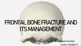
  • 3. CONTENTS 1 Introduction 2 embryology 3 SURGICAL ANATOMY 4 DIAGNOSIS 5 TREATMENT PLANNING 19th Century skull showing sword-blade trauma on frontal bone.
  • 4. INTRODUCTION Fontal sinus is most frequently damaged as a result of high velocity motor vehicle accidents. 70% of frontal sinus fractures were due to automobile accidents and 20% due to assaults.. Fractures involving frontal bone is rather rare because of its protected location 1 . It is basically protected from trauma by the prominence formed by the nasal pyramid. 2 . Incidence of fractures involving this area ranges between 5-15% 3 . Fractures involving this bone is considered to be rather dangerous because of its proximity to brain as well as due to the cosmetic defects it can produce. The proximity of this bone to the orbit and naso frontal duct doesn’t help matters either. Fractures involving this area if not treated promptly can lead to : 1. Meningitis 2. Mucopyocele 3. Encephalitis 4. Cerebral abscess 4 F r o n t a l b o n e f a c t u r e a n d i t s m a n a g e m e n t 2 0 X X
  • 6. EMBRYOLOGY OF FRONTAL BONE 6 p r e s e n t a t i o n t i t l e 2 0 X X Structure of the frontal bone The frontal bone is one of the skull bones enclosing the brain (neurocranium) and it consists of the following parts: •The squamous part; •The nasal part; •Two orbital plates; •Two zygomatic processes. Begin as an outgrowth from nasal chamber in utero Absent at birth Do not develop until 2" year of life Develop from ethmoidal infundibular air cells by invagination of frontal bone through frontal recess or from superior meatus Cannot be identified radiographically until about age of 8 years Development Orbital surface of frontal bone
  • 7. EMBRYOLOGY OF FRONTAL BONE 7 p r e s e n t a t i o n t i t l e 2 0 X X
  • 8. EMBRYOLOGY OF FRONTAL BONE 8 p r e s e n t a t i o n t i t l e 2 0 X X Structure of the frontal bone The frontal bone is made up of two main parts. These are the squamous part, and the orbital part. The squamous part marks the vertical, flat, and also the biggest part, and the main region of the forehead. The orbital part is the horizontal and second biggest region of the frontal bone. It enters into the formation of the roofs of the orbital and nasal cavities. Sometimes a third part is included as the nasal part of the frontal bone, and sometimes this is included with the squamous part. The nasal part is between the brow ridges, and ends in a serrated nasal notch that articulates with the nasal bones inferiorly, and with the lacrimal and maxilla bones laterally The frontal bone is presumed to be derived from neural crest cells.[3] The frontal bone is ossified in membrane from two primary centers, one for each half, which appear toward the end of the second month of fetal life, one above each supraorbital margin. From each of these centers, ossification extends upward to form the corresponding half of the squama, and backwards to form the orbital plate. The spine is ossified from a pair of secondary centers, on either side of the middle line; similar centers appear in the nasal part and zygomatic processes. At birth the bone consists of two pieces, separated by the frontal suture, which is usually obliterated by Intramembranous ossification, except at its lower part, by the eighth year, but occasionally persists throughout life. It is generally maintained that the development of the frontal sinuses begins at the end of the first or beginning of the second year, but may begin at birth. The sinuses are of considerable size by the seventh or eighth year, but do not attain their full proportions until after puberty. Development
  • 9. 9 p r e s e n t a t i o n t i t l e 2 0 X X SURGICAL ANATOMY From the Nasion the bone extends approximately 12.5cm superiorly, 8.0 cm laterally and 5.5 cm posteriorly. Two frontal tuberosities are noted lateral to the midline and superior to the supraorbial rim. The thickest area of the bone is the supraorbial rim from the frontozygomatic process to the nasal bones. The supraorbital foramen are located at the most superior portion of orbital rim. supratrochlear foramen is located medial to the supraorbital foramen or notch and lateral to the nasal bones. A spine or concavity exists on the frontal bone along the medial orbital roof; the trocha of the superior oblique muscle is attached to this spine.
  • 10. 1 0 p r e s e n t a t i o n t i t l e 2 0 X X FRONTAL SINUS Anatomy Evolution over age The frontal sinus is generally absent at birth. At one year the anterior ethmoid air cells begin to invade the frontal bone. Frontal sinus growth is then complete at approximately 15 years of age. The frontal sinus is irregularly shaped and scalloped at its margins. Asymmetry of the sinus is the rule rather the exception (10% of individuals have a unilateral sinus, 9% of individuals have a rudimentary or absent sinus). Applied Anatomy of Frontal Sinus
  • 11. 1 1 p r e s e n t a t i o n t i t l e 2 0 X X FRONTAL SINUS Functions of Frontal Sinus Following are the various functions of the frontal sinus: 1. Production and storage of mucus 2. Resonator for voice 3. Humidification and warming of inhaled air 4. Accessory area of olfaction 5. Conservation of heat from the nasal fossae 6. Definition of facial contours 7.“ Surge Tank” to dampen the pressure differential that develops during inspiration
  • 12. 1 2 p r e s e n t a t i o n t i t l e 2 0 X X FRONTAL SINUS Anatomic relationships The frontal sinus has several critical anatomic relationships. These include: Sinus floor -> orbital roof/anterior ethmoid air cells Posterior table -> anterior cranial fossa Anterior table -> frontal contour Drainage The frontal sinus drains via a small outflow tract into the ethmoid sinus/nasal cavity. The outflow tract is hour-glass shaped with the true ostium (3-4 mm) at the narrowest portion.
  • 13. Epidemiolog y 1 3 p r e s e n t a t i o n t i t l e 2 0 X X Frontal sinus fractures are relatively uncommon and account for only 5-15% of maxillofacial fractures with a preponderance of male patients. The most common frontal sinus fractures involve a combination of the anterior and posterior tables with or without frontal recess involvement (about 2/3). Isolated posterior table fractures are extremely uncommon (< 1%). This 3D CT reconstruction shows an isolated anterior table fracture.
  • 14. CLASSIFICATION 1 4 2 0 X X Frontal sinus fractures are usually classified based on: •Location •Extent of injury •Involvement of frontonasal duct •Current injury to the dura Stanley’s Classification of Frontal Sinus Fracture •Type I: Anterior Table Fracture • Isolated to anterior table • Accompanied by supraorbital rim fracture • Accompanied by naso-ethmoid complex fracture •Type II: Anterior and Posterior Table Fractures • It is a linear fracture either on transverse direction or in vertical direction •Type III: Comminuted Fractures • Isolated to both tables • Accompanied by naso-ethmoid complex Gonty Et al. Classification of Frontal Sinus Fracture •Type I: Anterior Table Fracture • Isolated to anterior table • Accompanied by supraorbital rim fracture • Accompanied by naso-ethmoid complex fracture •Type II: Anterior and Posterior Table Fractures • A linear fracture either on transverse direction or in vertical direction • Comminuted fracture either isolated to both tables or accompanied by naso-ethmoid complex fracture •Type III: Posterior Table Fracture •Type IV: Through and Through Frontal Sinus Fracture
  • 15. CLASSIFICATION 1 5 Type I : Anterior table fracture with minimum comminution Type II : Anterior Wall comminuted fracture with - Possible NOE or Orbital rim fracture Type III : Anterior & Posterior Wall fracture (Posterior wall fracture without significant displacement or ductal injury) Type IV : Anterior & Posterior Wall fracture with Dural injury & CSF leak Type V :Anterior & Posterior Wall fracture with Dural injury, CSF leak, soft tissue or bone loss and/or severe disruption of anterior cranial fossa Gerbino G, Roccia F, Benoch A, ot al. Analyss of 158 fontalsinus fractures: Current surgical management and complications. J Craniomaxilofac Surg 2000; 28133
  • 16. CLASSIFICATION 1 Frontal recess involvement A frontal recess injury involves the floor of the frontal sinus and the outflow tract. It may also involve the anterior skull base. Anterior table fractures Fractures of the anterior table of the frontal sinus vary from minimally displaced to severely displaced/comminuted depending upon the severity of the trauma and size of the sinus. Minimal displacement Less severe trauma may result in fractures of the anterior table which are minimally (as illustrated) or nondisplaced. 2 3 4 Posterior table fracture Posterior table fractures carry a higher risk of intracranial injury because they create a communication with the intracranial space. The degree of fracture displacement gives some insight into the severity of the injury. While there is no commonly agreed upon classification for displacement, many authors have used the width of the posterior table (0.1 mm – 4 mm) as a “measuring stick” to describe displacement because it is readily visible on axial CT-scans. 3 1 6 p r e s e n t a t i o n t i t l e 2 0 X X
  • 17. DIAGNOSIS  The diagnosis of frontal bone fracture is based upon proper history and physical examination of the patient  This includes Inspection and palpation of the affected area Detailed History includes:  Information about events  Visual difficulties  Numbness  Pain  Rhinorrhea  Sense of smell  Previous history of nasal or sinus disease surgery 1 7 p r e s e n t a t i o n t i t l e 2 0 X X
  • 18. CLINICAL FEATURES Forehead laceration (58%) Forehead pain (82%) swelling 1 8 p r e s e n t a t i o n t i t l e 2 0 X X Frontal bone depression (25%) Csf rhinorrhea (1/3rd patients Periorbital ecchymosis
  • 19. 1 9 p r e s e n t a t i o n t i t l e 2 0 X X Clinical evaluation General Facial pain Forehead paraesthesia or anaesthesia Forehead laceration Visible and/or palpable frontal bone depression CSF Rhinorrhoea Neurological injuries Cerebral contusion Subdural & epidural haematoma. Ophthalmic injuries Pupillary defect Optic neuropathy Hyphaema Disc oedema Corneal defect Loss of globe integrity Associated maxillofacial Injuries NOE fracture Orbital fracture Zygomatic fracture
  • 20. RADIOGRAPHIC EVALUATION 2 0 p r e s e n t a t i o n t i t l e 2 0 X X PLAIN RADIOGRAPHS are of little use in the diagnosis of frontal sinus fractures Caldwell's view EVIDENCE OF AIR FLUID LEVEL, CLOUDING OF FRONTAL SINUS, PNEUMOCEPHALUS Lateral view High Resolution CT Scan CT Scans are the GOLD standard for imaging of these fracture
  • 21. CT SCANS 2 1 p r e s e n t a t i o n t i t l e 2 0 X X AXIAL CT VIEW Axial images reveals location, severity and degree of comminution of anterior and posterior table fractures. A thin cut (1.0 to 1.5 mm), axial computed tomography (CT) scan is the gold standard. It is recommended to obtain coronal, sagittal, and three-dimensional (3-D) reconstructions for diagnostic accuracy Coronal images for the Frontal sinus floor and orbital Roof fracture Coronal CT VIEW
  • 22. CT SCANS 2 2 p r e s e n t a t i o n t i t l e 2 0 X X Sagittal images can be useful in assessing the patency of the frontal recess while 3-D reconstructions help define the shape, location, and orientation of individual bone fragments that are seen less clearly on 2-D views sagittal CT VIEW The 3-D information can reduce the need for surgical dissection, because the surgeon knows the number, location, and orientation of the larger bone fragments. It can also help the patient and/or family to understand the bony anatomy and severity of the injury.
  • 23. MANAGEMENT. FRONTAL BONE FRACTURE 2 3 p r e s e n t a t i o n t i t l e 2 0 X X
  • 24. PRINCIPLES OF FRONTAL BONE FRACTURE MANAGEMENT 2 4 p r e s e n t a t i o n t i t l e 2 0 X X SAFE SINUS RESTORE FACIAL CONTOUR AVOID SHORT & LONG TERM COMPLICATIONS
  • 25. Objectives of management • To avoid immediate and short-term complications such as CSF leak, meningitis, spreading infection. • To avoid long-term complications such as frontal bone osteomyelitis, chronic frontal sinusitis, mucocele, mycopyocele, and brain abscess. • To provide adequate exposure for anatomic reduction of naso- orbito-ethmoidal (NEO) fractures. • To restore proper aesthetic contour of the forehead. 2 5 p r e s e n t a t i o n t i t l e 2 0 X X TREATMENT OPTIONS Conservative Fracture reduction & fixation Sinus obliteration Sinus cranialization
  • 26. Management generally based on Three clinical factors: 2 6 p r e s e n t a t i o n t i t l e 2 0 X X • Fracture location and displacement • Dural and cerebral involvement • Damage to the frontal sinus drainage systems
  • 27. Guiding Principles for Frontal Sinus Management 2 7 p r e s e n t a t i o n t i t l e 2 0 X X •To separate nasal cavity from sinus •To eliminate dead space •To separate the frontonasal duct from frontal sinus by obstructing the duct •To eliminate a functional sinus, sinus mucosa is removed, and sinus is obliterated
  • 28. MANAGEMENT Surgical Access 2 8 p r e s e n t a t i o n t i t l e 2 0 X X 1. Through existing laceration 2. Butterfly incision 3. Gullwing or eyeglass incision 4. Bicoronal approach suggests sterile surgical glove tourniquet intraoperatively to get the hemorrhage control and blood less surgical field in elevation of bicoronal flap for the surgical management of frontal sinus fracture. Gullwing or eyeglass incision
  • 29. Surgical Access 2 9 p r e s e n t a t i o n t i t l e 2 0 X X Use of existing laceration to access left-sided frontal sinus fracture. Extensive transverse laceration of the forehead with easy access to the frontal sinus. The fractured segments have been removed to gain access to the nasofrontal duct. Through existing laceration
  • 30. Surgical Access 3 0 p r e s e n t a t i o n t i t l e 2 0 X X Irregular outline of a coronal incision in two different patients
  • 31. Surgical Access 3 1 p r e s e n t a t i o n t i t l e 2 0 X X Bicoronal approach Access areas The following areas can be exposed: •Entire calvarial vault •Anterior and lateral skull base •Frontal sinus/Ethmoid •Zygoma •Zygomatic arch •Orbit (lateral/cranial/medial) •Nasal dorsum •Temporomandibular joint (TMJ) and condylar/subcondylar region
  • 32. 3 2 p r e s e n t a t i o n t i t l e 2 0 X X Indications of Surgery in Frontal Sinus Fractures 1. To avoid immediate complications such as CSF leak, meningitis 2. To avoid long-term complications such as frontal sinusitis, meningitis and brain abscess formation 3. To provide aesthetic contour to the forehead 4. To provide exposure for anatomic reduction of NOE fractures
  • 33. 3 3 p r e s e n t a t i o n t i t l e 2 0 X X History of frontal sinus surgery  Surgical approaches to frontal sinus • Frontal sinus surgery was first described in the 18th century. It is noted that as early as 1750 Runge performed an obliteration procedure of the frontal sinus. • The first published report in 1870 by Wells described an external and intracranial drainage procedure for a frontal sinus mucocele. • In 1884 Alexander Ogston described a trephination procedure through the anterior table to evacuate the frontal sinus • Kuhnt in 1895 described removing the anterior wall of the frontal sinus in an attempt to clear disease In 1898 Riedel/Schenke described the first procedure for obliteration of the frontal sinus the Lynch and Howarth operation Lynch incision (A) with resulting access to frontal sinus and ethmoid sinuses (B)
  • 34. 3 4 p r e s e n t a t i o n t i t l e 2 0 X X History of frontal sinus surgery  Surgical approaches to frontal sinus
  • 35. MANAGEMENT. 3 5 p r e s e n t a t i o n t i t l e 2 0 X X
  • 36. 3 6 p r e s e n t a t i o n t i t l e 2 0 X X Management of Anterior Table Fracture Decision-Making •Simple greenstick or undisplaced fracture does not require surgical intervention. •In depressed anterior wall fracture, frontal sinus explored, careful irrigation carried out, fragments reduced and stabilize by internal fixation. In posterior wall fracture without CSF leak or pneumocephalus, reconstruction of only anterior wall is done. • Other than surgical intervention, antibiotics, sinus decongestants and analgesics are prescribed to keep the frontonasal duct patent and to prevent infection. Rai et al. suggested bone mapping/sketching in management of anterior table frontal sinus fracture with great success Bone mapping/sketching in management of anterior table frontal sinus fracture Yoo MH et al. suggested endoscopic trans nasal reduction of anterior table fracture To support the reduced fragments, they advocated use of custom made latex glove balloon to be inserted into the frontal sinus then expanded and maintained for 3 weeks
  • 37. ANTERIOR table fracture FRONTAL BONE FRACTURE 3 7 p r e s e n t a t i o n t i t l e 2 0 X X (Nondisplaced, linear/isolated) Conservative treatment • Local wound care • Antibiotics, nasal decongestant, analgesics • Follow up evaluation
  • 38. Operative Indication of FS injury FRONTAL BONE FRACTURE 3 8 p r e s e n t a t i o n t i t l e 2 0 X X • Nasofrontal duct involvement/obstruction • Displacement of posterior table with underlying neurological injury • Aesthetic forehead deformity
  • 39. ANTERIOR table fracture 3 9 p r e s e n t a t i o n t i t l e 2 0 X X Modified treatment algorithm for highly selected patients
  • 40. ANTERIOR table fracture 4 0 p r e s e n t a t i o n t i t l e 2 0 X X (Displaced) Approaches Intraoperative assessment • Any kind of fluid • Nasofrontal duct patency Nasofrontal duct injury • Nasofrontal duct filling with frontal sinus obliteration Grafting Reduction & fixation • Existing laceration • Butterfly/seagull/open sky approaches, coronal flap
  • 41. Isolated Anterior Table Fracture FRONTAL BONE FRACTURE 4 1 p r e s e n t a t i o n t i t l e 2 0 X X • Open Reduction Internal Fixation If the degree of displacement, as visualized on axial or sagittal sections of CT scans, is >1–2 mm, that is, more than a table’s width, it warrants an open reduction and fixation within 7–10 days*
  • 42. Open reduction internal fixation 4 2 Principle s The goal of open reduction is to reapproximate the bone fragments into their premorbid position and use internal fixation hardware to maintain them in their position. Approac h coronal approach Reduction General considerations The anterior table of the frontal sinus is normally convex. Compressive forces on the frontal bone deform the convexity into a concavity. This may or may not result in fracture comminution. Mobilization of the depressed bone fragment(s) may require significant effort to overcome compressive forces between bone fragments.
  • 43. Reduction techniques 4 3 Mobilizing the bone fragments using an elevator There are several techniques to reduce the fragments: Inserting a screw into a bony fragment and reducing it by pulling it outwards Fixation Simple reduction
  • 44. Postop care following management of frontal sinus fractures 4 4 Evaluation of the patient’s vision performed as soon as they are awakened from anesthesia and then at regular intervals until they are discharged from the hospital. Postoperative positioning Keeping the patient’s head in a raised position both preoperatively and postoperatively may significantly improve edema and pain. Nose- blowing Nose-blowing should be avoided for at least 3 weeks following frontal sinus and skull base repair.
  • 45. Frontal sinus Obliteration FRONTAL BONE FRACTURE 4 5 p r e s e n t a t i o n t i t l e 2 0 X X • Steps • Sinus Exploration • Mucosal Exenteration • Nasofrontal Duct obturation • Frontal sinus Obliteration • Fracture Reduction
  • 46. Frontal sinus FRONTAL BONE FRACTURE 4 6 p r e s e n t a t i o n t i t l e 2 0 X X • Filling materials for obturation and obliteration • Autogenous Material Synthetic materials - Autologous Fat Polyfluorotetraethylene, ePTFE - Cancellous bone’ Methylmethacrylate, MMA - Muscle - Pericranial flap - Banked cadaveric tissue
  • 47. Indication for Bone Grafting FRONTAL BONE FRACTURE 4 7 p r e s e n t a t i o n t i t l e 2 0 X X 1. Extensive loss of support at the skull base over the fovea ethmoidalis and cribriform plate, in combination with a peri cranial flap. 2. Superior orbital roof fractures, to avoid pulsatile exophthalmos and orbital deformity. 3. Extensive bone loss of the anterior table, which cannot be replaced with elements of the posterior table. 4. In combination with NOE and orbital reconstruction as layered bone grafts to obliterate the ethmoids and reconstruct the medial orbital wall.
  • 48. posterior table fracture FRONTAL BONE FRACTURE 4 8 p r e s e n t a t i o n t i t l e 2 0 X X CSF leak Absent and No displacement Frontal Sinus Obliteration CSF leak Absent and/or Displacement Frontal Sinus Cranialization
  • 49. Frontal Sinus Cranialization FRONTAL BONE FRACTURE 4 9 p r e s e n t a t i o n t i t l e 2 0 X X • It is another technique to minimize the dead space in sinus. It involves removing posterior table thus permitting brain to expand into frontal sinus resulting in confuence between sinus cavity and anterior cranial fossa. • It is done in cases with CSF leak and neurological injury due to displaced posterior table fracture. To isolate the splanchnocranium from the frontal sinus, a pericranial fap is used.
  • 50. Frontal Sinus Cranialization FRONTAL BONE FRACTURE 5 0 p r e s e n t a t i o n t i t l e 2 0 X X Although obliteration has been touted as the gold standard and safest method to treat the injured frontal sinus, there are many disadvantages including :
  • 51. Complications FRONTAL BONE FRACTURE 5 1 p r e s e n t a t i o n t i t l e 2 0 X X
  • 52. Recent Advancements FRONTAL BONE FRACTURE 5 2 p r e s e n t a t i o n t i t l e 2 0 X X Virtual Surgery Augmented Reality Stereolithography or 3D printing Virtual Endoscopy
  • 53. Preoperative Photographs and radiographs 5 3 A 48-year-old female patient who sustained a frontal bone fracture right side having a H/O fall from a hill
  • 54. Intraoperative & postoperative Photographs 5 4 A 48-year-old female patient who sustained a frontal bone fracture right side having a H/O fall from a hill
  • 55. Preoperative Photographs and radiographs 5 5 A 22-year-old male patient who sustained a frontal bone having a H/O RSA, having fallen form his bike
  • 56. Intraoperative & postoperative Photographs 5 6 A 22-year-old male patient who sustained a frontal bone having a H/O RSA, having fallen form his bike
  • 57. Follow up Photographs 5 7 A 22-year-old male patient who sustained a frontal bone having a H/O RSA, having fallen form his bike
  • 58. Conclusion The management of NOE and frontal sinus fracture is always a challenging task .The proper handling of MCL and nasofrontal duct is mandatory to get good post- operative results. Proper treatment planning in the form of incision selection, method of fixation and use of bone graft should be done to avoid unaesthetic results. 5 8 p r e s e n t a t i o n t i t l e 2 0 X X
  • 59. references 5 9 N O E F R A C T U R E S 2 0 X X • 1. Milad Etemadi SH, Shahnaseri S, Soltani P, Motamedi MRK. Management of naso-orbito-ethmoid fractures: a 10-year review. Trauma Mon. 2017;22:292–30. • 2. Brasileiro BF, Passeri LA. Epidemiological analysis of maxillofacial fractures in Brazil: a 5-year prospective study. Oral Surg Oral Med Oral Pathol Oral Radiol Endod. 2006;102(1):28–34. • 3. Rosenberger E, Kriet JD, Humphrey C. Management of nasoethmoid fractures. Curr Opin Otolaryngol Head Neck Surg. 2013;21:410–6. (Anatomy of MCL, physical examination of the complex and displacement) • 4. Poh E, Kakizaki H, Selva D, Leibovitch I. Anatomy of medial canthal tendon in Caucasians. Clin Exp Ophthalmol. 2012;40:170–3. • 5. Merkx MA, Freihofer HP, Borstlap WA, van’t Hoff MA. Effectiveness of primary correction of traumatic telecanthus. Int J Oral Maxillofac Surg. 1995;24:344–7. • 6. Heine D, Catone GA, Bavitz B, Grenadier MR. Naso-orbitalethmoid injury: report of a case and review of the literature. Oral Surg Oral Med Oral Path. 2005;69(5):542–9. • 7. Rowe NL. Rowe and Williams’ maxillofacial injuries, vol. 2. 2nd ed. Edinburgh: Churchill Livinstone; 1994. • 8. Markowitz BL, Manson PN, Sargent L, et al. Management of the medial canthal tendon in nasoethmoid orbital fractures: the importance of the central fragment in classifcation and treatment. Plast Reconstr Surg. 1991;87:843–853. • 9. Burstein F, Cohen S, Hudgins R, Boydston W. Frontal basilar trauma: classifcation and treatment. Plast Reconstr Surg. 1997;99(5):1314–21. • 10. Balaraman K, Ramani V, Bharathi R, Venkataramani H, Sabapathy SR. Outcome assessment of Nasoethmoid fractures. Int J Oral Maxillofac Surg. 2013;42(10):1220. • 11. Ellis E III. Sequencing treatment for naso-orbito-ethmoid fractures. J Oral Maxillofac Surg. 1993;51:543–558. • 12. Nguyen M, Koshy JC, Hollier LH Jr. Pearls of nasoorbitoethmoid trauma management. Semin Plast Surg. 2010;24:383–8. • 13. Dingman RO, Grabb WC, Oneal RM. Management of injuries of the naso-orbital complex. Arch Surg. 1969;98:566–71. • 14. Mustarde JC. Epicanthus and telecanthus. Int Opthalmol Clin. 1964;4:359. • 15. Cruse C, Blevins PK, Luce EA. Naso-ethmoid-orbital fractures. J Trauma. 1980;20:551.