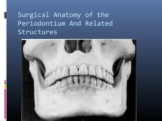
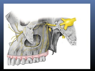
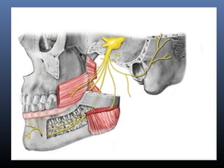
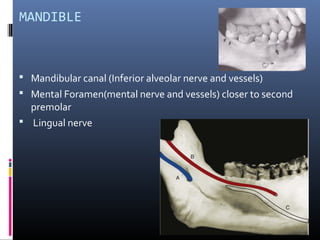
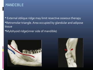
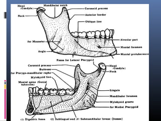
Surgical Anatomy of Periodontium and Related Structures
Surgical Anatomy of the
Periodontium And Related
Structures
Knowledge of the anatomy of the Periodontium and
the hard and soft structures is essential.
 Mandible.
 Maxilla.
 Muscles.
 Anatomic Spaces.
MANDIBLE
 Mandibular canal (Inferior alveolar nerve and vessels)
 Mental Foramen(mental nerve and vessels) closer to second
premolar
 Lingual nerve
MANDIBLE
 External oblique ridge may limit resective osseous therapy
Retromolar triangle. Area occupied by glandular and adipose
tissue
Mylohyoid ridge(inner side of mandible)
The Mandibular Canal
 Occupied by inferior alveolar nerve and vessels
 Begins at the mandibular foramen on the medial
surface of the mandibular ramus
 Curves downward and forward and then horizontally
below the apices of molars
 In the premolar area divides into two; incisive canal
and mental canal
The mental foramen
 Mental nerve and vessels emerge
 Located on buccal surface of mandible below the apices of
premolars
 Sometimes closer to the second premolar
 Surgical trauma of the mental nerve can produce
paresthesia of the lip, which recovers slowly
 Implants placement in totally edentulous jaws, the
distance between the canal and superior surface of
the bone must be carefully determined to avoid nerve
injury
Lingual nerve
 Branch of the posterior division of mandibular nerve
 Close to the surface of oral mucosa in third molar area
 Can be damaged during anesthetic injections and during third
molar extractions
 In periodontal surgery, it can be injured during raising partial
thickness flap in third molar region or releasing incision is made.
Inner side of the mandible
External oblique ridge.
 Arrows show the attachment of the buccinator
muscle.
 may limit resective osseous therapy
Maxilla
 The alveolar process
 The Palatine process
 The zygomatic Process
 The frontal Process.
MAXILLA
 Incisive canal (incisive papilla and vessels emerging through the
canal).
 Greater Palatine foramen( opens 3-4mm anterior to post.
border of the hard palate) including all the nerves and vessels
emerging from it and run anteriorly.
 Submucosa contains palatal glands which protects the
underlying vessels and nerve.
MAXLIIA
 Maxillary tuberosity.
 Maxillary Sinus.
 Maxillary or mandibular tori.
Tori or exostosis
 Mandible
 Lingual to canine & premolars above mylohyoid muscle
 Maxilla
 Midline of hard palate
 Buccal exostosis in the maxillary arch
Muscles
Several muscles may be encountered during
performing flaps particullary in mucogingival surgery.
 Mentalis,
 incisve labii inferioris,
 depressor anguli oris,
 incisive labii superioris and
 Buccinator m/s
 They have bony attachment and they provide mobility
to lips and cheeks
MUSCLES
 1, Nasalis;
 2,Levator anguli oris
 3,Buccinator
 4,Depressor anguli oris
 5,Depressor labii inferioris
 6,Mentalis.
Anatomic spaces
 Surgical invasion of these areas may result in
dangerous infections because these spaces contain
loose connective tissues and can be distended by
inflammatory fluid and infection.
ANATOMIC SPACES
Surgical invasion may result in dangerous
infections and should be avoided.
 Canine fossa
 Buccal Space
 Mental space
 Masticator space
 Sublingual, submental and submandibular space
 Canine fossa
-------- swelling of the upper lip and upper and lower
eyelid closing the eye.
 Buccal space (between buccinator and masseter
muscle).
-------leads to swelling of the cheek.
 Mental space
------lead to swelling of chin
 Masticator space
------infection result in swelling of the face and
trismus and pain.
 Sublingual space
-----results in pain around the tongue & swallowing.
Anatomic spaces
Infection is dangerous in submental and submandibular spaces
Conclusions
In the mandible,
 the position of the mandibular nerve must be
ascertained before implant placement so that there is
no risk of damage.
 2mm space should be present between the coronal
border of the nerve and the apex of the implant.
 Mental nerve should be considered during implant
placement and mucogingival surgery.
Conclusion
In the maxilla,
 the nasopalatine nerves and vessels are of little
important if they are included in the surgery.
 involvement of the greater palatine artery should be
avoided because of significant hemorrhage that could
occur if it is severed.
 Maxillary sinus extent has to evaluated during
treatment planning for implants.
MCQs
I. In the followings;
(A)Knowledge of the anatomy of the Periodontium
and the adjacent structures is not necessary
(B) Mental foramen lies closer to second premolar
(C) 12 mm space should be present between the
coronal border of the nerve and the apex of the
implant.
(D) Infection in sublingual space will results in pain
around the tongue & swallowing.
(E) Lingual nerve can be damaged during LA
injections and third molar extractions
II.(A) Mandibular canal ------- located on buccal
surface of mandible below the apices of premolars
(B) Mental foramen-------sometimes closer to the
second premolar
(C) Lingual nerve -------close to the surface of oral
mucosa in third molar area
(D) Canine fossa  swelling of the upper lip and upper
and lower eyelid closing the eye.
(E) Masticator space infection result in swelling of
the face and trismus and pain.
Sources of autograft for bone ------
(A) maxillary tuberosity.
(B) maxillary Sinus.
(C) mandibular tori.
(D) mental space
(E) maxillary tori
.

Periodontal Surgery

  • 1.
    Surgical Anatomy ofPeriodontium and Related Structures
  • 2.
    Surgical Anatomy ofthe Periodontium And Related Structures
  • 5.
    Knowledge of theanatomy of the Periodontium and the hard and soft structures is essential.  Mandible.  Maxilla.  Muscles.  Anatomic Spaces.
  • 6.
    MANDIBLE  Mandibular canal(Inferior alveolar nerve and vessels)  Mental Foramen(mental nerve and vessels) closer to second premolar  Lingual nerve
  • 7.
    MANDIBLE  External obliqueridge may limit resective osseous therapy Retromolar triangle. Area occupied by glandular and adipose tissue Mylohyoid ridge(inner side of mandible)
  • 9.
    The Mandibular Canal Occupied by inferior alveolar nerve and vessels  Begins at the mandibular foramen on the medial surface of the mandibular ramus  Curves downward and forward and then horizontally below the apices of molars  In the premolar area divides into two; incisive canal and mental canal
  • 10.
    The mental foramen Mental nerve and vessels emerge  Located on buccal surface of mandible below the apices of premolars  Sometimes closer to the second premolar
  • 11.
     Surgical traumaof the mental nerve can produce paresthesia of the lip, which recovers slowly  Implants placement in totally edentulous jaws, the distance between the canal and superior surface of the bone must be carefully determined to avoid nerve injury
  • 12.
    Lingual nerve  Branchof the posterior division of mandibular nerve  Close to the surface of oral mucosa in third molar area  Can be damaged during anesthetic injections and during third molar extractions  In periodontal surgery, it can be injured during raising partial thickness flap in third molar region or releasing incision is made.
  • 13.
    Inner side ofthe mandible
  • 14.
    External oblique ridge. Arrows show the attachment of the buccinator muscle.  may limit resective osseous therapy
  • 15.
    Maxilla  The alveolarprocess  The Palatine process  The zygomatic Process  The frontal Process.
  • 16.
    MAXILLA  Incisive canal(incisive papilla and vessels emerging through the canal).  Greater Palatine foramen( opens 3-4mm anterior to post. border of the hard palate) including all the nerves and vessels emerging from it and run anteriorly.  Submucosa contains palatal glands which protects the underlying vessels and nerve.
  • 17.
    MAXLIIA  Maxillary tuberosity. Maxillary Sinus.  Maxillary or mandibular tori.
  • 18.
    Tori or exostosis Mandible  Lingual to canine & premolars above mylohyoid muscle  Maxilla  Midline of hard palate  Buccal exostosis in the maxillary arch
  • 20.
    Muscles Several muscles maybe encountered during performing flaps particullary in mucogingival surgery.  Mentalis,  incisve labii inferioris,  depressor anguli oris,  incisive labii superioris and  Buccinator m/s  They have bony attachment and they provide mobility to lips and cheeks
  • 21.
    MUSCLES  1, Nasalis; 2,Levator anguli oris  3,Buccinator  4,Depressor anguli oris  5,Depressor labii inferioris  6,Mentalis.
  • 26.
    Anatomic spaces  Surgicalinvasion of these areas may result in dangerous infections because these spaces contain loose connective tissues and can be distended by inflammatory fluid and infection.
  • 27.
    ANATOMIC SPACES Surgical invasionmay result in dangerous infections and should be avoided.  Canine fossa  Buccal Space  Mental space  Masticator space  Sublingual, submental and submandibular space
  • 28.
     Canine fossa --------swelling of the upper lip and upper and lower eyelid closing the eye.  Buccal space (between buccinator and masseter muscle). -------leads to swelling of the cheek.  Mental space ------lead to swelling of chin
  • 29.
     Masticator space ------infectionresult in swelling of the face and trismus and pain.  Sublingual space -----results in pain around the tongue & swallowing.
  • 30.
    Anatomic spaces Infection isdangerous in submental and submandibular spaces
  • 31.
    Conclusions In the mandible, the position of the mandibular nerve must be ascertained before implant placement so that there is no risk of damage.  2mm space should be present between the coronal border of the nerve and the apex of the implant.  Mental nerve should be considered during implant placement and mucogingival surgery.
  • 32.
    Conclusion In the maxilla, the nasopalatine nerves and vessels are of little important if they are included in the surgery.  involvement of the greater palatine artery should be avoided because of significant hemorrhage that could occur if it is severed.  Maxillary sinus extent has to evaluated during treatment planning for implants.
  • 33.
  • 34.
    I. In thefollowings; (A)Knowledge of the anatomy of the Periodontium and the adjacent structures is not necessary (B) Mental foramen lies closer to second premolar (C) 12 mm space should be present between the coronal border of the nerve and the apex of the implant. (D) Infection in sublingual space will results in pain around the tongue & swallowing. (E) Lingual nerve can be damaged during LA injections and third molar extractions
  • 35.
    II.(A) Mandibular canal------- located on buccal surface of mandible below the apices of premolars (B) Mental foramen-------sometimes closer to the second premolar (C) Lingual nerve -------close to the surface of oral mucosa in third molar area (D) Canine fossa  swelling of the upper lip and upper and lower eyelid closing the eye. (E) Masticator space infection result in swelling of the face and trismus and pain.
  • 36.
    Sources of autograftfor bone ------ (A) maxillary tuberosity. (B) maxillary Sinus. (C) mandibular tori. (D) mental space (E) maxillary tori .