Downloaded 567 times


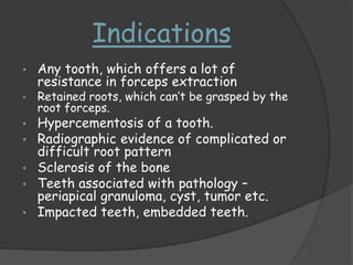


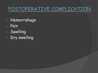


This document discusses transalveolar extraction, also known as surgical extraction. It involves reflecting a muco-periosteal flap, cutting bone if needed, sectioning tooth roots, and removing the tooth. The document outlines the indications, contraindications, advantages, and steps of the procedure including incisions, bone removal, tooth elevation, debridement, suturing, and post-operative instructions. Potential intraoperative and postoperative complications are also listed.


