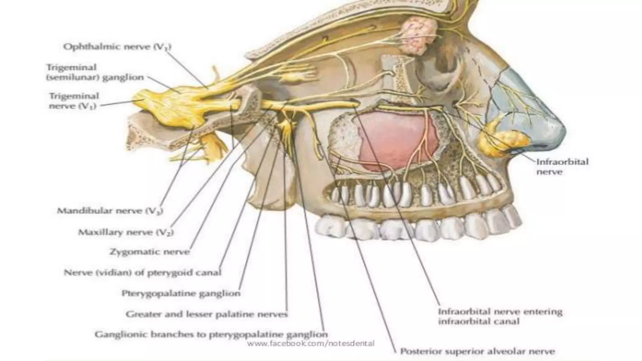
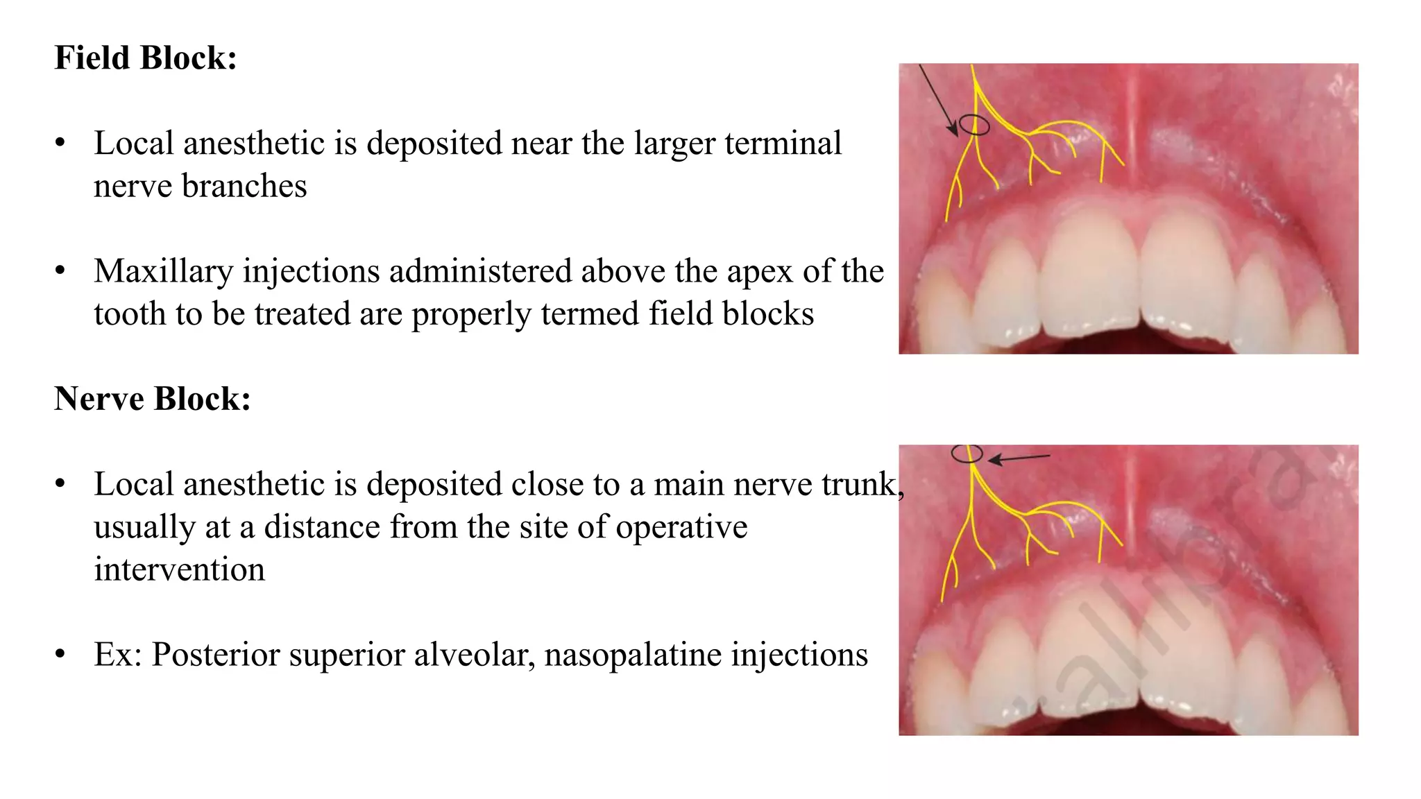
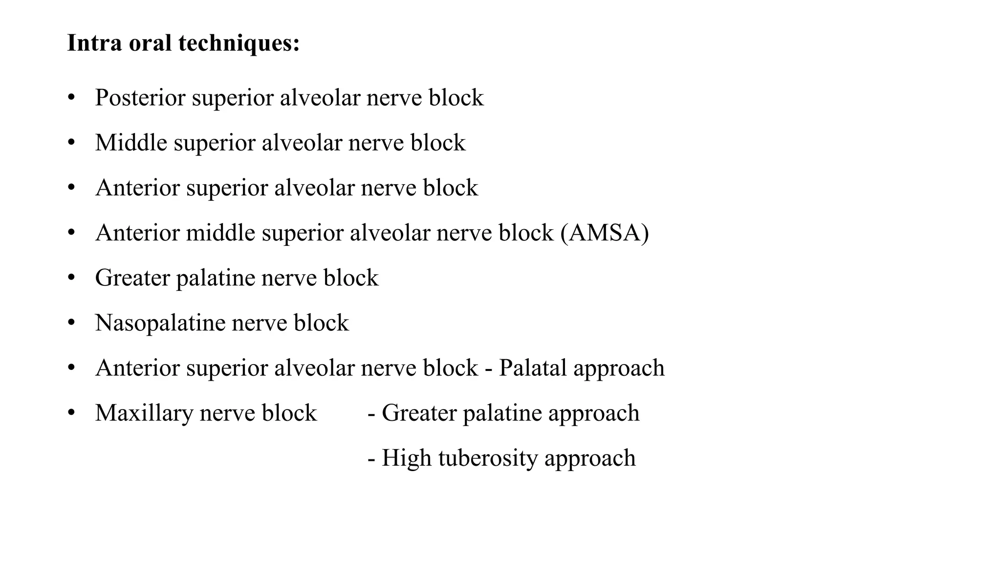
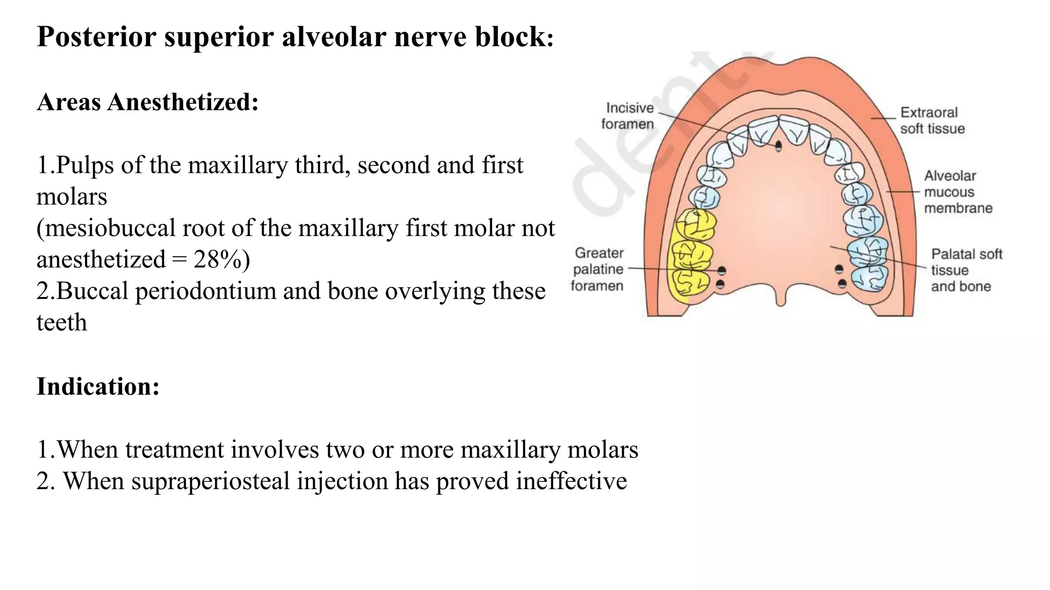
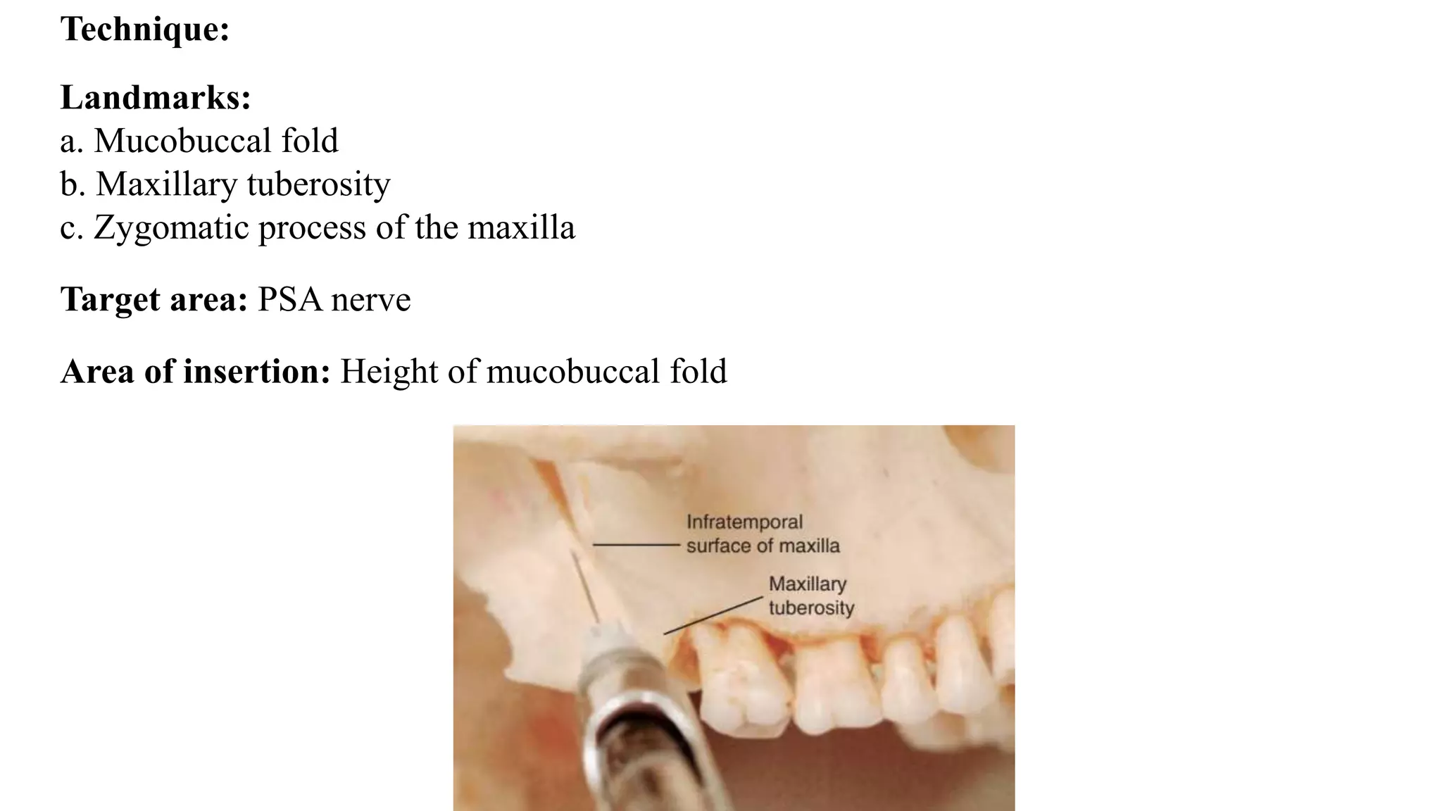
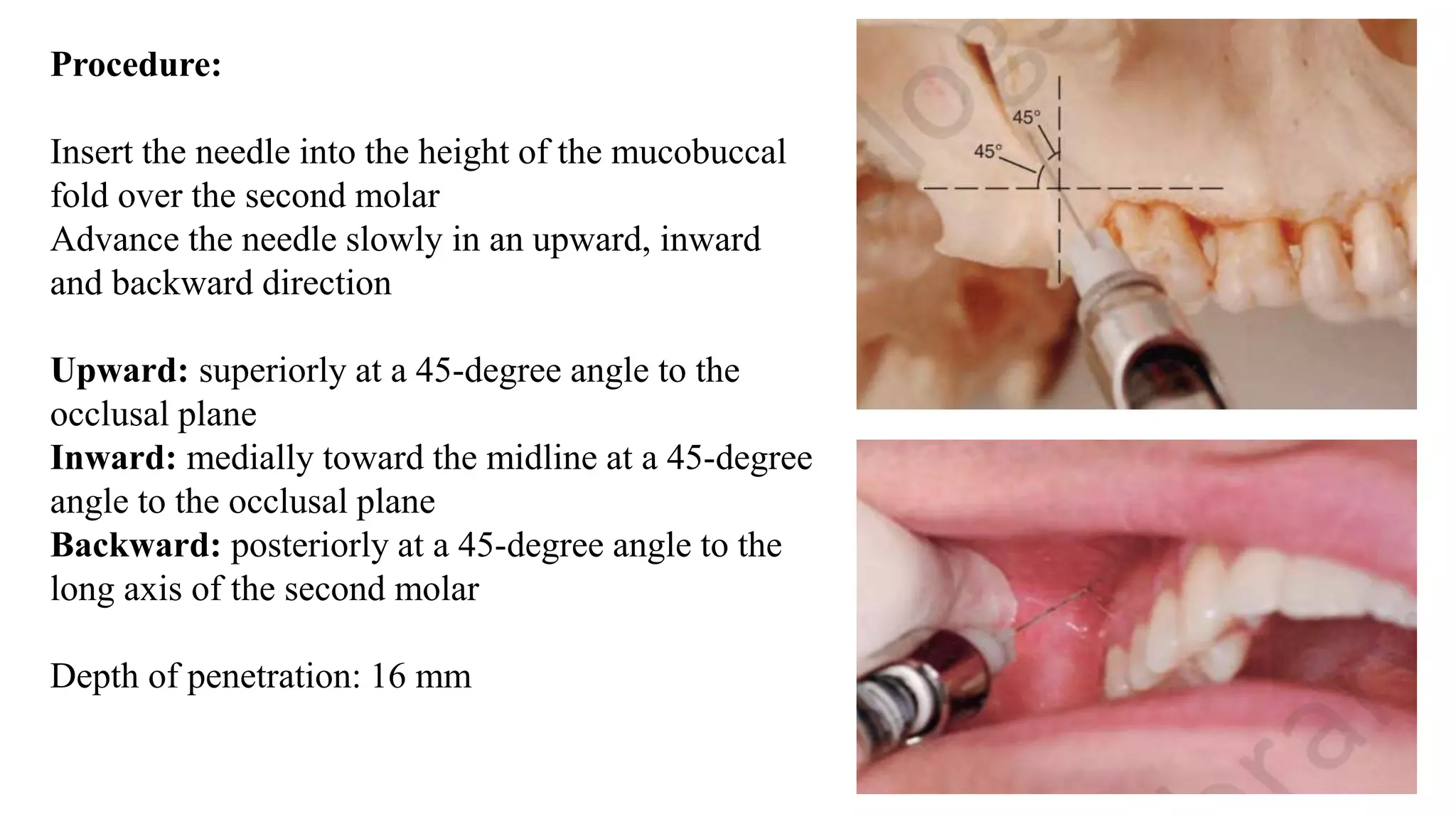
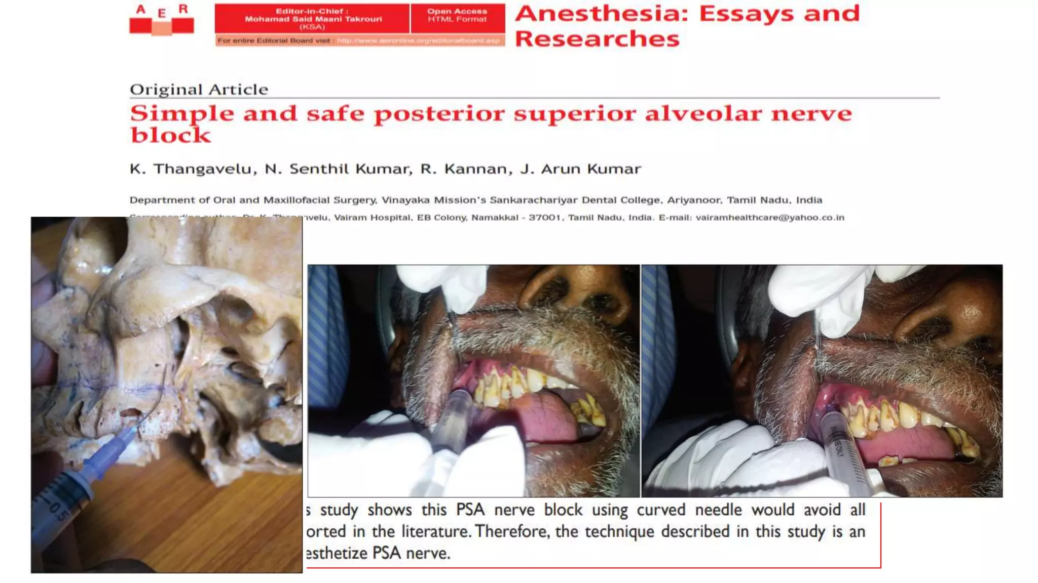
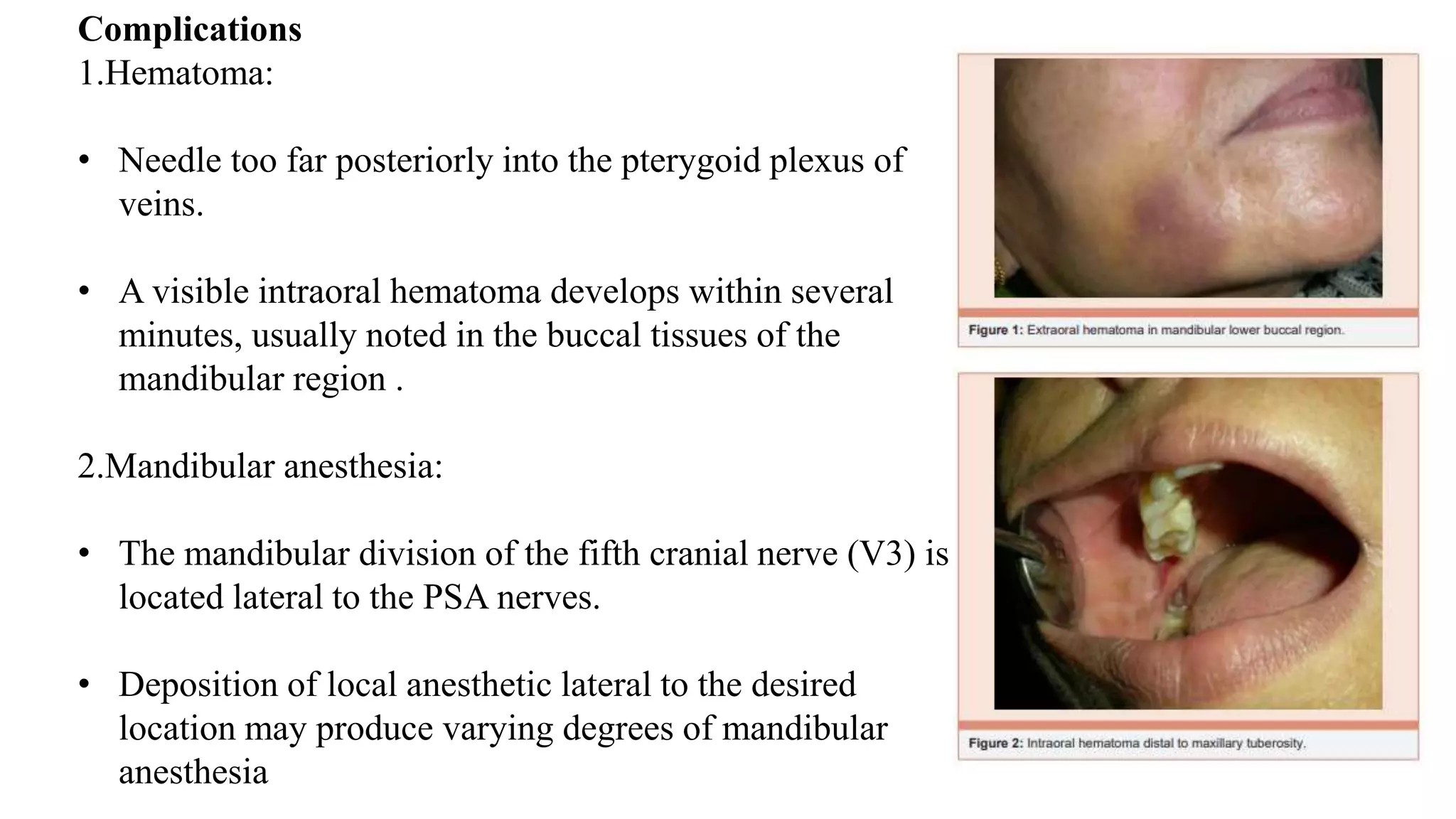
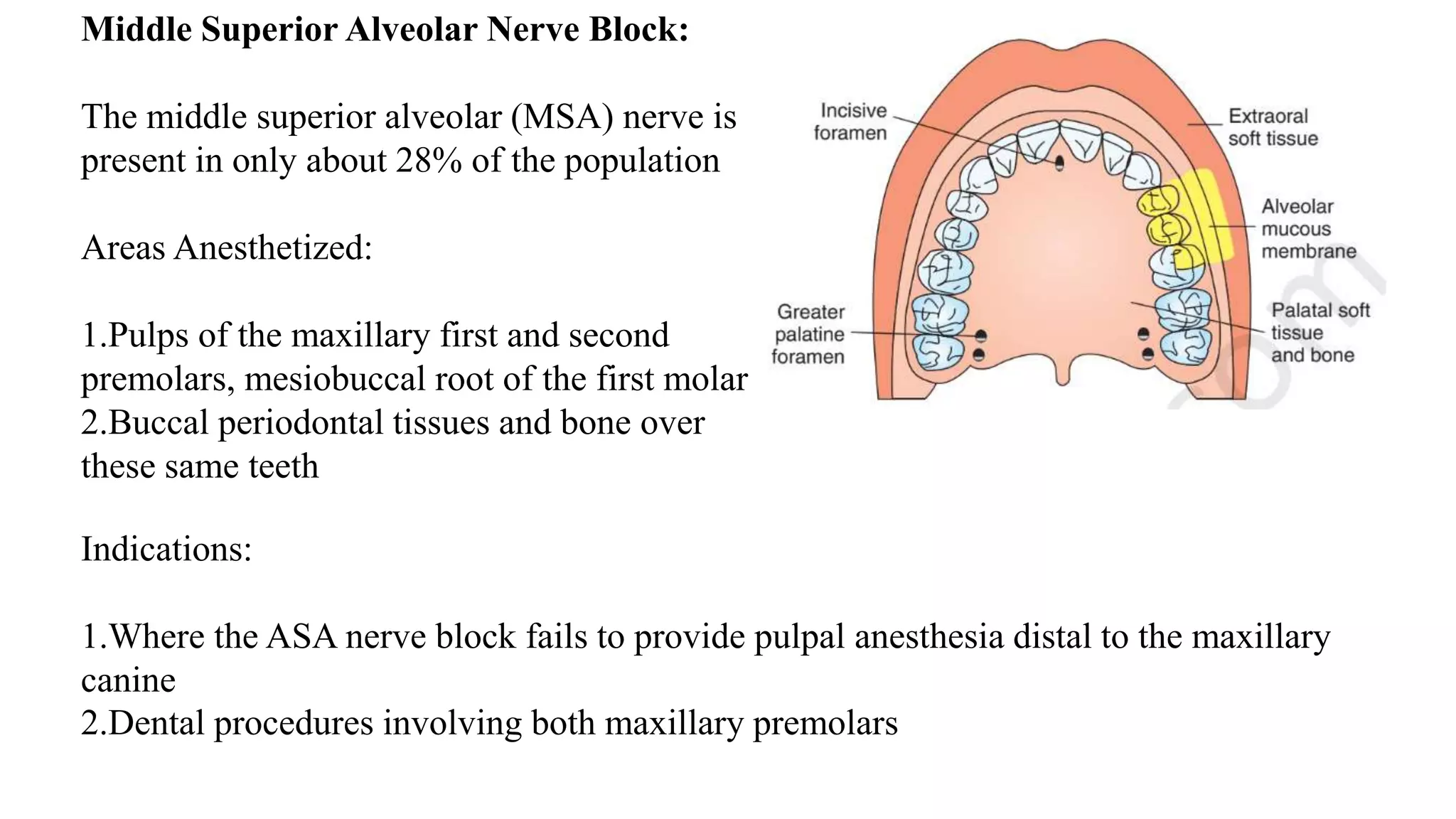
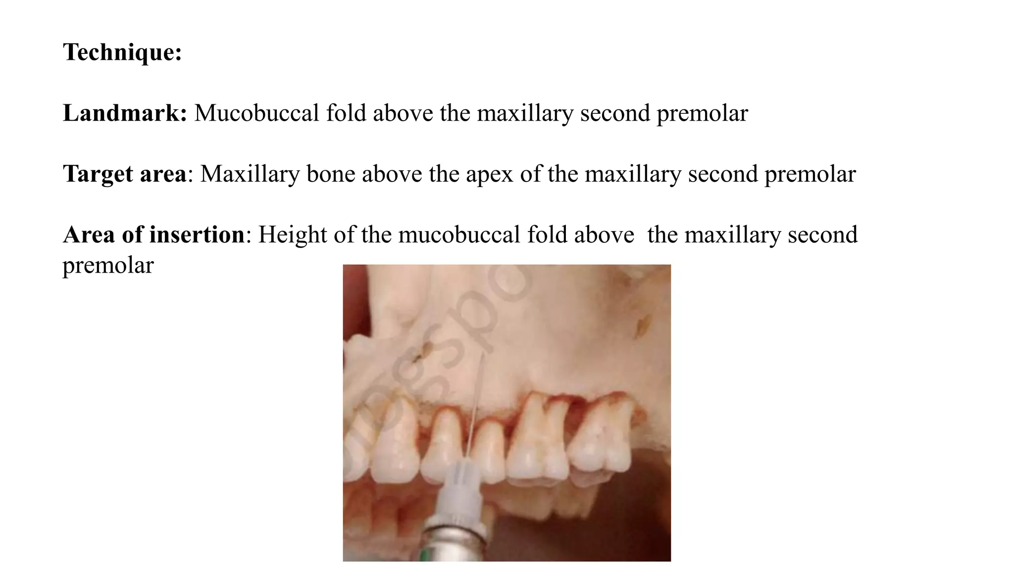
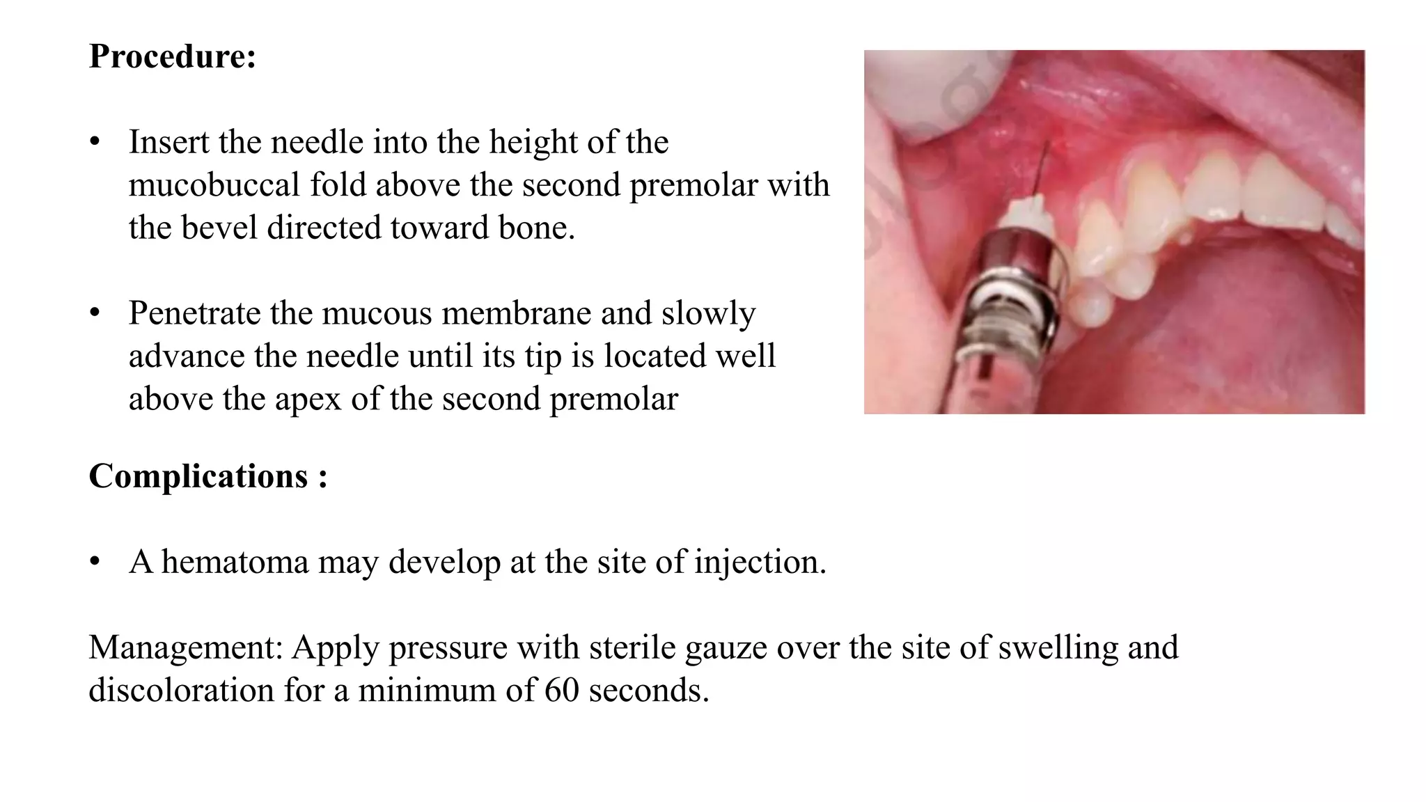
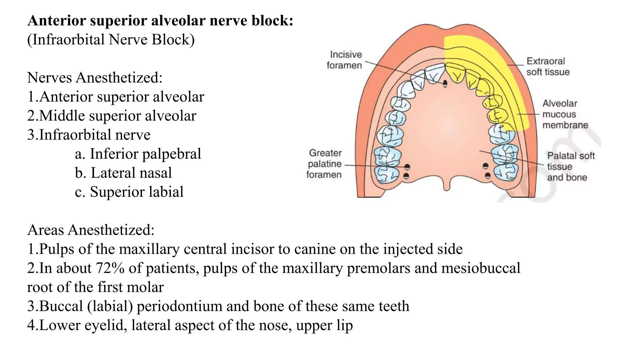
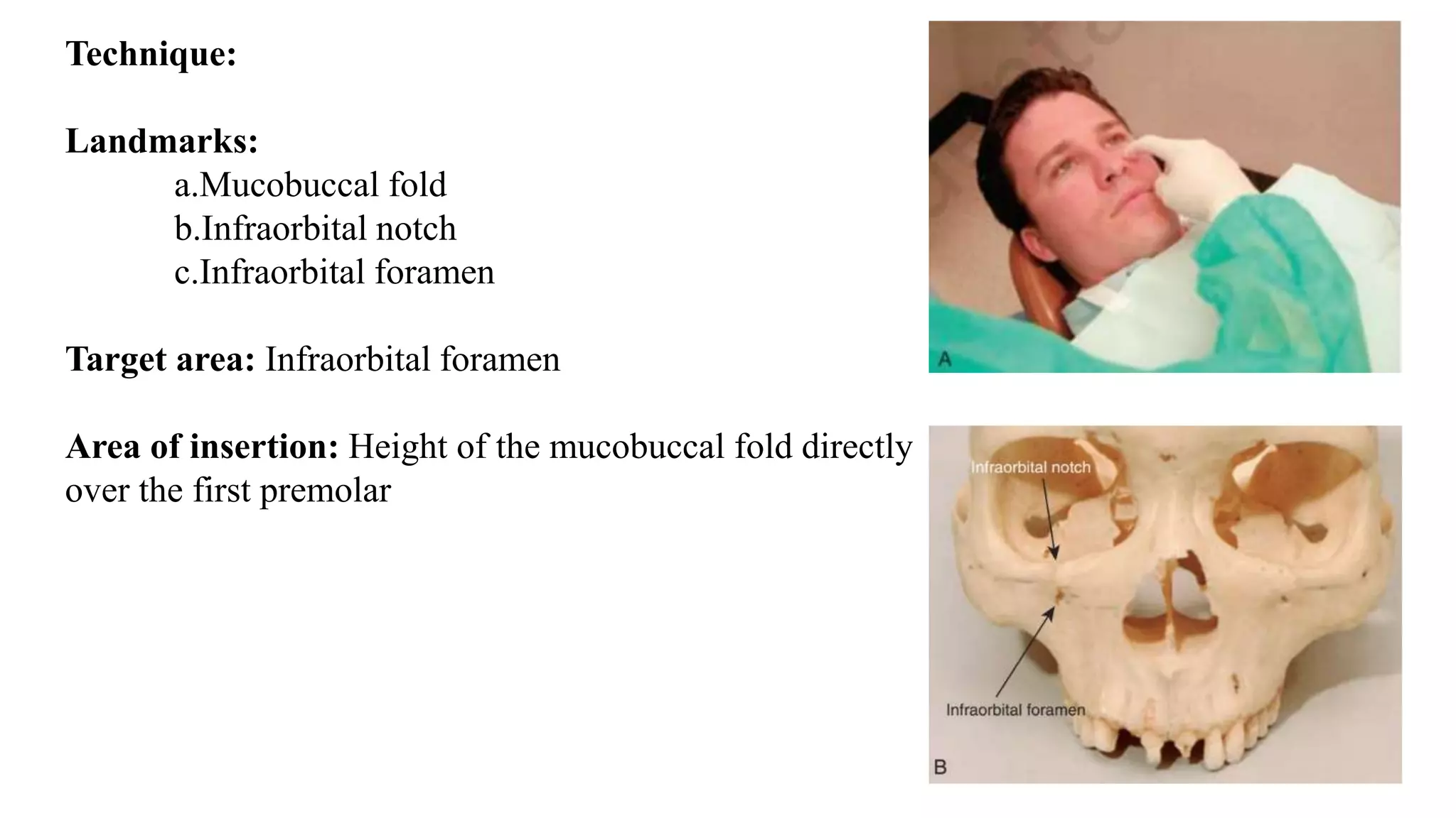
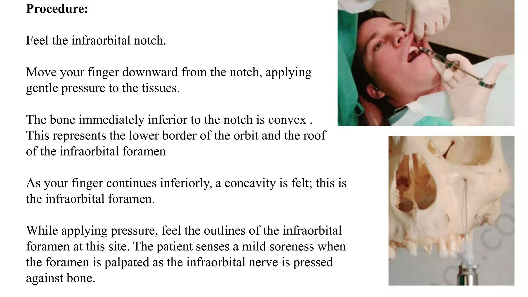
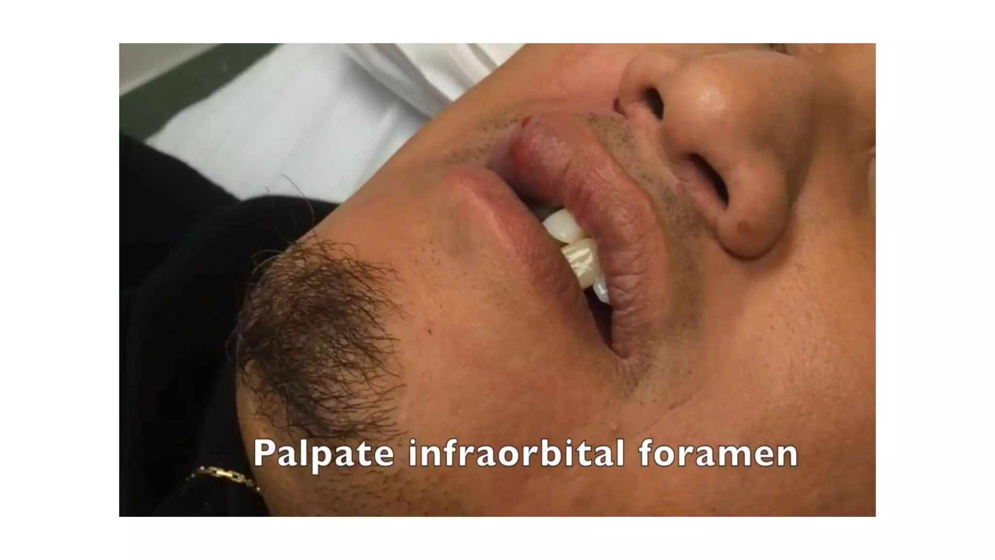
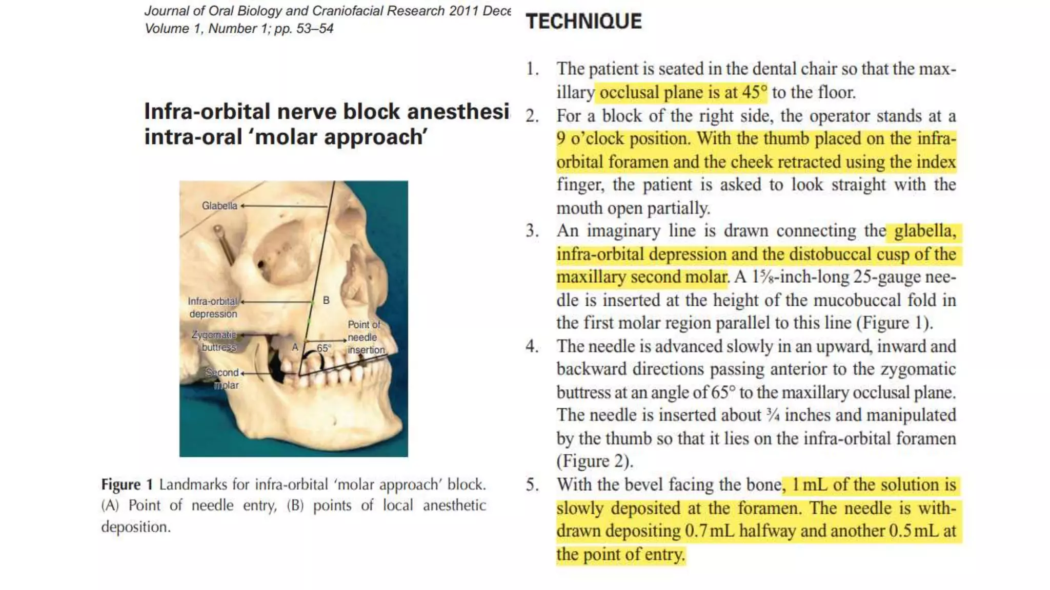
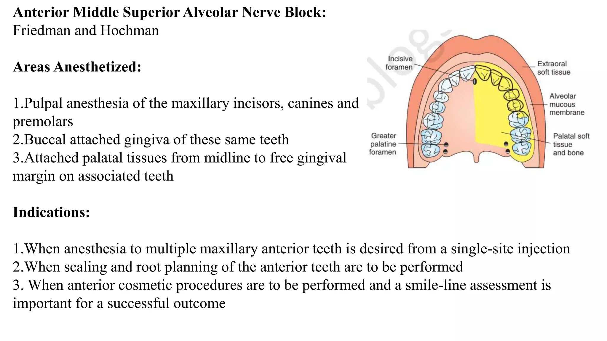
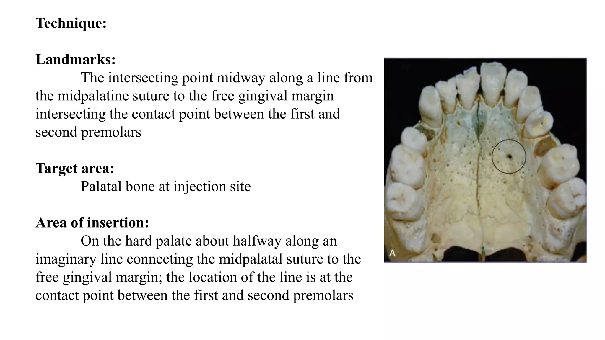
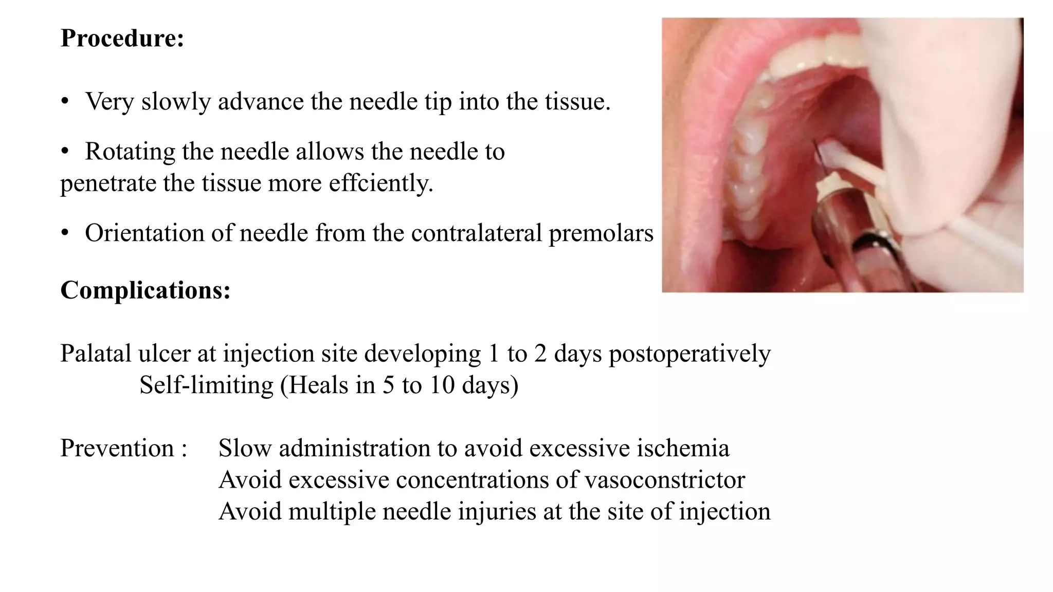
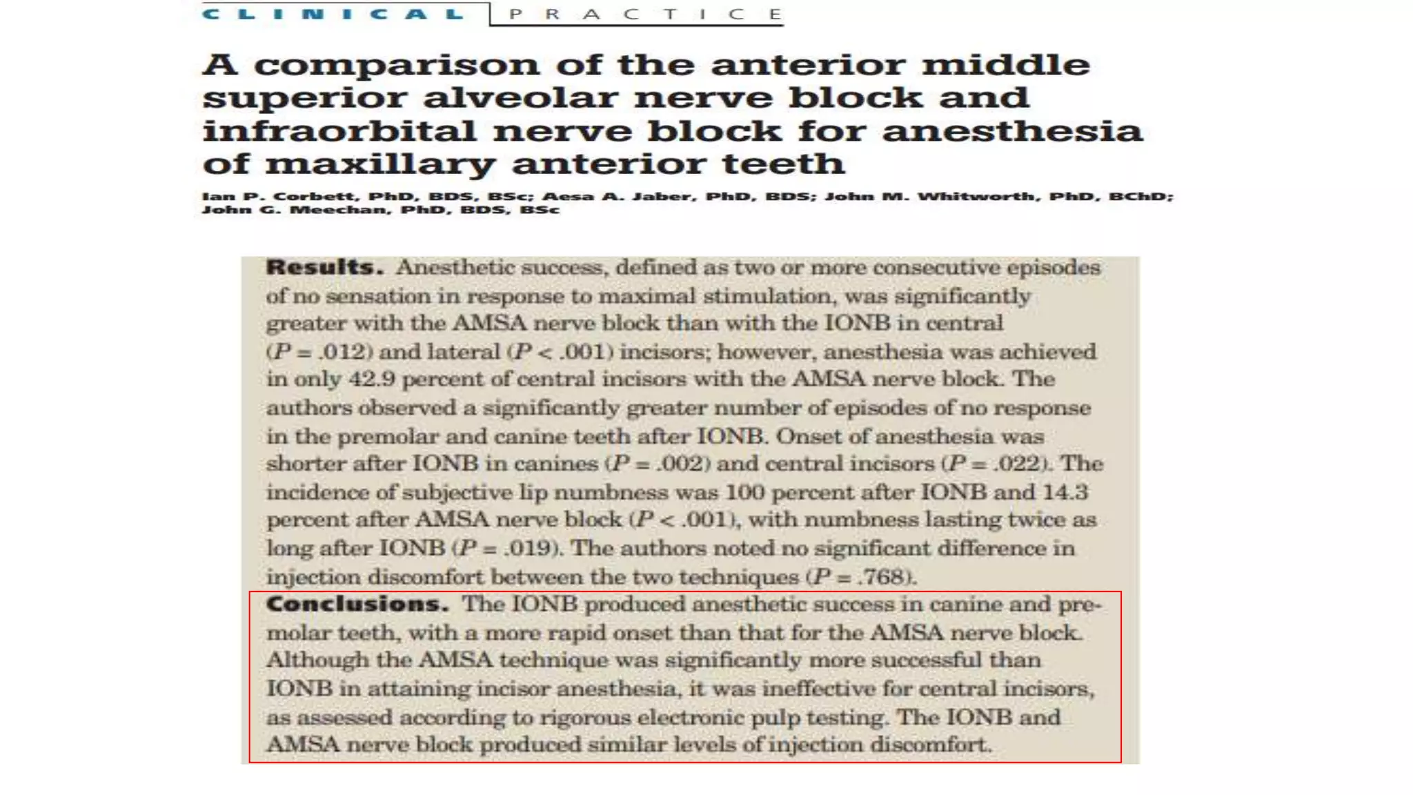
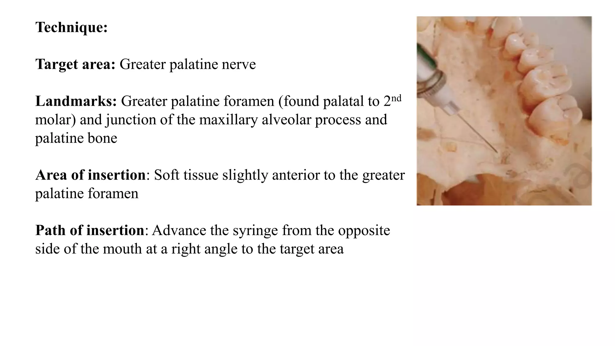
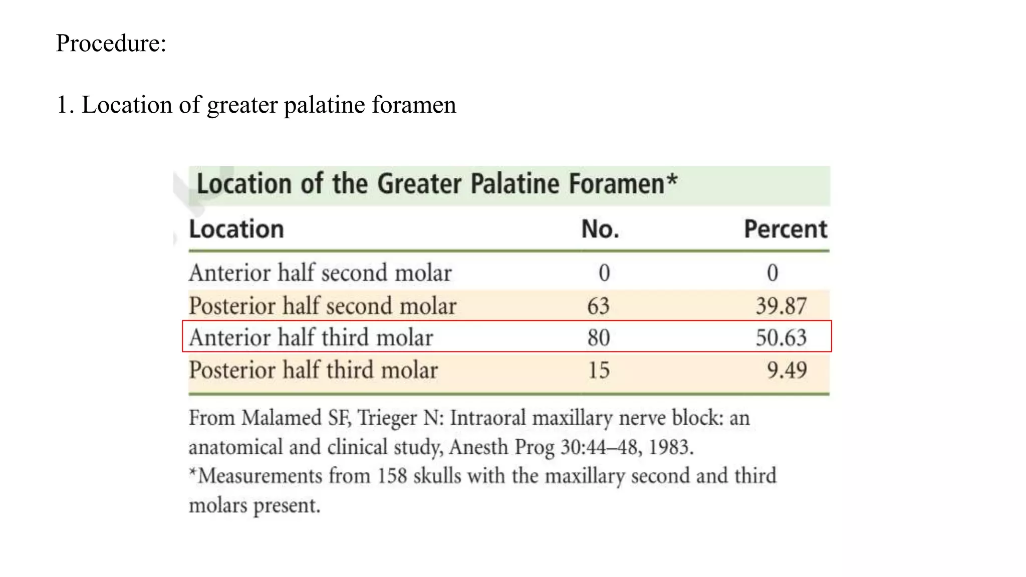
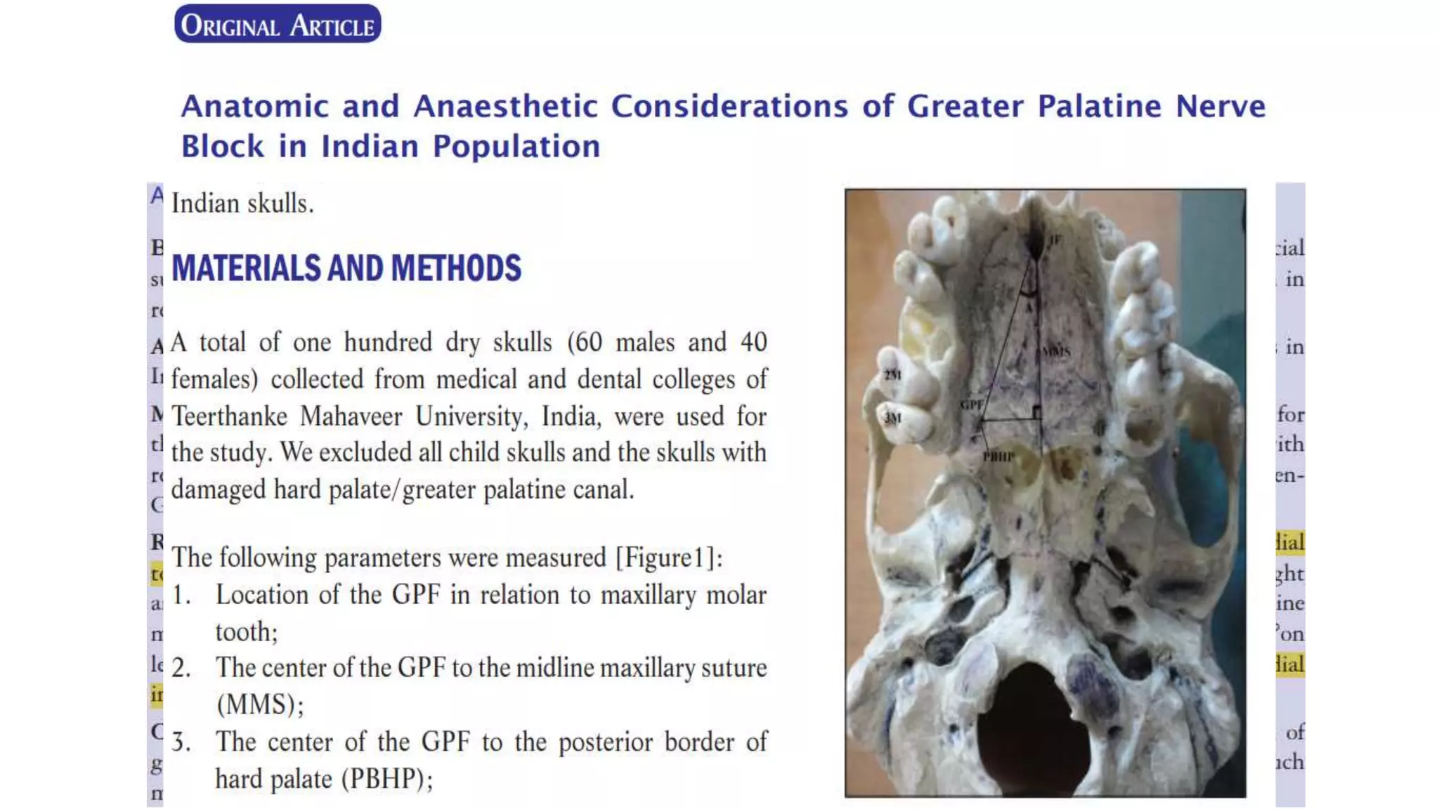
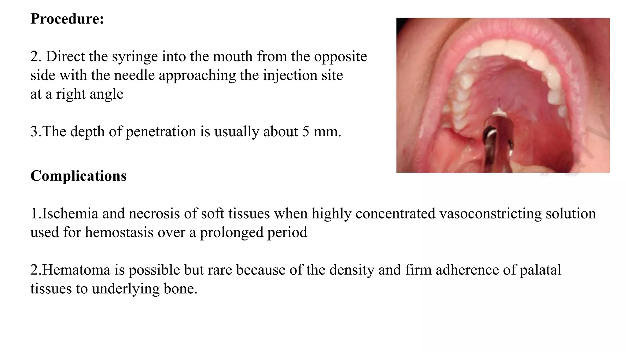
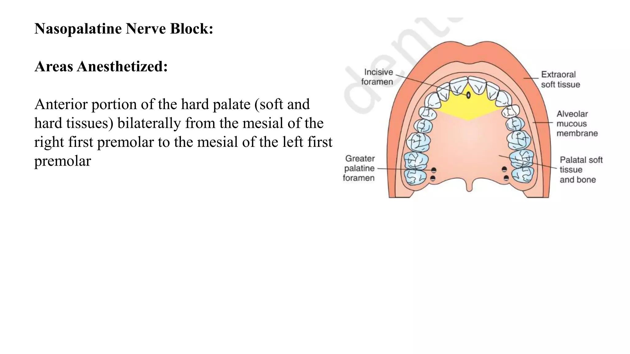
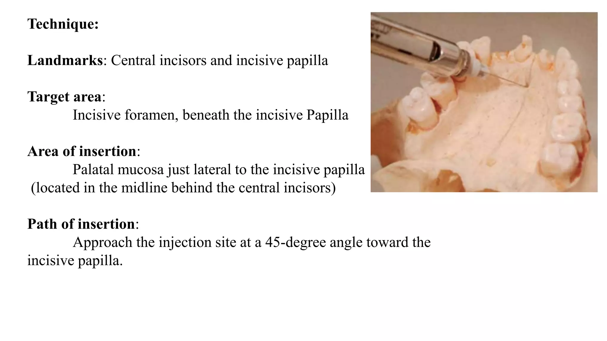
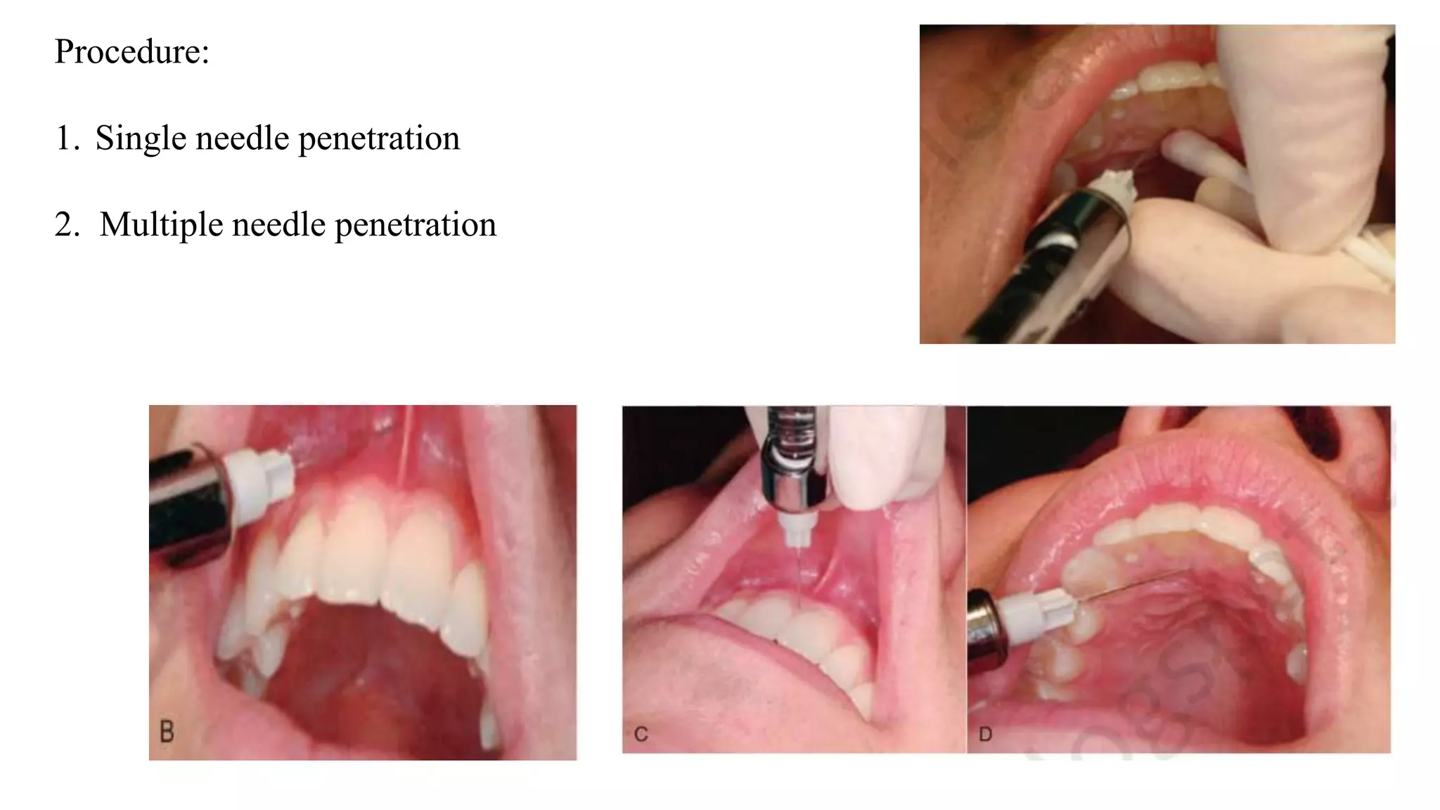
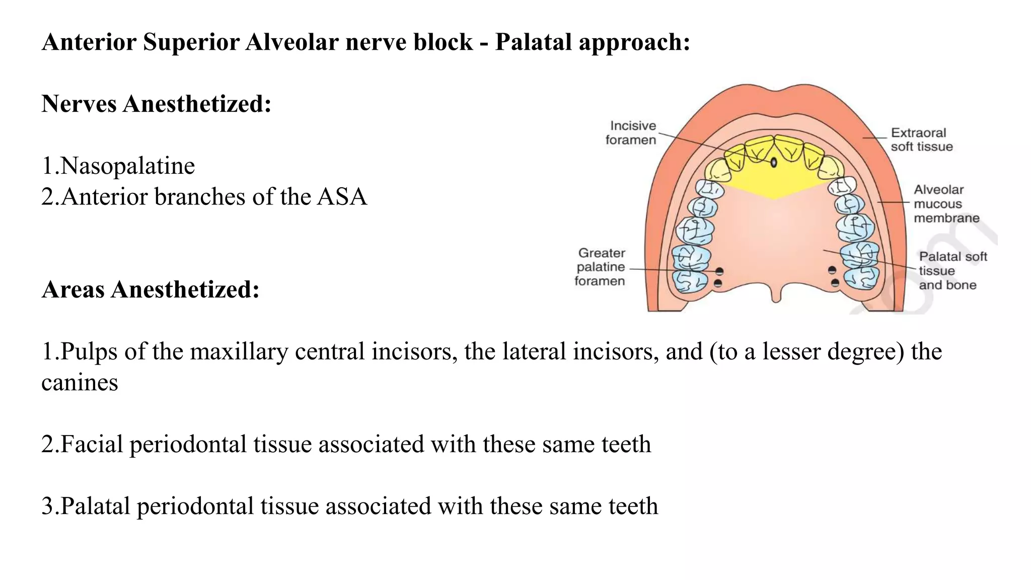
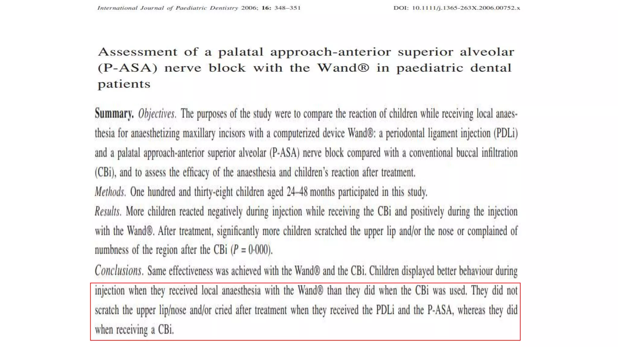
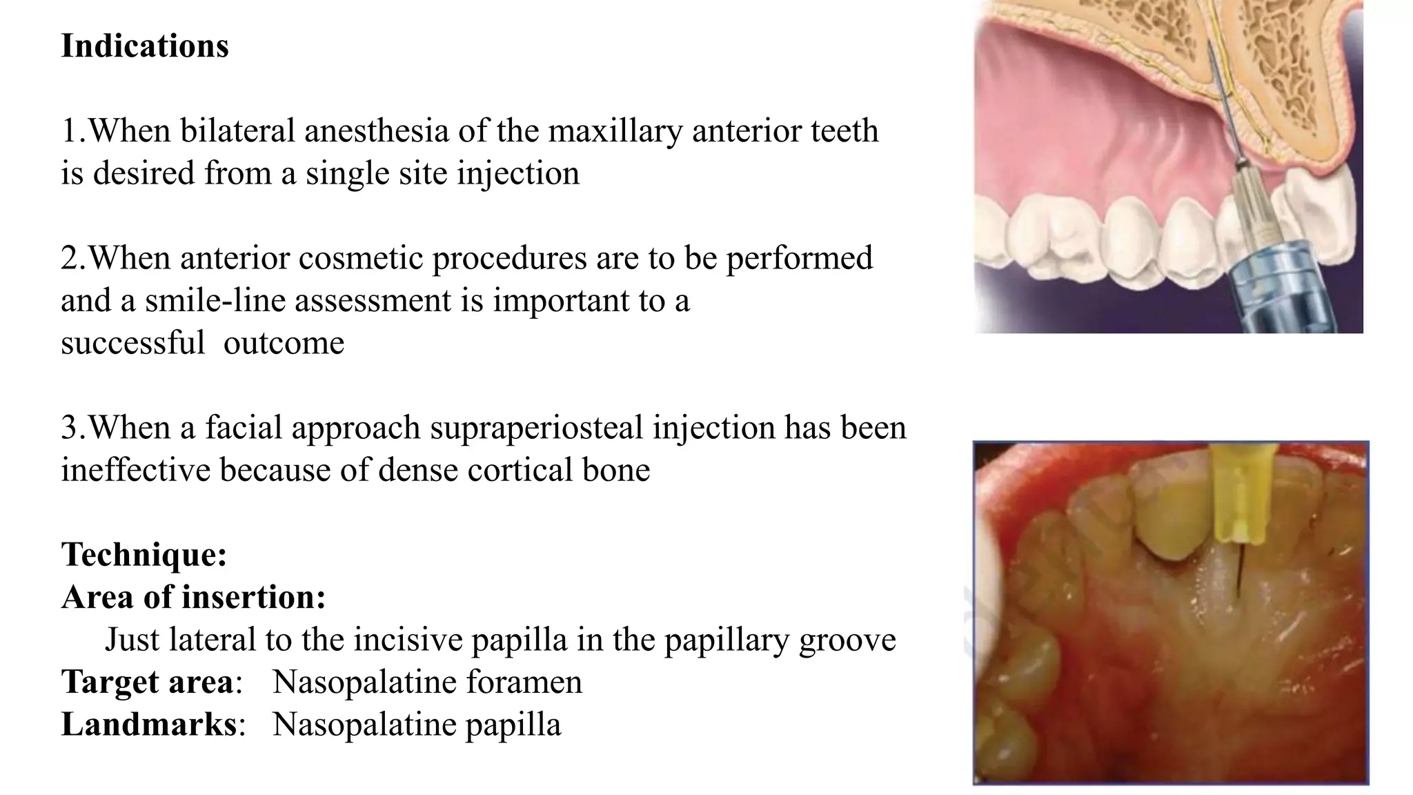
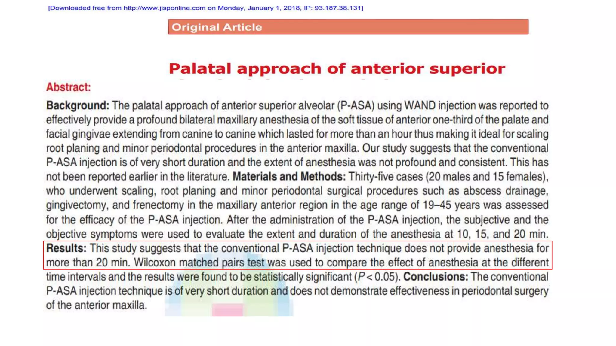
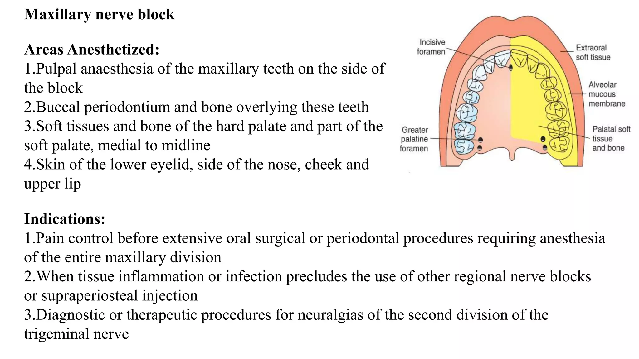
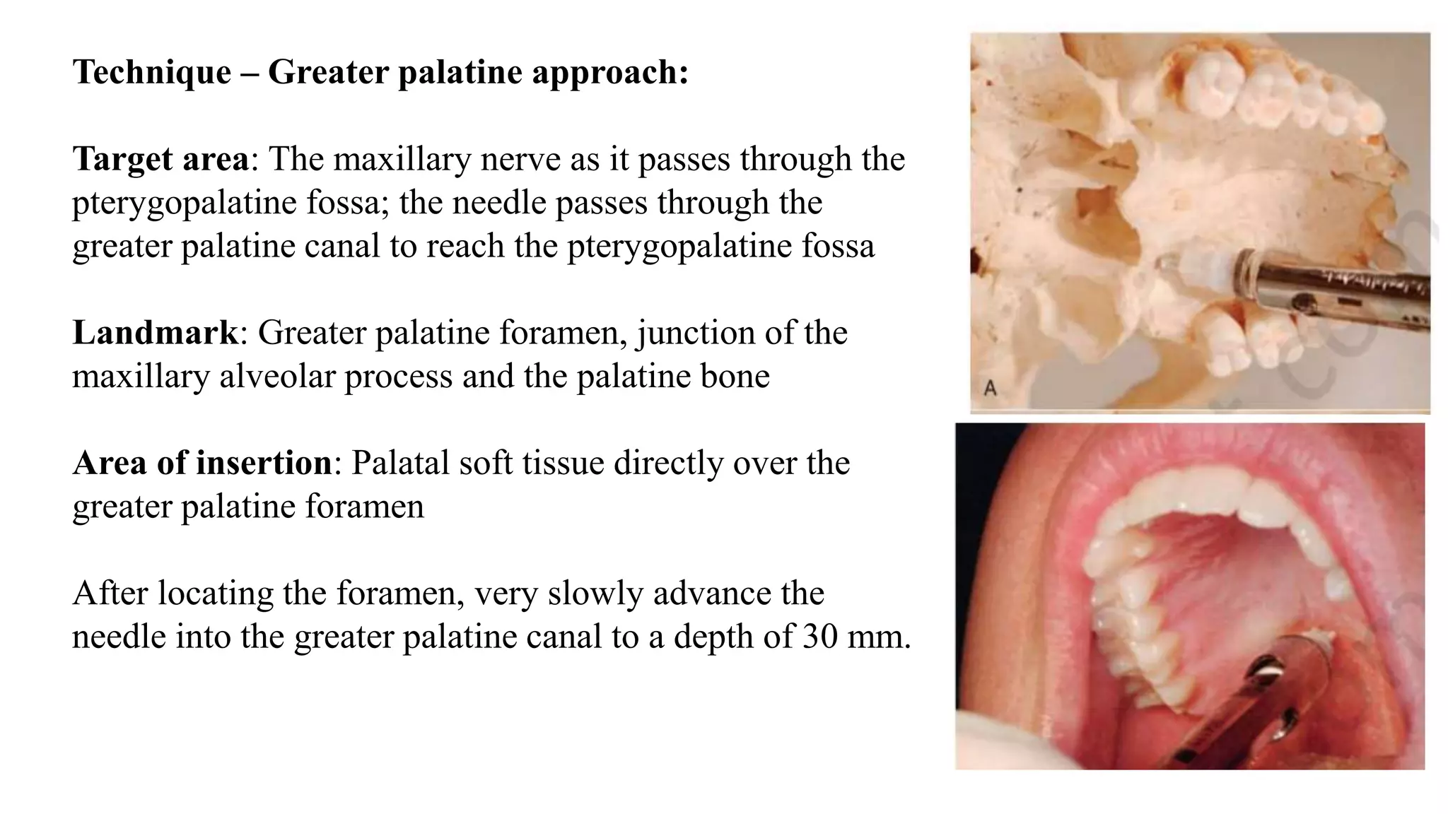
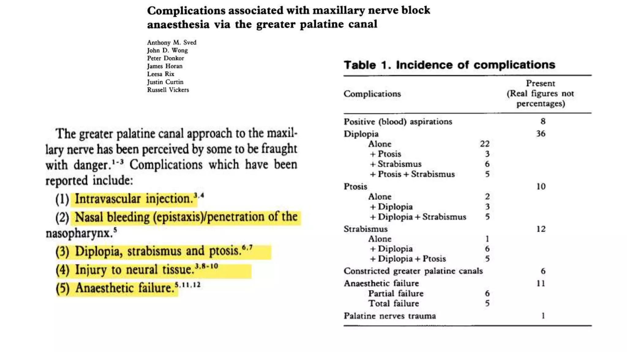
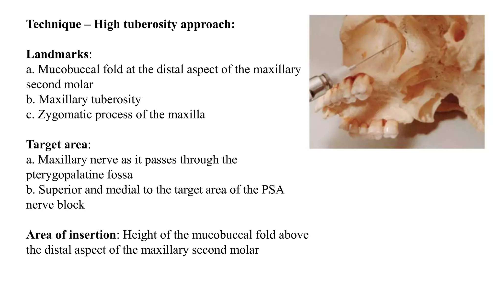
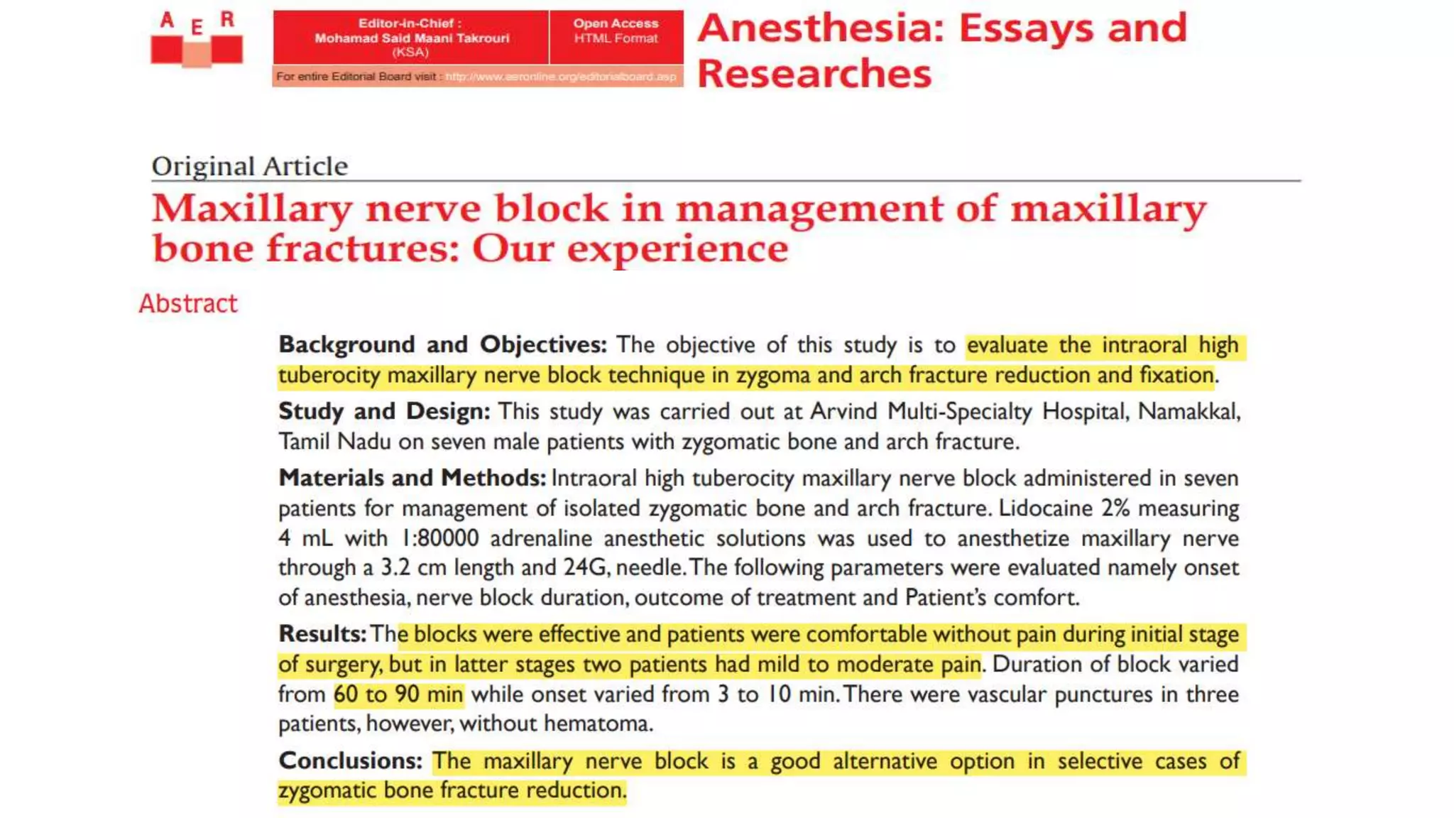
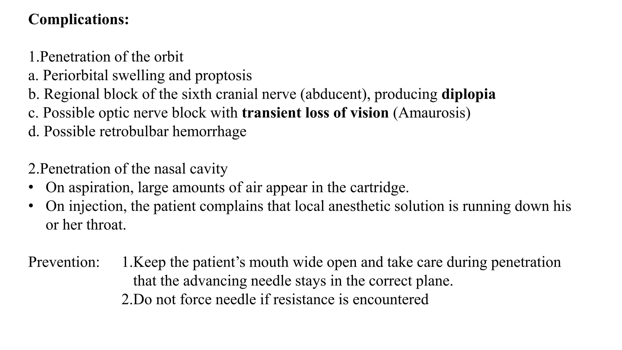
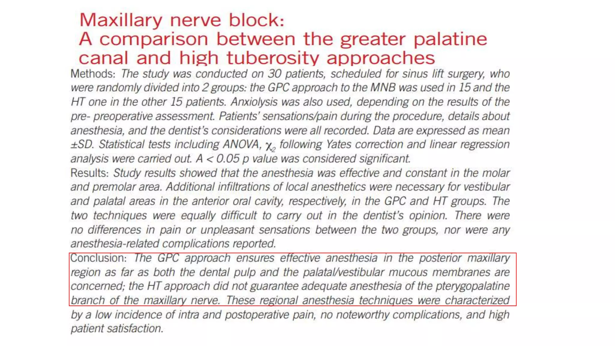
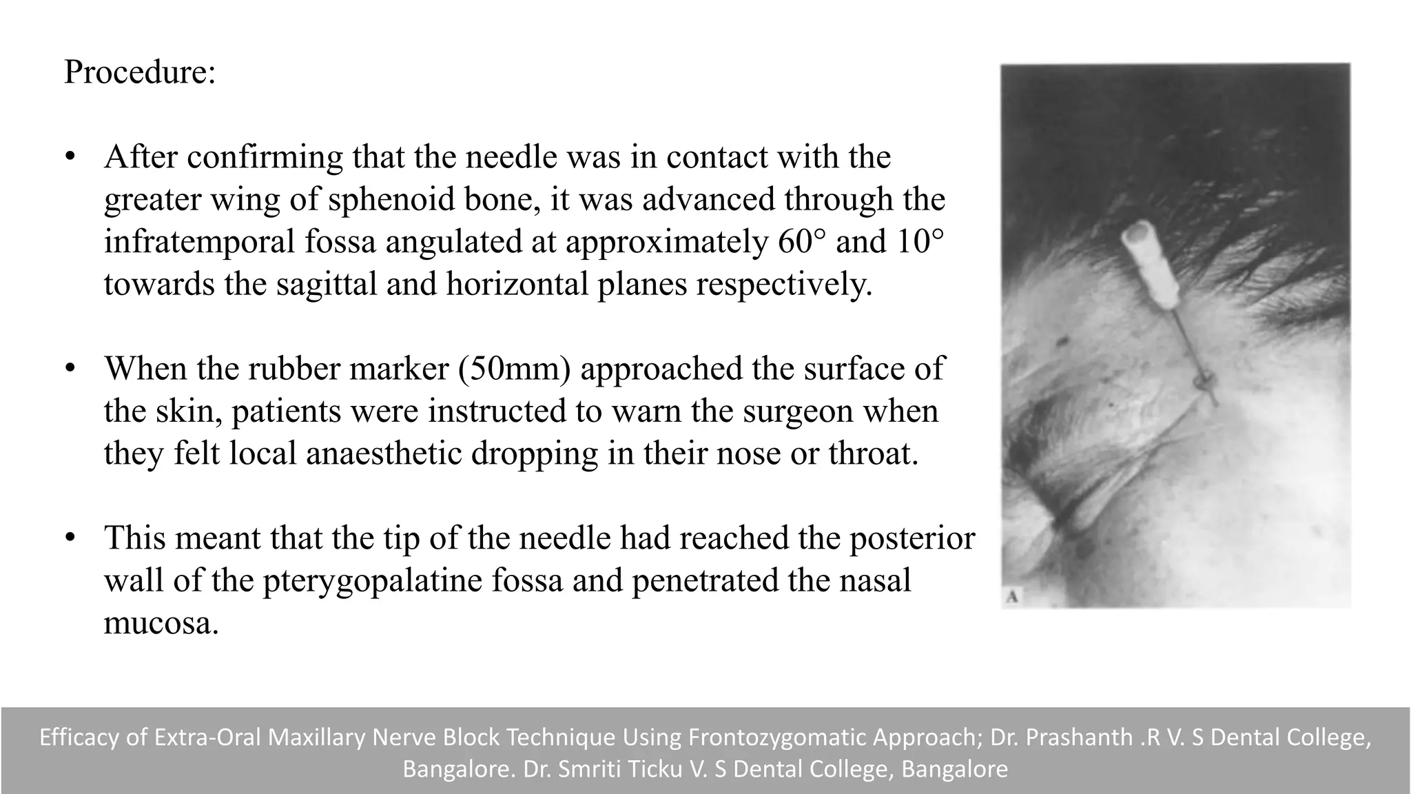
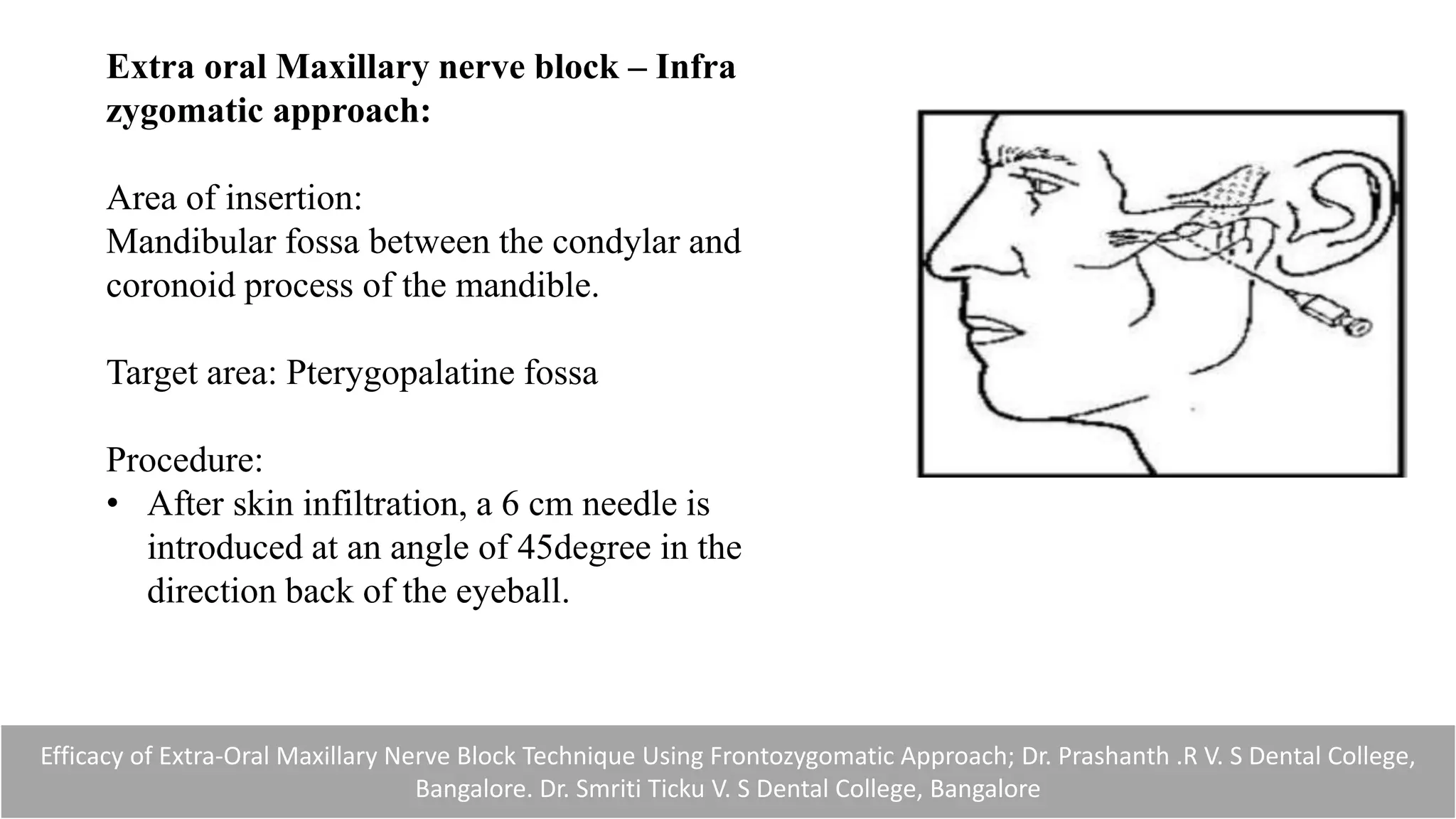
MAXILLARY NERVE BLOCK
Dr. Kathirvel G
PG OMFS
CONTENTS:
• Introduction
• Anatomy of maxilla
• Nerve supply
• Local anesthesia
• Intra-oral techniques
• Extra-oral techniques
MAXILLA:
OSSEOUS ANATOMY:
• Alveolar process
• Palatine process
• Frontal process
• Zygomatic process
NERVE SUPPLY:
Maxillary division of trigeminal nerve
Origin : Trigeminal ganglion located in the meckel's cave
In Middle Cranial Fossa : Meningeal branch
In Pterygopalatine Fossa : Ganglionic branches
Zygomatic
Posterior superior alveolar
Sphenopalatine ganglion
Greater and lesser palatine nerves
Nasal branch
Nasopalatine nerve
Pharyngeal nerve
In Infraorbital Canal : Middle superior alveolar
Anterior superior alveolar
On Face: Inferior Palpebral branch
Superior Labial branch
Nasal branch
Local anesthesia:
Definition:
“Local anesthesia is defined as a reversible loss of sensation in a circumscribed
area of the body caused by depression of excitation in nerve endings or an inhibition of
the conduction process in peripheral nerves”
STANLEY F.MALAMED
Local Infiltration:
Local anesthetic solution deposited over small
terminal nerve endings in the area of the dental
treatment
Field Block:
• Local anesthetic is deposited near the larger terminal
nerve branches
• Maxillary injections administered above the apex of the
tooth to be treated are properly termed field blocks
Nerve Block:
• Local anesthetic is deposited close to a main nerve trunk,
usually at a distance from the site of operative
intervention
• Ex: Posterior superior alveolar, nasopalatine injections
Intra oral techniques:
• Posterior superior alveolar nerve block
• Middle superior alveolar nerve block
• Anterior superior alveolar nerve block
• Anterior middle superior alveolar nerve block (AMSA)
• Greater palatine nerve block
• Nasopalatine nerve block
• Anterior superior alveolar nerve block - Palatal approach
• Maxillary nerve block - Greater palatine approach
- High tuberosity approach
Posterior superior alveolar nerve block:
Areas Anesthetized:
1.Pulps of the maxillary third, second and first
molars
(mesiobuccal root of the maxillary first molar not
anesthetized = 28%)
2.Buccal periodontium and bone overlying these
teeth
Indication:
1.When treatment involves two or more maxillary molars
2. When supraperiosteal injection has proved ineffective
Technique:
Landmarks:
a. Mucobuccal fold
b. Maxillary tuberosity
c. Zygomatic process of the maxilla
Target area: PSA nerve
Area of insertion: Height of mucobuccal fold
Procedure:
Insert the needle into the height of the mucobuccal
fold over the second molar
Advance the needle slowly in an upward, inward
and backward direction
Upward: superiorly at a 45-degree angle to the
occlusal plane
Inward: medially toward the midline at a 45-degree
angle to the occlusal plane
Backward: posteriorly at a 45-degree angle to the
long axis of the second molar
Depth of penetration: 16 mm
Complications
1.Hematoma:
• Needle too far posteriorly into the pterygoid plexus of
veins.
• A visible intraoral hematoma develops within several
minutes, usually noted in the buccal tissues of the
mandibular region .
2.Mandibular anesthesia:
• The mandibular division of the fifth cranial nerve (V3) is
located lateral to the PSA nerves.
• Deposition of local anesthetic lateral to the desired
location may produce varying degrees of mandibular
anesthesia
Middle Superior Alveolar Nerve Block:
The middle superior alveolar (MSA) nerve is
present in only about 28% of the population
Areas Anesthetized:
1.Pulps of the maxillary first and second
premolars, mesiobuccal root of the first molar
2.Buccal periodontal tissues and bone over
these same teeth
Indications:
1.Where the ASA nerve block fails to provide pulpal anesthesia distal to the maxillary
canine
2.Dental procedures involving both maxillary premolars
Technique:
Landmark: Mucobuccal fold above the maxillary second premolar
Target area: Maxillary bone above the apex of the maxillary second premolar
Area of insertion: Height of the mucobuccal fold above the maxillary second
premolar
Procedure:
• Insert the needle into the height of the
mucobuccal fold above the second premolar with
the bevel directed toward bone.
• Penetrate the mucous membrane and slowly
advance the needle until its tip is located well
above the apex of the second premolar
Complications :
• A hematoma may develop at the site of injection.
Management: Apply pressure with sterile gauze over the site of swelling and
discoloration for a minimum of 60 seconds.
Anterior superior alveolar nerve block:
(Infraorbital Nerve Block)
Nerves Anesthetized:
1.Anterior superior alveolar
2.Middle superior alveolar
3.Infraorbital nerve
a. Inferior palpebral
b. Lateral nasal
c. Superior labial
Areas Anesthetized:
1.Pulps of the maxillary central incisor to canine on the injected side
2.In about 72% of patients, pulps of the maxillary premolars and mesiobuccal
root of the first molar
3.Buccal (labial) periodontium and bone of these same teeth
4.Lower eyelid, lateral aspect of the nose, upper lip
Technique:
Landmarks:
a.Mucobuccal fold
b.Infraorbital notch
c.Infraorbital foramen
Target area: Infraorbital foramen
Area of insertion: Height of the mucobuccal fold directly
over the first premolar
Procedure:
Feel the infraorbital notch.
Move your finger downward from the notch, applying
gentle pressure to the tissues.
The bone immediately inferior to the notch is convex .
This represents the lower border of the orbit and the roof
of the infraorbital foramen
As your finger continues inferiorly, a concavity is felt; this is
the infraorbital foramen.
While applying pressure, feel the outlines of the infraorbital
foramen at this site. The patient senses a mild soreness when
the foramen is palpated as the infraorbital nerve is pressed
against bone.
Complications:
• Hematoma (rare) may develop across the lower eyelid and the tissues between it and
the infraorbital foramen.
Management: Apply pressure on the soft tissue over the foramen for 2 to 3 minutes.
Anterior Middle Superior Alveolar Nerve Block:
Friedman and Hochman
Areas Anesthetized:
1.Pulpal anesthesia of the maxillary incisors, canines and
premolars
2.Buccal attached gingiva of these same teeth
3.Attached palatal tissues from midline to free gingival
margin on associated teeth
Indications:
1.When anesthesia to multiple maxillary anterior teeth is desired from a single-site injection
2.When scaling and root planning of the anterior teeth are to be performed
3. When anterior cosmetic procedures are to be performed and a smile-line assessment is
important for a successful outcome
Technique:
Landmarks:
The intersecting point midway along a line from
the midpalatine suture to the free gingival margin
intersecting the contact point between the first and
second premolars
Target area:
Palatal bone at injection site
Area of insertion:
On the hard palate about halfway along an
imaginary line connecting the midpalatal suture to the
free gingival margin; the location of the line is at the
contact point between the first and second premolars
Procedure:
• Very slowly advance the needle tip into the tissue.
• Rotating the needle allows the needle to
penetrate the tissue more effciently.
• Orientation of needle from the contralateral premolars
Complications:
Palatal ulcer at injection site developing 1 to 2 days postoperatively
Self-limiting (Heals in 5 to 10 days)
Prevention : Slow administration to avoid excessive ischemia
Avoid excessive concentrations of vasoconstrictor
Avoid multiple needle injuries at the site of injection
Greater Palatine Nerve Block:
Areas Anesthetized:
The posterior portion of the hard palate
and its overlying soft tissues, anteriorly as far as
the first premolar and medially to the midline
Indications:
1.When palatal soft tissue anesthesia is necessary for restorative therapy on more
than two teeth
2.For pain control during periodontal or oral surgical procedures involving the
palatal soft and hard tissue
Technique:
Target area: Greater palatine nerve
Landmarks: Greater palatine foramen (found palatal to 2nd
molar) and junction of the maxillary alveolar process and
palatine bone
Area of insertion: Soft tissue slightly anterior to the greater
palatine foramen
Path of insertion: Advance the syringe from the opposite
side of the mouth at a right angle to the target area
Procedure:
1. Location of greater palatine foramen
Procedure:
2. Direct the syringe into the mouth from the opposite
side with the needle approaching the injection site
at a right angle
3.The depth of penetration is usually about 5 mm.
Complications
1.Ischemia and necrosis of soft tissues when highly concentrated vasoconstricting solution
used for hemostasis over a prolonged period
2.Hematoma is possible but rare because of the density and firm adherence of palatal
tissues to underlying bone.
Nasopalatine Nerve Block:
Areas Anesthetized:
Anterior portion of the hard palate (soft and
hard tissues) bilaterally from the mesial of the
right first premolar to the mesial of the left first
premolar
Technique:
Landmarks: Central incisors and incisive papilla
Target area:
Incisive foramen, beneath the incisive Papilla
Area of insertion:
Palatal mucosa just lateral to the incisive papilla
(located in the midline behind the central incisors)
Path of insertion:
Approach the injection site at a 45-degree angle toward the
incisive papilla.
Procedure:
1. Single needle penetration
2. Multiple needle penetration
Complications
1.Ischemia and necrosis of soft tissues while using high concentration of
vasoconstrictor
2.Hematoma is possible but rare because of the density and firm adherence of palatal
tissues to underlying bone.
3.Interdental papillae between the maxillary incisors sometimes are tender for several
days after injection.
Anterior Superior Alveolar nerve block - Palatal approach:
Nerves Anesthetized:
1.Nasopalatine
2.Anterior branches of the ASA
Areas Anesthetized:
1.Pulps of the maxillary central incisors, the lateral incisors, and (to a lesser degree) the
canines
2.Facial periodontal tissue associated with these same teeth
3.Palatal periodontal tissue associated with these same teeth
Indications
1.When bilateral anesthesia of the maxillary anterior teeth
is desired from a single site injection
2.When anterior cosmetic procedures are to be performed
and a smile-line assessment is important to a
successful outcome
3.When a facial approach supraperiosteal injection has been
ineffective because of dense cortical bone
Technique:
Area of insertion:
Just lateral to the incisive papilla in the papillary groove
Target area: Nasopalatine foramen
Landmarks: Nasopalatine papilla
Procedure:
• Rotating the needle allows the needle to penetrate the tissue more efficiently.
• Continue the slow insertion technique into the nasopalatine canal.
• Orientation of the needle should be parallel to the long axis of the central incisors.
• The needle is advanced to a depth of 6 to 10 mm
Complication:
1. Palatal ulcer at the site of injection postoperatively
• Prevention : Slow administration to avoid excessive ischemia.
Avoid excessive concentrations of vasoconstrictor
2. Density of soft tissues at injection site causing squirt back LA
Maxillary nerve block
Areas Anesthetized:
1.Pulpal anaesthesia of the maxillary teeth on the side of
the block
2.Buccal periodontium and bone overlying these teeth
3.Soft tissues and bone of the hard palate and part of the
soft palate, medial to midline
4.Skin of the lower eyelid, side of the nose, cheek and
upper lip
Indications:
1.Pain control before extensive oral surgical or periodontal procedures requiring anesthesia
of the entire maxillary division
2.When tissue inflammation or infection precludes the use of other regional nerve blocks
or supraperiosteal injection
3.Diagnostic or therapeutic procedures for neuralgias of the second division of the
trigeminal nerve
Technique – Greater palatine approach:
Target area: The maxillary nerve as it passes through the
pterygopalatine fossa; the needle passes through the
greater palatine canal to reach the pterygopalatine fossa
Landmark: Greater palatine foramen, junction of the
maxillary alveolar process and the palatine bone
Area of insertion: Palatal soft tissue directly over the
greater palatine foramen
After locating the foramen, very slowly advance the
needle into the greater palatine canal to a depth of 30 mm.
Technique – High tuberosity approach:
Landmarks:
a. Mucobuccal fold at the distal aspect of the maxillary
second molar
b. Maxillary tuberosity
c. Zygomatic process of the maxilla
Target area:
a. Maxillary nerve as it passes through the
pterygopalatine fossa
b. Superior and medial to the target area of the PSA
nerve block
Area of insertion: Height of the mucobuccal fold above
the distal aspect of the maxillary second molar
Complications:
1.Penetration of the orbit
a. Periorbital swelling and proptosis
b. Regional block of the sixth cranial nerve (abducent), producing diplopia
c. Possible optic nerve block with transient loss of vision (Amaurosis)
d. Possible retrobulbar hemorrhage
2.Penetration of the nasal cavity
• On aspiration, large amounts of air appear in the cartridge.
• On injection, the patient complains that local anesthetic solution is running down his
or her throat.
Prevention: 1.Keep the patient’s mouth wide open and take care during penetration
that the advancing needle stays in the correct plane.
2.Do not force needle if resistance is encountered
Extra-oral technique
• Extra oral Infra orbital nerve block
• Extra oral Maxillary nerve block – Supra zygomatic approach
• Extra oral Maxillary nerve block – Infra zygomatic approach
Extraoral infra orbital nerve block:
Area anesthetized:
Complete anesthesia to the teeth and facial structures of the anterior part of the maxilla
Indications :
• Multiple Extractions in the anterior part of the maxilla
• Surgical removal of impacted teeth
• Surgical exploration in the anterior part of the maxilla
• Endodontic surgery in the anterior part of the maxilla
• Treatment of acute trauma requiring suturing of soft tissues or repositioning of alveolar
bone and associated teeth, or both
The extraoral approach to the infraorbital nerve block; Donald J. Kleier, DMD Deborah K. Deeg, DDS Robert E. Averbach, DDS
Technique:
Target area : Infraorbital foramen, which is
located 5 to 10 mm inferior to the
zygomaticomaxillary suture.
Area of insertion : Just below the operator’s
palpating infraorbital foramen
Procedure: The needle was inserted through the
skin and directed upward at a 45° angle
The extraoral approach to the infraorbital nerve block; Donald J. Kleier, DMD Deborah K. Deeg, DDS Robert E. Averbach, DDS
Complications:
• Bruising - Bruising in the infraorbital area would be highly visible
• Post injection soreness
• If anesthetic solution should inadvertently enter the orbit, a transient
diplopia or loss of vision could occur.
The extraoral approach to the infraorbital nerve block; Donald J. Kleier, DMD Deborah K. Deeg, DDS Robert E. Averbach, DDS
Efficacy of Extra-Oral Maxillary Nerve Block Technique Using Frontozygomatic Approach; Dr. Prashanth .R V. S Dental College,
Bangalore. Dr. Smriti Ticku V. S Dental College, Bangalore
Extra oral Maxillary nerve block – Supra
zygomatic approach:
Nerve anesthetized: Maxillary branch of
trigeminal nerve
Point of insertion: Skin projection of the
frontozygomatic angle
Target area: Foramen rotundum
Procedure:
• After confirming that the needle was in contact with the
greater wing of sphenoid bone, it was advanced through the
infratemporal fossa angulated at approximately 60° and 10°
towards the sagittal and horizontal planes respectively.
• When the rubber marker (50mm) approached the surface of
the skin, patients were instructed to warn the surgeon when
they felt local anaesthetic dropping in their nose or throat.
• This meant that the tip of the needle had reached the posterior
wall of the pterygopalatine fossa and penetrated the nasal
mucosa.
Efficacy of Extra-Oral Maxillary Nerve Block Technique Using Frontozygomatic Approach; Dr. Prashanth .R V. S Dental College,
Bangalore. Dr. Smriti Ticku V. S Dental College, Bangalore
Efficacy of Extra-Oral Maxillary Nerve Block Technique Using Frontozygomatic Approach; Dr. Prashanth .R V. S Dental College,
Bangalore. Dr. Smriti Ticku V. S Dental College, Bangalore
• The needle was then withdrawn for 3–5 mm to avoid intranasal
injection
• The remaining drug was slowly deposited into the vicinity of the
foramen rotundum where the maxillary nerve leaves the base of the
skull to enter the pterygopalatine fossa.
COMPLICATIONS
• Hematoma
• Ecchymosis
• Visual disturbance
• Limitation of mouth opening
• Deviation of the lower jaw
Efficacy of Extra-Oral Maxillary Nerve Block Technique Using Frontozygomatic Approach; Dr. Prashanth .R V. S Dental College,
Bangalore. Dr. Smriti Ticku V. S Dental College, Bangalore
Extra oral Maxillary nerve block – Infra
zygomatic approach:
Area of insertion:
Mandibular fossa between the condylar and
coronoid process of the mandible.
Target area: Pterygopalatine fossa
Procedure:
• After skin infiltration, a 6 cm needle is
introduced at an angle of 45degree in the
direction back of the eyeball.
• 4-4.5 cm the lateral part of the pterygoid process is reached and the needle is
withdrawn slightly in the pterygopalatine fossa (about 0.5 cm medial to
pterygoid)
• Aspiration in various levels, local anesthesia is carefully injected in several
doses.
COMPLICATIONS
• Transient visual weakness (Rarely)
• Horner’s syndrome (Extremely rare usually occurs due to administration of
local anesthesia solution in higher doses)
• Hematoma over the cheek or in the orbital cavity due to blood vessel puncture
Efficacy of Extra-Oral Maxillary Nerve Block Technique Using Frontozygomatic Approach; Dr. Prashanth .R V. S Dental College,
Bangalore. Dr. Smriti Ticku V. S Dental College, Bangalore
Thank you

Maxillary nerve block

  • 1.
    MAXILLARY NERVE BLOCK Dr.Kathirvel G PG OMFS
  • 2.
    CONTENTS: • Introduction • Anatomyof maxilla • Nerve supply • Local anesthesia • Intra-oral techniques • Extra-oral techniques
  • 3.
    MAXILLA: OSSEOUS ANATOMY: • Alveolarprocess • Palatine process • Frontal process • Zygomatic process
  • 4.
    NERVE SUPPLY: Maxillary divisionof trigeminal nerve Origin : Trigeminal ganglion located in the meckel's cave In Middle Cranial Fossa : Meningeal branch In Pterygopalatine Fossa : Ganglionic branches Zygomatic Posterior superior alveolar Sphenopalatine ganglion Greater and lesser palatine nerves Nasal branch Nasopalatine nerve Pharyngeal nerve In Infraorbital Canal : Middle superior alveolar Anterior superior alveolar On Face: Inferior Palpebral branch Superior Labial branch Nasal branch
  • 6.
    Local anesthesia: Definition: “Local anesthesiais defined as a reversible loss of sensation in a circumscribed area of the body caused by depression of excitation in nerve endings or an inhibition of the conduction process in peripheral nerves” STANLEY F.MALAMED Local Infiltration: Local anesthetic solution deposited over small terminal nerve endings in the area of the dental treatment
  • 7.
    Field Block: • Localanesthetic is deposited near the larger terminal nerve branches • Maxillary injections administered above the apex of the tooth to be treated are properly termed field blocks Nerve Block: • Local anesthetic is deposited close to a main nerve trunk, usually at a distance from the site of operative intervention • Ex: Posterior superior alveolar, nasopalatine injections
  • 8.
    Intra oral techniques: •Posterior superior alveolar nerve block • Middle superior alveolar nerve block • Anterior superior alveolar nerve block • Anterior middle superior alveolar nerve block (AMSA) • Greater palatine nerve block • Nasopalatine nerve block • Anterior superior alveolar nerve block - Palatal approach • Maxillary nerve block - Greater palatine approach - High tuberosity approach
  • 9.
    Posterior superior alveolarnerve block: Areas Anesthetized: 1.Pulps of the maxillary third, second and first molars (mesiobuccal root of the maxillary first molar not anesthetized = 28%) 2.Buccal periodontium and bone overlying these teeth Indication: 1.When treatment involves two or more maxillary molars 2. When supraperiosteal injection has proved ineffective
  • 10.
    Technique: Landmarks: a. Mucobuccal fold b.Maxillary tuberosity c. Zygomatic process of the maxilla Target area: PSA nerve Area of insertion: Height of mucobuccal fold
  • 11.
    Procedure: Insert the needleinto the height of the mucobuccal fold over the second molar Advance the needle slowly in an upward, inward and backward direction Upward: superiorly at a 45-degree angle to the occlusal plane Inward: medially toward the midline at a 45-degree angle to the occlusal plane Backward: posteriorly at a 45-degree angle to the long axis of the second molar Depth of penetration: 16 mm
  • 13.
    Complications 1.Hematoma: • Needle toofar posteriorly into the pterygoid plexus of veins. • A visible intraoral hematoma develops within several minutes, usually noted in the buccal tissues of the mandibular region . 2.Mandibular anesthesia: • The mandibular division of the fifth cranial nerve (V3) is located lateral to the PSA nerves. • Deposition of local anesthetic lateral to the desired location may produce varying degrees of mandibular anesthesia
  • 15.
    Middle Superior AlveolarNerve Block: The middle superior alveolar (MSA) nerve is present in only about 28% of the population Areas Anesthetized: 1.Pulps of the maxillary first and second premolars, mesiobuccal root of the first molar 2.Buccal periodontal tissues and bone over these same teeth Indications: 1.Where the ASA nerve block fails to provide pulpal anesthesia distal to the maxillary canine 2.Dental procedures involving both maxillary premolars
  • 16.
    Technique: Landmark: Mucobuccal foldabove the maxillary second premolar Target area: Maxillary bone above the apex of the maxillary second premolar Area of insertion: Height of the mucobuccal fold above the maxillary second premolar
  • 17.
    Procedure: • Insert theneedle into the height of the mucobuccal fold above the second premolar with the bevel directed toward bone. • Penetrate the mucous membrane and slowly advance the needle until its tip is located well above the apex of the second premolar Complications : • A hematoma may develop at the site of injection. Management: Apply pressure with sterile gauze over the site of swelling and discoloration for a minimum of 60 seconds.
  • 18.
    Anterior superior alveolarnerve block: (Infraorbital Nerve Block) Nerves Anesthetized: 1.Anterior superior alveolar 2.Middle superior alveolar 3.Infraorbital nerve a. Inferior palpebral b. Lateral nasal c. Superior labial Areas Anesthetized: 1.Pulps of the maxillary central incisor to canine on the injected side 2.In about 72% of patients, pulps of the maxillary premolars and mesiobuccal root of the first molar 3.Buccal (labial) periodontium and bone of these same teeth 4.Lower eyelid, lateral aspect of the nose, upper lip
  • 19.
    Technique: Landmarks: a.Mucobuccal fold b.Infraorbital notch c.Infraorbitalforamen Target area: Infraorbital foramen Area of insertion: Height of the mucobuccal fold directly over the first premolar
  • 20.
    Procedure: Feel the infraorbitalnotch. Move your finger downward from the notch, applying gentle pressure to the tissues. The bone immediately inferior to the notch is convex . This represents the lower border of the orbit and the roof of the infraorbital foramen As your finger continues inferiorly, a concavity is felt; this is the infraorbital foramen. While applying pressure, feel the outlines of the infraorbital foramen at this site. The patient senses a mild soreness when the foramen is palpated as the infraorbital nerve is pressed against bone.
  • 23.
    Complications: • Hematoma (rare)may develop across the lower eyelid and the tissues between it and the infraorbital foramen. Management: Apply pressure on the soft tissue over the foramen for 2 to 3 minutes.
  • 24.
    Anterior Middle SuperiorAlveolar Nerve Block: Friedman and Hochman Areas Anesthetized: 1.Pulpal anesthesia of the maxillary incisors, canines and premolars 2.Buccal attached gingiva of these same teeth 3.Attached palatal tissues from midline to free gingival margin on associated teeth Indications: 1.When anesthesia to multiple maxillary anterior teeth is desired from a single-site injection 2.When scaling and root planning of the anterior teeth are to be performed 3. When anterior cosmetic procedures are to be performed and a smile-line assessment is important for a successful outcome
  • 25.
    Technique: Landmarks: The intersecting pointmidway along a line from the midpalatine suture to the free gingival margin intersecting the contact point between the first and second premolars Target area: Palatal bone at injection site Area of insertion: On the hard palate about halfway along an imaginary line connecting the midpalatal suture to the free gingival margin; the location of the line is at the contact point between the first and second premolars
  • 26.
    Procedure: • Very slowlyadvance the needle tip into the tissue. • Rotating the needle allows the needle to penetrate the tissue more effciently. • Orientation of needle from the contralateral premolars Complications: Palatal ulcer at injection site developing 1 to 2 days postoperatively Self-limiting (Heals in 5 to 10 days) Prevention : Slow administration to avoid excessive ischemia Avoid excessive concentrations of vasoconstrictor Avoid multiple needle injuries at the site of injection
  • 28.
    Greater Palatine NerveBlock: Areas Anesthetized: The posterior portion of the hard palate and its overlying soft tissues, anteriorly as far as the first premolar and medially to the midline Indications: 1.When palatal soft tissue anesthesia is necessary for restorative therapy on more than two teeth 2.For pain control during periodontal or oral surgical procedures involving the palatal soft and hard tissue
  • 29.
    Technique: Target area: Greaterpalatine nerve Landmarks: Greater palatine foramen (found palatal to 2nd molar) and junction of the maxillary alveolar process and palatine bone Area of insertion: Soft tissue slightly anterior to the greater palatine foramen Path of insertion: Advance the syringe from the opposite side of the mouth at a right angle to the target area
  • 30.
    Procedure: 1. Location ofgreater palatine foramen
  • 32.
    Procedure: 2. Direct thesyringe into the mouth from the opposite side with the needle approaching the injection site at a right angle 3.The depth of penetration is usually about 5 mm. Complications 1.Ischemia and necrosis of soft tissues when highly concentrated vasoconstricting solution used for hemostasis over a prolonged period 2.Hematoma is possible but rare because of the density and firm adherence of palatal tissues to underlying bone.
  • 34.
    Nasopalatine Nerve Block: AreasAnesthetized: Anterior portion of the hard palate (soft and hard tissues) bilaterally from the mesial of the right first premolar to the mesial of the left first premolar
  • 35.
    Technique: Landmarks: Central incisorsand incisive papilla Target area: Incisive foramen, beneath the incisive Papilla Area of insertion: Palatal mucosa just lateral to the incisive papilla (located in the midline behind the central incisors) Path of insertion: Approach the injection site at a 45-degree angle toward the incisive papilla.
  • 36.
    Procedure: 1. Single needlepenetration 2. Multiple needle penetration
  • 37.
    Complications 1.Ischemia and necrosisof soft tissues while using high concentration of vasoconstrictor 2.Hematoma is possible but rare because of the density and firm adherence of palatal tissues to underlying bone. 3.Interdental papillae between the maxillary incisors sometimes are tender for several days after injection.
  • 39.
    Anterior Superior Alveolarnerve block - Palatal approach: Nerves Anesthetized: 1.Nasopalatine 2.Anterior branches of the ASA Areas Anesthetized: 1.Pulps of the maxillary central incisors, the lateral incisors, and (to a lesser degree) the canines 2.Facial periodontal tissue associated with these same teeth 3.Palatal periodontal tissue associated with these same teeth
  • 41.
    Indications 1.When bilateral anesthesiaof the maxillary anterior teeth is desired from a single site injection 2.When anterior cosmetic procedures are to be performed and a smile-line assessment is important to a successful outcome 3.When a facial approach supraperiosteal injection has been ineffective because of dense cortical bone Technique: Area of insertion: Just lateral to the incisive papilla in the papillary groove Target area: Nasopalatine foramen Landmarks: Nasopalatine papilla
  • 42.
    Procedure: • Rotating theneedle allows the needle to penetrate the tissue more efficiently. • Continue the slow insertion technique into the nasopalatine canal. • Orientation of the needle should be parallel to the long axis of the central incisors. • The needle is advanced to a depth of 6 to 10 mm Complication: 1. Palatal ulcer at the site of injection postoperatively • Prevention : Slow administration to avoid excessive ischemia. Avoid excessive concentrations of vasoconstrictor 2. Density of soft tissues at injection site causing squirt back LA
  • 44.
    Maxillary nerve block AreasAnesthetized: 1.Pulpal anaesthesia of the maxillary teeth on the side of the block 2.Buccal periodontium and bone overlying these teeth 3.Soft tissues and bone of the hard palate and part of the soft palate, medial to midline 4.Skin of the lower eyelid, side of the nose, cheek and upper lip Indications: 1.Pain control before extensive oral surgical or periodontal procedures requiring anesthesia of the entire maxillary division 2.When tissue inflammation or infection precludes the use of other regional nerve blocks or supraperiosteal injection 3.Diagnostic or therapeutic procedures for neuralgias of the second division of the trigeminal nerve
  • 45.
    Technique – Greaterpalatine approach: Target area: The maxillary nerve as it passes through the pterygopalatine fossa; the needle passes through the greater palatine canal to reach the pterygopalatine fossa Landmark: Greater palatine foramen, junction of the maxillary alveolar process and the palatine bone Area of insertion: Palatal soft tissue directly over the greater palatine foramen After locating the foramen, very slowly advance the needle into the greater palatine canal to a depth of 30 mm.
  • 47.
    Technique – Hightuberosity approach: Landmarks: a. Mucobuccal fold at the distal aspect of the maxillary second molar b. Maxillary tuberosity c. Zygomatic process of the maxilla Target area: a. Maxillary nerve as it passes through the pterygopalatine fossa b. Superior and medial to the target area of the PSA nerve block Area of insertion: Height of the mucobuccal fold above the distal aspect of the maxillary second molar
  • 49.
    Complications: 1.Penetration of theorbit a. Periorbital swelling and proptosis b. Regional block of the sixth cranial nerve (abducent), producing diplopia c. Possible optic nerve block with transient loss of vision (Amaurosis) d. Possible retrobulbar hemorrhage 2.Penetration of the nasal cavity • On aspiration, large amounts of air appear in the cartridge. • On injection, the patient complains that local anesthetic solution is running down his or her throat. Prevention: 1.Keep the patient’s mouth wide open and take care during penetration that the advancing needle stays in the correct plane. 2.Do not force needle if resistance is encountered
  • 51.
    Extra-oral technique • Extraoral Infra orbital nerve block • Extra oral Maxillary nerve block – Supra zygomatic approach • Extra oral Maxillary nerve block – Infra zygomatic approach
  • 52.
    Extraoral infra orbitalnerve block: Area anesthetized: Complete anesthesia to the teeth and facial structures of the anterior part of the maxilla Indications : • Multiple Extractions in the anterior part of the maxilla • Surgical removal of impacted teeth • Surgical exploration in the anterior part of the maxilla • Endodontic surgery in the anterior part of the maxilla • Treatment of acute trauma requiring suturing of soft tissues or repositioning of alveolar bone and associated teeth, or both The extraoral approach to the infraorbital nerve block; Donald J. Kleier, DMD Deborah K. Deeg, DDS Robert E. Averbach, DDS
  • 53.
    Technique: Target area :Infraorbital foramen, which is located 5 to 10 mm inferior to the zygomaticomaxillary suture. Area of insertion : Just below the operator’s palpating infraorbital foramen Procedure: The needle was inserted through the skin and directed upward at a 45° angle The extraoral approach to the infraorbital nerve block; Donald J. Kleier, DMD Deborah K. Deeg, DDS Robert E. Averbach, DDS
  • 54.
    Complications: • Bruising -Bruising in the infraorbital area would be highly visible • Post injection soreness • If anesthetic solution should inadvertently enter the orbit, a transient diplopia or loss of vision could occur. The extraoral approach to the infraorbital nerve block; Donald J. Kleier, DMD Deborah K. Deeg, DDS Robert E. Averbach, DDS
  • 55.
    Efficacy of Extra-OralMaxillary Nerve Block Technique Using Frontozygomatic Approach; Dr. Prashanth .R V. S Dental College, Bangalore. Dr. Smriti Ticku V. S Dental College, Bangalore Extra oral Maxillary nerve block – Supra zygomatic approach: Nerve anesthetized: Maxillary branch of trigeminal nerve Point of insertion: Skin projection of the frontozygomatic angle Target area: Foramen rotundum
  • 56.
    Procedure: • After confirmingthat the needle was in contact with the greater wing of sphenoid bone, it was advanced through the infratemporal fossa angulated at approximately 60° and 10° towards the sagittal and horizontal planes respectively. • When the rubber marker (50mm) approached the surface of the skin, patients were instructed to warn the surgeon when they felt local anaesthetic dropping in their nose or throat. • This meant that the tip of the needle had reached the posterior wall of the pterygopalatine fossa and penetrated the nasal mucosa. Efficacy of Extra-Oral Maxillary Nerve Block Technique Using Frontozygomatic Approach; Dr. Prashanth .R V. S Dental College, Bangalore. Dr. Smriti Ticku V. S Dental College, Bangalore
  • 57.
    Efficacy of Extra-OralMaxillary Nerve Block Technique Using Frontozygomatic Approach; Dr. Prashanth .R V. S Dental College, Bangalore. Dr. Smriti Ticku V. S Dental College, Bangalore • The needle was then withdrawn for 3–5 mm to avoid intranasal injection • The remaining drug was slowly deposited into the vicinity of the foramen rotundum where the maxillary nerve leaves the base of the skull to enter the pterygopalatine fossa. COMPLICATIONS • Hematoma • Ecchymosis • Visual disturbance • Limitation of mouth opening • Deviation of the lower jaw
  • 58.
    Efficacy of Extra-OralMaxillary Nerve Block Technique Using Frontozygomatic Approach; Dr. Prashanth .R V. S Dental College, Bangalore. Dr. Smriti Ticku V. S Dental College, Bangalore Extra oral Maxillary nerve block – Infra zygomatic approach: Area of insertion: Mandibular fossa between the condylar and coronoid process of the mandible. Target area: Pterygopalatine fossa Procedure: • After skin infiltration, a 6 cm needle is introduced at an angle of 45degree in the direction back of the eyeball.
  • 59.
    • 4-4.5 cmthe lateral part of the pterygoid process is reached and the needle is withdrawn slightly in the pterygopalatine fossa (about 0.5 cm medial to pterygoid) • Aspiration in various levels, local anesthesia is carefully injected in several doses. COMPLICATIONS • Transient visual weakness (Rarely) • Horner’s syndrome (Extremely rare usually occurs due to administration of local anesthesia solution in higher doses) • Hematoma over the cheek or in the orbital cavity due to blood vessel puncture Efficacy of Extra-Oral Maxillary Nerve Block Technique Using Frontozygomatic Approach; Dr. Prashanth .R V. S Dental College, Bangalore. Dr. Smriti Ticku V. S Dental College, Bangalore
  • 60.