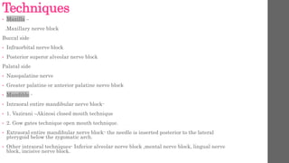
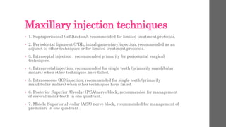
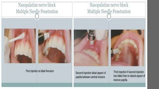
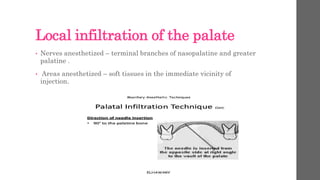
This document discusses various techniques for local anesthesia in dentistry. It describes different nerve blocks for anesthetizing specific regions of the maxilla and mandible, including the posterior superior alveolar nerve block, middle superior alveolar nerve block, and anterior superior alveolar (infraorbital) nerve block. It also covers maxillary injection techniques such as supraperiosteal injections and palatal anesthesia techniques like the greater palatine nerve block.
a brief for local anesthesia techniques in dentistry; practical for after perusing the techniques before. Used reference was MALAMED handbook of oral anesthesia.
This document discusses various local anesthesia techniques used in dentistry. It begins by introducing regional anesthesia techniques like field blocks, nerve blocks, and local infiltration. It then describes specific injection techniques for the maxilla including posterior superior alveolar nerve blocks, middle superior alveolar nerve blocks, anterior superior alveolar/infraorbital nerve blocks, nasopalatine nerve blocks, greater palatine nerve blocks, and maxillary nerve blocks. Mandibular injection techniques discussed include inferior alveolar nerve blocks and mental nerve blocks. The document provides details on appropriate needle size, insertion points, depths, and areas anesthetized for each technique.
This document provides information on various techniques of local anesthesia. It begins by classifying local anesthesia techniques into topical, infiltration, and nerve block. For infiltration anesthesia, it describes soft tissue and intra-bony infiltration techniques for the maxilla and mandible. For nerve block anesthesia, it details techniques for blocking specific nerves of the maxilla like the posterior superior alveolar nerve and mandible like the inferior alveolar nerve. Factors influencing choice of technique and contraindications are also summarized. The document provides in-depth descriptions of performing local anesthesia techniques for the maxilla and mandible.
This document provides information on the maxillary nerve block technique. It begins with an overview of the trigeminal nerve and its branches, including the maxillary nerve. It then describes the course and branches of the maxillary nerve in detail. It discusses the pterygopalatine ganglion and its branches. The document outlines different maxillary nerve block techniques including posterior superior alveolar, anterior superior alveolar, greater palatine, and nasopalatine nerve blocks. It concludes with a description of a maxillary nerve block and intraligamentary anesthesia technique.
This document discusses various techniques for blocking nerves that innervate the oral cavity, including both intraoral and extraoral approaches. Intraoral techniques include local infiltration of nerve endings, blocking terminal branches through paraperiosteal or intraosseous injections, and blocking specific nerves like the anterior and middle superior alveolar nerves. Extraoral techniques allow blocking of nerves like the anterior and middle superior alveolar nerves from outside the mouth. Each technique aims to anesthetize specific nerves, areas, and teeth while avoiding pain and complications from the injections.
This document describes various techniques for mandibular nerve blocks, including the inferior alveolar nerve block, buccal nerve block, Gow-Gates technique, Vazirani-Akinosi closed mouth mandibular block, mental nerve block, and incisive nerve block. It provides details on the target sites, techniques, and areas of anesthesia achieved for each type of mandibular nerve block.
This document discusses various techniques for maxillary nerve blocks and anesthesia. It begins by outlining the maxillary nerve and its branches, then describes 10 different injection techniques in detail. These include supraperiosteal, posterior superior alveolar, anterior superior alveolar, middle superior alveolar, greater palatine, nasopalatine, and maxillary nerve blocks. Each technique section explains the nerves anesthetized, areas anesthetized, anatomical landmarks, advantages and disadvantages, and procedural steps. Images are provided to illustrate the injection sites and anatomical relationships.
Local aneasthesia techniques which are to be performed extraorally when the conventional intraoral approches for local anaesthesia cant be performed.
Very useful for dental Practioners
This document provides information on various nerve block techniques used in dentistry. It discusses the history of nerve blocks and describes techniques such as local infiltration, field blocks, and nerve blocks. It then covers specific nerve blocks like posterior superior alveolar nerve block, anterior superior alveolar nerve block, greater palatine nerve block, nasopalatine nerve block, maxillary nerve block, inferior alveolar nerve block, and mental nerve block. For each block, it outlines the areas anesthetized, landmarks, target area, needle placement, amount of solution used, and potential complications. The document aims to inform dental practitioners on best practices for different nerve block techniques.
The document discusses mandibular infiltration anesthetic techniques. It outlines the necessary equipment, including the chair, dentist position, syringe and needle. It describes the areas and nerves to be anesthetized, including landmarks. It explains the point of needle insertion and direction of injection, as well as how to check that the anesthesia was effective.
This document discusses techniques for mandibular anesthesia. It begins by noting the lower success rate of mandibular blocks compared to maxillary blocks, due to bone density and access to nerve trunks. It then reviews different types of mandibular nerve blocks and focuses on the inferior alveolar nerve block technique. This technique anesthetizes multiple nerves but has the highest failure rate. Proper deposition of local anesthetic solution within 1mm of the nerve trunk is needed for success. Precautions, alternatives, indications, contraindications and complications are also outlined.
Local anesthesia in dentistry : RECENT ADVANCESPooja Jayan
This document provides an overview of local anesthesia. It begins with definitions of local anesthesia and discusses its history from the isolation of cocaine in 1859 to the development of modern local anesthetics like lidocaine. It describes the ideal properties, theories of action, classification, composition, maximum doses, armamentarium, techniques for maxillary and mandibular injections, and potential complications. The key information is that local anesthesia temporarily interrupts nerve conduction to produce loss of sensation in a circumscribed area, allowing for painless dental procedures.
This document provides information on various local anesthesia techniques used in dentistry. It begins with an introduction to regional anesthesia, including field blocks, nerve blocks, and local infiltration. It then describes different local anesthesia injection techniques such as supraperiosteal, intraligamentary, intraosseous, and intraseptal injections. The document proceeds to explain specific maxillary and mandibular injection techniques including posterior superior alveolar nerve block, anterior superior alveolar nerve block, greater palatine nerve block, and others. It concludes with a brief section on recent advancements in local anesthesia.
This document discusses various nerve blocks for anesthetizing different areas innervated by branches of the mandibular nerve, including the inferior alveolar nerve block. It provides details on the nerves anesthetized, areas anesthetized, techniques, indications, and complications for the inferior alveolar nerve block as well as mental nerve block, incisive nerve block, long buccal nerve block, and lingual nerve block. Modifications to the classical inferior alveolar nerve block technique including those of Clarke and Holmes and Gow Gates are also summarized.
The document discusses various techniques for mandibular nerve anesthesia, including both intraoral and extraoral approaches. Intraoral techniques covered include the inferior alveolar nerve block (both direct and indirect techniques), lingual nerve block, buccinator nerve block, mental nerve block, incisive nerve block, infiltration of terminal branches, and submucosal infiltration. Extraoral techniques discussed are the mandibular nerve block, mental nerve block, infraorbital nerve block, and inferior alveolar nerve block. The document then provides more detailed descriptions and illustrations of specific techniques such as the inferior alveolar nerve block, Vazirani-Akinosi closed mouth technique, Gow-Gates mandibular
This document discusses various techniques for local anesthesia in the maxilla. It begins by defining local infiltration, field block, and nerve block injections. It then describes specific maxillary injections including supraperiosteal, intraligamentary, intrapulpal, intraosseous, intraseptal, and various nerve blocks of the anterior superior alveolar, middle superior alveolar, and greater palatine nerves. For each technique, it provides indications, contraindications, anatomy anesthetized, complications and failure causes. The anterior superior alveolar nerve block is described in detail as the most common and effective maxillary nerve block.
This document discusses local anesthesia and pain control techniques for pediatric dentistry. It defines pain and anesthesia and covers various local anesthesia techniques including topical anesthesia, infiltration, nerve blocks, and supplemental injection techniques. It provides details on the contents of local anesthesia carpules, how local anesthetics are metabolized in the body, recommended dosages, and complications. The goal is to effectively manage pain for dental procedures in children.
This document discusses methods for managing dental pain, including through local anesthesia. It describes techniques for local infiltration anesthesia and regional nerve blocks to block painful stimuli. When inflammation is present, supplemental techniques may be needed like interseptal injections between teeth to complete anesthesia of pulp tissue so vital pulp can be removed painlessly. Intrapulpal injections directly into exposed pulp tissue can also profoundly anesthetize it.
Indian Dental Academy: will be one of the most relevant and exciting training center with best faculty and flexible training programs for dental professionals who wish to advance in their dental practice,Offers certified courses in Dental implants,Orthodontics,Endodontics,Cosmetic Dentistry, Prosthetic Dentistry, Periodontics and General Dentistry.
The document discusses techniques for mandibular anesthesia. It focuses on the inferior alveolar nerve block, which has the highest success rate of mandibular nerve blocks but still a failure rate of 15-20%. The inferior alveolar nerve block anesthetizes multiple areas including the mandibular teeth and soft tissues, but does not provide complete mandibular anesthesia and may require supplemental techniques. Precise deposition of the local anesthetic solution within 1mm of the inferior alveolar nerve is required for success.
This document outlines emergency drugs used in dentistry. It discusses different medical emergencies that may occur during dental procedures like syncope, hypoglycemia, and anaphylactic shock. It categorizes emergency drugs into injectable and non-injectable types. Injectable drugs are further divided into primary (essential) and secondary (non-essential) categories. Primary injectables include epinephrine, antihistamines, anticonvulsants, and narcotic antagonists. Secondary injectables include analgesics, vasopressors, corticosteroids, and antihypoglycemics. Non-injectable emergency drugs discussed are oxygen, vasodilators, respiratory stimulants, antihypoglyce
Techniques for local anasthesia in dentistryMohammed Rhael
This document discusses various techniques for local anesthesia in dentistry. It describes topical, infiltration, and regional (block) techniques. For the maxilla, it outlines infiltration and block techniques for different tooth regions, including posterior superior alveolar nerve block and infraorbital block. For the mandible, it focuses on inferior alveolar nerve block and long buccal nerve block. It provides guidance on performing various block techniques and notes variations between patients.
Technique of maxillary anesthesia which includes Greater Palatine Nerve Block and Incisive Nerve Block. The reference is of LA Book by Malamed.
Hope you find it useful.
Please like and share.
This document discusses techniques for maxillary nerve blocks. It begins by describing the anatomy of the maxillary nerve and its branches that supply sensation to the upper teeth and gums. It then explains in detail the posterior superior alveolar nerve block technique, including patient and dentist positioning, needed equipment, landmarks, injection technique, and confirming anesthesia. Finally, it provides a brief overview of the maxillary nerve block technique to anesthetize the main trunk of the maxillary nerve.
This document provides information on local anesthetics used in dentistry. It discusses the components of the local anesthetic armamentarium including syringes, needles, and cartridges. It describes different types of syringes and needles and their appropriate uses. It also discusses the components and handling of local anesthetic cartridges. The document outlines the mechanisms of local anesthetics and vasoconstrictors as well as considerations for maximum safe dosing.
The document discusses various mandibular anesthetic techniques including infiltration anesthesia of the anterior labial, anterior lingual, and long buccal regions as well as inferior alveolar and lingual nerve block. It describes the relevant anatomy, patient positioning, needle selection, injection sites, and steps for each technique. The goal is to effectively anesthetize the nerves supplying sensation to the mandibular teeth and surrounding tissues.
This document summarizes the anterior middle superior alveolar nerve block and palatal approach anterior superior alveolar nerve block techniques. It describes the nerves anesthetized, areas anesthetized, indications, contraindications, advantages, disadvantages, techniques, and signs of successful anesthesia for each block. Both techniques anesthetize the anterior maxillary teeth using a single injection site in the palate and have advantages of minimizing anesthetic volume and needle punctures compared to individual injections.
The inferior alveolar nerve block anesthetizes the inferior alveolar nerve and its branches, providing anesthesia to the mandibular teeth, chin, lower lip, and parts of the tongue. A long needle is inserted into the mucosa on the lingual side of the ramus to anesthetize the inferior alveolar nerve as it passes downward. When bone is contacted, local anesthetic is slowly deposited over 60 seconds while aspirating. Successful anesthesia is confirmed through subjective symptoms like lip tingling and objective tests showing no response to pulp stimulation. Complications can include hematoma, trismus, or transient facial paralysis if the facial nerve is contacted.
oral surgery - techniques of local anesthesia powerpoint Mustafatj1
Credit goes to meryem hilal for making this powerpoint
Check this youtube channel if you want to help me back.
https://youtube.com/user/mustafabk1
Make sure to spread love.
I hope this powerpoint helps you in your research.
Thanks
This document provides information on three types of superior alveolar nerve blocks: posterior, middle, and anterior. It describes the nerves anesthetized, areas anesthetized, indications, contraindications, techniques, and complications for each type of block. The posterior block anesthetizes the posterior superior alveolar nerve and its branches, providing anesthesia for maxillary molars. The middle block targets the middle superior alveolar nerve for premolars. The anterior block anesthetizes the anterior superior alveolar, middle superior alveolar, and infraorbital nerves, providing wide anesthesia from incisors to premolars and surrounding tissues. All three techniques aim to deposit local anesthetic in close proximity to the
This document discusses various techniques for local anesthesia in the maxillary and mandibular regions. It provides detailed descriptions of nerve blocks including the posterior superior alveolar nerve block, middle superior alveolar nerve block, anterior superior alveolar nerve block, greater palatine nerve block, nasopalatine nerve block, and maxillary nerve block. It also briefly mentions techniques used for mandibular anesthesia such as the inferior alveolar nerve block. The goal of these techniques is to anesthetize specific teeth, gums, lips, and other tissues by blocking select nerves that innervate the target areas. Precise needle placement and aspiration are important to minimize risks like hematoma formation.
This document discusses techniques for local anesthesia in dentistry. It describes four main types of local anesthesia: topical, infiltration, field block, and regional block. Infiltration anesthesia involves depositing the anesthetic solution near terminal nerve fibers to infiltrate the tissue and anesthetize a localized area. It is commonly used for the front of the maxilla and mandible. Regional block anesthesia deposits the solution near the main nerve trunk to anesthetize a larger area. The document provides details on specific infiltration and regional block techniques for anesthetizing different areas of the maxilla and palate.
This document discusses various techniques for mandibular anesthesia. It begins by introducing the classical inferior alveolar nerve block and its limitations. Alternative techniques are then presented, including the indirect technique, Clarke and Holmes' method, Sargenti's method, the Gow-Gates technique, the Vazirani-Akinosi closed mouth block, lingual nerve anesthesia, and extra-oral approaches. Each technique is described in detail, outlining the relevant anatomy, indications, contraindications, advantages, disadvantages, and procedural steps. Common causes of failure are also reviewed, such as deposition of anesthetic in the wrong site due to anatomical variations or faulty technique.
The document provides information on various types of maxillary anesthesia techniques:
1. Supraperiosteal injection is commonly used to anesthetize one or two maxillary teeth and soft tissues. It has a low risk of intravascular administration.
2. Posterior superior alveolar nerve block anesthetizes maxillary molars and buccal tissues through the posterior superior alveolar nerve. It has a high success rate but risks hematoma formation.
3. Nasopalatine nerve block provides wide palatal soft tissue anesthesia using a minimum volume of local anesthetic, minimizing the need for multiple injections.
Gow gates & vazirani akinosi technique of nervePOOJAKUMARI277
The document summarizes two techniques for mandibular nerve blocks - the Gow-Gates technique and the Vazirani-Akinosi closed mouth technique.
The Gow-Gates technique involves injecting the anesthetic at the neck of the condyle using intraoral and extraoral landmarks to block the mandibular nerve. It provides anesthesia of the mandibular teeth and surrounding soft tissues with a single injection. The Vazirani-Akinosi technique is done with the patient's mouth closed by inserting the needle through the mucosa at the level of the maxillary molar junction to block the mandibular nerve. Both techniques effectively anesthetize the mandibular region for dental
brief description on posterior superior alveolar nerve block.
its uses in dentistry, technique and action. locating PSA nerve is easy and this is the most used nerve block in dentistry.
This document discusses various nerve blocks used for mandibular anesthesia, including the inferior alveolar nerve block, Gow-Gates technique, Vazirani-Akinosi closed-mouth block, and mental nerve block. It describes the nerves anesthetized, areas anesthetized, indications, contraindications, landmarks, techniques, and potential complications for each block. The inferior alveolar nerve block and Gow-Gates technique provide the most extensive mandibular anesthesia while the mental nerve block specifically targets the area innervated by the mental nerve. Proper administration of these blocks requires identifying the correct anatomical landmarks and depositing the local anesthetic in the appropriate location near the target nerve.
This document discusses various nerve blocks of the maxillary nerve including the posterior superior alveolar nerve block, middle superior alveolar nerve block, anterior superior alveolar nerve block, anterior middle superior alveolar nerve block, maxillary nerve block, greater palatine nerve block, and nasopalatine nerve block. It provides details on the nerves anesthetized, anatomical landmarks, techniques, advantages and disadvantages for each nerve block. The goal of these nerve blocks is to provide pulpal and soft tissue anesthesia of the maxillary teeth and surrounding structures for dental procedures.
This document discusses various nerve block techniques used in dentistry. It describes the areas anesthetized, indications, contraindications, landmarks, and complications for different types of nerve blocks including:
- Maxillary nerve blocks (supraperiosteal, posterior superior alveolar, anterior superior alveolar)
- Palatal nerve blocks (greater palatine, nasopalatine)
- Mandibular nerve blocks (inferior alveolar, Gow-Gates, mental, lingual)
- Extraoral techniques for maxillary and mandibular nerve blocks
It also defines complications of local anesthesia as any deviation from the normal expected outcome, classifying them as local, systemic,
Mandibular Nerve Block - By Dr Saikat Saha Dr Saikat Saha
Mandibular nerve block techniques in short for Dental Surgeons. Mandibular nerve blocks are very important for all dental surgeons as it becomes a part and parcel of all dental and oral surgeons. This presentation will be useful for students of dentistry and doctors.
This document summarizes various local anesthesia techniques used in dentistry. It describes the basic atraumatic injection technique in 19 steps and provides details on various local anesthesia techniques including topical anesthetics, local infiltration, field blocks, and nerve blocks. It also discusses newer trends in pain control during local anesthesia administration such as safety syringes, computerized delivery systems, and vibration devices. Complications of local anesthesia and contraindications are briefly covered as well.
This document provides information on maxillary anesthesia techniques. It discusses the anatomy of the maxillary nerve and its branches that innervate the maxilla. Several injection techniques are described including posterior superior alveolar nerve block, middle superior alveolar nerve block, anterior superior alveolar nerve block, and greater palatine nerve block. Each technique lists the nerves anesthetized, areas anesthetized, indications, contraindications, and steps for administration. Complications and alternatives are also noted for some techniques.
Dr, Kathirvel Gopalakrishnan
M.D.S (OMFS)
Presentation on Maxillary nerve block which helps for a quick refresh.
Applied aspects described well and slides contains images for easy understanding of the subject.
The document discusses various regional anesthesia techniques including infiltration, field block, nerve block, and nerve blocks for specific nerves of the head and neck. It covers the basic injection technique, evaluation of patients, contents to be covered, and descriptions of different regional anesthesia methods with their indications, contraindications, techniques, and complications.
Similar to Techniques of local anesthesia [autosaved] (20)
Strategies for Effective Upskilling is a presentation by Chinwendu Peace in a Your Skill Boost Masterclass organisation by the Excellence Foundation for South Sudan on 08th and 09th June 2024 from 1 PM to 3 PM on each day.
Leveraging Generative AI to Drive Nonprofit InnovationTechSoup
In this webinar, participants learned how to utilize Generative AI to streamline operations and elevate member engagement. Amazon Web Service experts provided a customer specific use cases and dived into low/no-code tools that are quick and easy to deploy through Amazon Web Service (AWS.)
Walmart Business+ and Spark Good for Nonprofits.pdfTechSoup
"Learn about all the ways Walmart supports nonprofit organizations.
You will hear from Liz Willett, the Head of Nonprofits, and hear about what Walmart is doing to help nonprofits, including Walmart Business and Spark Good. Walmart Business+ is a new offer for nonprofits that offers discounts and also streamlines nonprofits order and expense tracking, saving time and money.
The webinar may also give some examples on how nonprofits can best leverage Walmart Business+.
The event will cover the following::
Walmart Business + (https://business.walmart.com/plus) is a new shopping experience for nonprofits, schools, and local business customers that connects an exclusive online shopping experience to stores. Benefits include free delivery and shipping, a 'Spend Analytics” feature, special discounts, deals and tax-exempt shopping.
Special TechSoup offer for a free 180 days membership, and up to $150 in discounts on eligible orders.
Spark Good (walmart.com/sparkgood) is a charitable platform that enables nonprofits to receive donations directly from customers and associates.
Answers about how you can do more with Walmart!"
it describes the bony anatomy including the femoral head , acetabulum, labrum . also discusses the capsule , ligaments . muscle that act on the hip joint and the range of motion are outlined. factors affecting hip joint stability and weight transmission through the joint are summarized.
This document provides an overview of wound healing, its functions, stages, mechanisms, factors affecting it, and complications.
A wound is a break in the integrity of the skin or tissues, which may be associated with disruption of the structure and function.
Healing is the body’s response to injury in an attempt to restore normal structure and functions.
Healing can occur in two ways: Regeneration and Repair
There are 4 phases of wound healing: hemostasis, inflammation, proliferation, and remodeling. This document also describes the mechanism of wound healing. Factors that affect healing include infection, uncontrolled diabetes, poor nutrition, age, anemia, the presence of foreign bodies, etc.
Complications of wound healing like infection, hyperpigmentation of scar, contractures, and keloid formation.
Reimagining Your Library Space: How to Increase the Vibes in Your Library No ...Diana Rendina
Librarians are leading the way in creating future-ready citizens – now we need to update our spaces to match. In this session, attendees will get inspiration for transforming their library spaces. You’ll learn how to survey students and patrons, create a focus group, and use design thinking to brainstorm ideas for your space. We’ll discuss budget friendly ways to change your space as well as how to find funding. No matter where you’re at, you’ll find ideas for reimagining your space in this session.
हिंदी वर्णमाला पीपीटी, hindi alphabet PPT presentation, hindi varnamala PPT, Hindi Varnamala pdf, हिंदी स्वर, हिंदी व्यंजन, sikhiye hindi varnmala, dr. mulla adam ali, hindi language and literature, hindi alphabet with drawing, hindi alphabet pdf, hindi varnamala for childrens, hindi language, hindi varnamala practice for kids, https://www.drmullaadamali.com
Exploiting Artificial Intelligence for Empowering Researchers and Faculty, In...Dr. Vinod Kumar Kanvaria
Exploiting Artificial Intelligence for Empowering Researchers and Faculty,
International FDP on Fundamentals of Research in Social Sciences
at Integral University, Lucknow, 06.06.2024
By Dr. Vinod Kumar Kanvaria
This presentation was provided by Steph Pollock of The American Psychological Association’s Journals Program, and Damita Snow, of The American Society of Civil Engineers (ASCE), for the initial session of NISO's 2024 Training Series "DEIA in the Scholarly Landscape." Session One: 'Setting Expectations: a DEIA Primer,' was held June 6, 2024.
A review of the growth of the Israel Genealogy Research Association Database Collection for the last 12 months. Our collection is now passed the 3 million mark and still growing. See which archives have contributed the most. See the different types of records we have, and which years have had records added. You can also see what we have for the future.
2. Techniques
• Maxilla –
.Maxillary nerve block
Buccal side
• Infraorbital nerve block
• Posterior superor alveolar nerve block
Palatal side
• Nasopalatine nerve
• Greater palatine or anterior palatine nerve block
• Mandible -
• Intraoral entire mandibular nerve block-
• 1. Vazirani –Akinosi closed mouth technique
• 2. Gow gates technique open mouth technique.
• Extraoral entire mandibular nerve block- the needle is inserted posterior to the lateral
pterygoid below the zygomatic arch.
• Other intraoral techniques- Inferior alveolar nerve block ,mental nerve block, lingual nerve
block, incisive nerve block.
3. Maxillary injection techniques
• 1. Supraperiosteal (infiltration), recommended for limited treatment protocols.
• 2. Periodontal ligament (PDL, intraligamentary)injection, recommended as an
adjunct to other techniques or for limited treatment protocols.
• 3. Intraseptal injection , recommended primarily for periodontal surgical
techniques.
• 4. Intracrestal injection, recommended for single teeth (primarily mandibular
molars) when other techniques have failed.
• 5. Intraosseous (IO) injection, recommended for single teeth (primarily
mandibular molars) when other techniques have failed.
• 6. Posterior Superior Alveolar (PSA)nerve block, recommended for management
of several molar teeth in one quadrant.
• 7. Middle Superior alveolar (ASA) nerve block, recommended for management of
premolars in one quadrant .
4. • 8. Anterior Superior Alveolar (ASA) nerve block, recommended for management of anerior
teeth in one quadrant .
• 9. Maxillary (V2, second division) nerve block , recommended for extensive buccal, palatal
and pulpal management in one quadrant.
• 10. Greater (anterior) palatine nerve block, recommended for palatal soft and osseous
tissue treatment distal to the canine in one quadrant .
• 11. Nasopalatine nerve block, recommended for palatal soft and osseous tissue
management from canine to canine bilaterally.
• 12. Anterior middle superior alveolar (AMSA) nerve block , recommended for extensive
management of anterior teeth , palatal and buccal soft and hard tissues.
• 13. Palatal approach – anterior superior alveolar nerve (P-ASA) nerve block ,
recommended for treatment of maxillary anterior teeth and their palatal and facial soft
and hard tissues.
5.
6. Supraperiosteal injection
• The supraperiosteal injection, more commonly (but incorrectly) called local infiltration.
• indications.:-
• 1. pulpal anesthesia of the maxillary teeth when treatment is limited to one or two teeth.
2. soft tissue anesthesia when indicated for surgical procedures in a circumscribed.
Contraindications :-
1. Infections or acute inflammation in the area of injection.
2. Dense bone covering the apices of teeth (can be determined only trial and error; most likely over the
permanent maxillary first molar in children, as its apex may be located beneath the zygomatic bone, which is
relatively dense). The apex of an adult’s central incisor may aso be located beneath denser bone(e.g., of the
nose), thereby increasing the failure rate (although not significantly).
Advantages:-
High success rate (>95%)
Technically easy injection.
Usually entirely atraumatic
Disadvantages:-
Not for large areas due to multiple needle insertions and necessity to administer larer total volumes of LA.
7. Positive aspiration- Negligible, but possible (1%)
Alternative – PDL, IO, regional nerve block
• A 27 gauge short needle is recommended.
• Area of insertion : height of the mucobuccal fold above the apex of the tooth being anesthetized
• Landmarks:-
• A. mucobuccal fold
• B. crown of the tooth
• C. root contour of the tooth.
• Orientation of the bevel – toward bone.
• Signs and symptoms
• 1. subjective :- feeling of numbness in the area of administration.
• 2. objective : use of electrical pulp testing (EPT) with n response from tooth with maximal EPT output (80/80)
• 3. absence of pain during treatment.
• Safety features –
• 1. minimal risk of intravascular administration.
• 2. slow injection of anesthetic ; aspiration.
• Complication – pain on needle insertion with the needle tip against the periosteum. To correct: withdraw and
reisnsert it farther from the periosteum.
8. Posterior Superior Alveolar Nerve Block
• The posterior superior alveolar (PSA) nerve block is a commonly used dental nerve block.
Although it is a highly successful technique (>95%).
• Other common names :- Tuberosity block, zygomatic block
• Nerves anesthetized :- posterior superior alveolar and branches.
• Area anesthetized.
1. Pulps of the maxillary third, second and first molars (entire tooth =72%; mesiobuccal root
of the mesiobuccal root of the maxillary first molar not anesthetized = 28%).
2. Buccal periodontium and bone overlying these teeth .
Indications
1. When treatment involves 2 or more maxillary molars.
2. When supraperiosteal injection is contraindicated (e.g., with infection or acute
inflammation ).
3. When supraperiosteal injection has proved ineffective.
9. • Advantage :-
1. atraumatic ; when administered properly , no pain is experienced by the patient receiving the
PSA because of the relatively large area soft tissue into which the local anesthetic is deposited
and the fact that bone is no contacted .
2. high success rate (>95%).
3. minimum number of necessary injections
• A. one injection compared with opinion of three infiltrations.
4. minimizes the total volume of local anesthetic solution administered
• A. equivalent volume of anesthetic solution necessary for three supraperiosteal injections.=1.8
mL.
• Disadvantages
• 1. Risk of hematoma,usully diffuse , discomforting and visually embarrassing to patient.
• 2. technique somewhat arbitrary: no bony landmarks during insertion.
• 3. second injection necessary for 1st molar (mesiobuccal root) in 28% of patients.
• Alternatives
• 1. supraperiosteal or PDL injections for pulpal and root anesthesia.
• 2. infiltration for the buccal periodontium and hard tissues.
• 3. maxillary nerve block.
10. Technique-
• A 27 gauge short needle recommended.
• Area of insertion: height of the mucobuccal old above the maxillary second molar.
• Target area: PSA nerve – posterior , superior and medial to the posterior border of the
maxilla.
Landmarks:
• A. mucobuccal fold
• B. maxillarytuberosity
• C. zygomatic process of the maxilla.
Orientation of the bevel : toward bone during the injection. If bone is accidentally touched
sensation is less unpleasant .
Procedure:- left PSA – right handed administrator should sit at 10 o’clock position facing the
patient
Right PSA – a right PSA – right handed administrator should sit at the 8 o’clock position
facing the patient.
• Insert the needle into the height of the mucobuccal fold over the second molar . Advance the
needle slowly in an upward, inward and backard direction in one movement not three.
11.
12. • A. aspirate in two planes .
• 1. rotate the syringe barrel (needle bevel) one fourth turn and reaspirate.
• 2. if both aspirations are negative :-
• A. slowly over 30-60 seconds , deposit 0.9 to 1.8mL of anesthetic solution.
• B. aspirate sveral additional times(in one plane )during drug administration.
• C. the PSA injection is normally atraumatic because of large tissue space available.
• Signs and symptoms
• 1. subjective : usually none.
• 2. objective :use of electrical pulptesting with no response form tooth with maximal EPT output (80/80)
• Absence of pain during treatment.
• Safety features.
• 1. slow injection, repeated aspirations.
• No anatomic safety features to prevent overinsertion of the needle; therefore careful observation is
necessary.
• Precaution – depth of needle penetration should be checked: due to hematoma.
• Failures:- needle too lateral, needle not high enough, needle too far posterior.
• Complications- hematoma , mandibular anesthesia ( the mandibular division of the fifth cranial nerve (V3 )
is located lateral to the PSA.
13. Middle Superior Alveolar Nerve
• The MSA block is indicated for procedures on premolars and on the mesiobuccal root of the
maxillary first molar. The success rate of MSA nerve block is high.
Nerves anesthetized – middle superior alveolar and terminal branches.
Areas anesthetized –
• 1. pulps of the maxillary first and second premolars.
• 2. buccal periodontal tissues and bone over these same teeth.
Indications-
• Where ASA fails.
• Dental procedures involving both maxillary premolars only.
Contraindications-
• Infection or inflammation
• Where MSA absent .
Advantages – minimizes number of injections and volume of solution.
Disadvantages-
• None.
14. • Positive aspiration- negligible (>3%).
• Alternative-
• 1. local infiltration (supraperiosteal), PDL, IO injections
• 2. ASA nerve block for 1st and 2nd premolar and the mesiobuccal root of the 1st molar.
• Technique
• 1. 27 gauge short or long needle is recommended.
• 2. area of insertion : height of mucobuccal fold above maxillary second premolar.
• 3. target area: maxillary bone above the apex of the maxillary second premolar.
• Landmark – mucobuccal fold above the maxillary second premolar.
• Orientation of the bevel : toward bevel.
• Procedure:-
• For right MSA nerve block , a right –handed administrator – 10 o’clock position.
• For left MSA nerve block, a right- handed administrator – 8 or 9 o’clock position.
• Slowly deposit 0.9 to 1.2 mL (1/2 to 2/3rd catridge) of solution( approximately 30-40 seconds).
• Signs and Symptoms-
• 1. Subjective – upper lip numb.
• 2. objective- use of electrical pulp testing with no response from tooth with maximal EPT output (80/80).
• 3. absence of pain during treatment.
15. • Safety Feature- relative avascular area, anatomically safe.
• Precautions- to prevent pain, do not insert too close to the periosteum and do not inject too
close to the periosteum and do not inject too rapidly;the MSA should be an atraumatic
injection.
• Failures of anesthesia-
• 1. anesthetic solution not deposited high above the apex of the second premolar , to correct-
check radiographs and increase the depth of penetration .
• 2.deposition of solution too far from maxillary bone with the needle placed in tissues lateral
to the height of mucobucal fold , to correct: reinsert at the height of the mucobuccal fold.
• 3. Bone of zygomatic arch at the site of injection preventing the diffusion of anesthetic , to
correct : superiosteal,ASA or PSA injection in place of the MSA.
• Complications – hematoma (rare).
16. Anterior Superior Alveolar Nerve Block
(infraorbital nerve block)
• There is a general lack of experience with this highly successful and extremely safe technique.
Dentists fear from using this block due to injury to the patient’s eye.
• Nerve anesthetized-
1. anterior superior alveolar.
2. middle superior alveolar .
3. infraorbital nerve
• A. inferior palpebral
• B. lateral nasal
• C. Superior labial.
• Areas anesthetized-
• 1. pulps of maxillary central incisor through the canine on the injected side.
• 2. in about 72% of pateints, pulps of the maxillary premolars and mesiobuccal root of the first
molar .
• 3. lower eyelid, lateral aspect of the nose, upper lip.
17. • Indications :-
• Dental procedures involving more than two maxillary teeth and their overlying buccal tissues.
• Inflammation or infection which contraindicates supraperiosteal injection .
• When superaperiosteal injections have been ineffective because of dense cortical bone.
• Contraindicates:-
• 1. discrete treatment areas (one or two teeth only ; supraperiosteal)
• 2. hemostasis of localized areas, when desirable, cannot be achieved with injection; local infiltration into the
treatment area is indicated.
• Advantages –
• 1. comparatively simple technique.
• 2. comparatively safe; minimizes the volume of solution used and no of needle punctures also.
• Disadvantages :-
• 1. psychological :-
• A. administer – initial fear of injury to the patient’s eye (experience with the technique leads to confidence.
• B. patient – extraoral approach t infraorbital nerve may prove disturbing; however, intraoral techniques are
rarely a problem.
• C. anatomic : difficulty defining landmarks.
• Positive aspiration – 0.7%
• Alternatives – 1. superaperiosteal, PDL or IO injection for each tooth., 2. infiltration 3. maxillary nerve block.
18. • Technique :-
• A 25 or 27 gauge long needle is recommended.
• Area of insertion : height of the mucobuccal fold directly over the first premolar (shortest route)
• Target area- infraorbital foramen (below the infraorbital notch).
• Landmarks – a. mucobuccal fold, b. infraorbital notch , c. infraorbital foramen.
• Orientation of the bevel – toward bone.
• Slowly deposit 0.9 to 1.2 mL over 30-40 seconds.
• Signs and symptoms
• 1. Subjective : tingling and numbness of lower eyelid, side of the nose and upper lip indicate
anesthesia of the infraorbital nerve , not the ASA or MSA nerve.(soft tissue anesthesia develops
almost instantly as the anesthetic is being administered).
• 2. subjective and objective : numbness in the teeth and soft tissues along the distribution of the
ASA and MSA nerves (developing within 3 to 5 minutes if pressure is maintained over the
injection site).
• 3. objective – use of electrical pulp testing with no response from tooth with maximal EPT
output (80/80).
• 4. absence of pain during treatment.
19. • Safety Features. 1. needle contact with bone at the roof of the infraorbital foramen prevents
inadvertent overisertion and possible puncture of the orbit.
• 2. a finger positioned over the infraorbital foramen helps direct the needle toward the
foramen.
• A. the needle should not be palpable . If it is felt , then its path is too superficial (away from
the bone). If this occurs , withdraw the needle slightly and redirect it towards the target
area.
• B. in most patients its not palpable unless its superficial, however with less well-developed
,its palpable.
• Precaution- pain on insertion, tearing of periosteum, to prevent overinsertion exert finger
pressure over the infraorbital foramen.
• Failures of anesthesia- needle contacting bone below (inferior to) the infraorbital foramen.
• Needle deviation medial or lateral to the infraorbital foramen.
• Complications.-
hematoma rarely , to manage apply pressure on soft tissue foramen for 2-3 minutes.
20. Palatal Anesthesia
• Anesthesia of the hard palate is necessary for dental procedures involving manipulation of
palatal soft or hard tissues but it proves to be a very traumatic experience.
• 1. Greater Palatine Nerve Block/ Anterior palatine nerve block - useful for procedures
involving the palatal soft tissues distal to the canine.
• Nerve anesthetized- Greater palatine
• Areas Anesthetized- the posterior position portion of the hard palate and its overlying soft
tissues, anteriorly as far as the first premolar and medially to the midline.
• Indications – 1. palatal soft tissue anesthesia, subgingival restoration.
• 2. for pain control during periodontal or oral surgical procedures involving palatal soft and
hard tissues.
• Contraindications- inflammation or infection , smaller areas of therapy .
• Advantages- minimize needle penetrations and volume of solution.
• Minimizes patient discomfort.
• Disadvantages- 1. no hemostasis except in immediate area of injections.
• 2. potentially traumatic.
21. • Positive aspiration- less than 1%.
• Alternatives- local infiltration into specific regions, maxillary nerve block.
• Technique-
• A 27 gauge short needle is recommended.
• Area of insertion – soft tissue slightly anterior to the greater palatine foramen.
• Target area: greater (anterior) palatine nerve as it passes anteriorly between soft tissues and
bone of the hard palate.
• Landmarks- greater palatine foramen and junction of the maxillary alveolar process and
palatine bone.
• Path of insertion: advance the syringe from the opposite side of mouth at a right angle to target
area.
• Orientation of the bevel- toward the palatal soft tissues
• Technique- 1) for right greater palatine nerve block, a right handed administrator – 7 or 8
o’clock. 2) left greater palatine nerve block, a right handed administrator – 11 0’clock.
• Slowly deposit not more than 1/4th to 1/3rd of catridge. (0.45 to 0.6 mL )
• Signs and symptoms – 1. subjective : numbness in the posterior portion of the palate.
• 2. objective: no pain during dental therapy.
22. • Safety Features – 1. contact with bone . 2. aspiration.
• Precautions- do nt enter the greater palatine canal. Although this is not hazardous, there is
no reason to enter the canal for this technique to be successful.
• Failures of anesthesia – 1) the greater palatine nerve block is not a technically difficult
injection to administer , incidence of success is well above 95%.
• 2) if local anesthetic is deposited too far anterior to the foramen, adequate soft tissue
anesthesia may not occur in the palatal tissues posterior to the site of injection (partial
success).
• 3) anesthesia on the palate in the area of the maxillary first premolar may prove inadequate
because of overlapping fibres from the nasopalatine nerve (partial success).
• A. to correct :- local infiltration may be necessary as a supplement in the area of inadequate
anesthesia.
• Complications- few of significance , ischemia and necrosis of soft tissues when highly
concentrated vasoconstricting solution used for hemostasis over a prolonged period bcoz
Norepinephrine should never be used for hemostasis on the palatal soft tissues.
• Hematoma is possible but rare due to firm adherence of palatal tissues to underlying bone.
• Uncomfortable to patients.
23.
24. Nasopalatine Nerve Block
• Potentially highly traumatic injections. 2 techniques- 1. single tissue penetration 2. multiple
needle penetration.
• Other common names- Incisive nerve block, sphenopalatine nerve block.
• Nerves anesthetized- anterior portion of the hard palate (soft and hard tissues) bilaterally
from the mesial of the right first premolar to the mesial of the left first premolar.
• Indications- palatal soft tissue anesthesia – for restorative treatment ,subgingival
restorations, insertion of matrix bands subgingivally).
• For pain control during periodontal or oral surgical procedures involving palatal soft and
hard tissues.
• Contraindications- inflammations or infection, smaller area of therapy .
• Advantages- minimizes needle penetrations and volume of solution.
• Minimal patient discomfort from multiple needle penetration.
• Disadvantages –no hemostasis except in the immediate area of injection.
• Potentially most traumatic intraoral injection.
25. Positive aspiration- less than 1%.
Alternatives- 1. local infiltration into specific regions. 2. maxillary nerve block, 3. anterior
middle superior alveolar (AMSA) nerve block (unilateral only).
Technique- 27 gauge short needle is recommended.
Area of insertion – palatal mucosa just lateral to incisive papilla (located in the midline
behind the central incisors); the tissue here is more sensitive than other palatal mucosa.
Target area – incisive foramen , beneath the incisive papilla .
Landmarks- central incisors and incisive papilla.
Path of insertion – approach the injection site at a 45 degree angle toward the incisive
papilla .
Orientation of the bevel- toward the palatal soft tissues (review procedure for the basic
palatal injection ).
Procedure – sit at the 9 or 10 o’clock position , slowly deposit not more than 1/4th of a
catridge (0.45 mL ).
Signs and symptoms – subjective – numbness in the anterior portion of the palate , Objective
– no pain during dental therapy.
Safety features- contact with bone , aspiration.
26.
27. • Precautions – 1) against pain- don’t insert directly into the incisive papilla (quite painful), don’t
deposit solution too rapidly , don’t deposit too much solution.
• 2) against infection – if needle advanced more than 5mm into the incisive canal and the floor of
the nose is entered accidentally , infection may result. There is no reason for the needle to enter
the incisive canal during nasopalatine.
• Failures of anesthesia – highly successful injection (>95% incidence of success) , unilateral
anesthesia , inadequate palatal soft tissue anesthesia in the area of the maxillary canine and
first premolar.
• Complications - few of significance , hematoma is possible but extremely rare , necrosis of soft
tissues is possible when highly concentrated vasoconstricting solution, because of density of soft
tissues , anesthetic solution may squirt back out the needle puncture site during administration
or after needle withdrawal.
• Technique (Multiple needle penetration) – 27 gauge short needle used.
• Areas of insertion :- labial frenum in the midline between the maxillary central incisors,
interdental papilla between the maxillary central incisors, if needed, palatal soft tissues lateral
to the incisive papilla .
• Landmarks- central incisors and incisive papilla .
• Path of insertion –a. 1st injection – infiltration 0.3 mL into the labial frenum.
• B. 2nd injection – needle held at right angle to the interdental papilla .
• C. 3rd injection – needle held at 45 degree angle to the incisive papilla.
28. • Signs and symptoms – 1. subjective – numbness of the upper lip and the anterior portion of
the palate. 2. objective – no pain during dental therapy.
• Safety features- aspiration , contact with bone.
• Advantages – entirely or relatively atraumatic.
• Disadvantages- 1. requires multiple injections(three). 2. difficult to stabilize the syringe
during the second injection .3. syringe barrel usually within the patient’s line of sight
during the second injection.
• Precautions – 1. against pain – entire technique is atraumatic if performed as above. 2.
against infection – on 3rd injection don’t advance the needle into incisive canal due to risk of
nasal floor penetration.
• Failures of anesthesia- highly successful (>95%) injection , inadequate anesthesia of soft
tissues around canine and first premolar due to overlapping of fibres from greater palatine
nerves.
• Complications – few of significance , necrosis of soft tissues is possible when a highly
concentrated vasoconstrictor solution used. , interdental papilla between maxillary incisors
sometimes are tender for several days after injection.
29.
30. Local infiltration of the palate
• Nerves anesthetized – terminal branches of nasopalatine and greater
palatine .
• Areas anesthetized – soft tissues in the immediate vicinity of
injection.