Mandibular Nerve Block Techniques
•Download as PPTX, PDF•
117 likes•52,275 views

Mandibular nerve blocks techniques

Recommended
Recommended
More Related Content
What's hot
What's hot (20)
Similar to Mandibular Nerve Block Techniques
Similar to Mandibular Nerve Block Techniques (20)
More from ANNOOR DENTAL COLLEGE,MUVATTUPUZHA
More from ANNOOR DENTAL COLLEGE,MUVATTUPUZHA (10)
Recently uploaded
Recently uploaded (20)
Mandibular Nerve Block Techniques
- 2. COURSE OF MANDIBULAR NERVE
- 5. NERVE SUPPLY FOR MANDIBULAR TEETH •INCISIVE NERVE : The Pulp & Investing structure Of the lower anterior teeth ( 1 , 2 , 3 ) •INFERIOR ALVEOLAR NERVE : The Pulp &Investing structure of the lower premolars (4 ,5 ) & molar teeth (6 ,7 ,8 ) •LONG BUCCAL NERVE : Buccal mucoperiosteum of the lower molars ( 6 ,7 , 8 ) . •LINGUAL NERVE : Lingual mucoperiosteum of the all lower teeth ( 1 , 2 , 3 , 4 , 5 , 6 , 7 ,8 ) .
- 6. Closed mouth Open mouth Vazirani- Akinosi block Direct Indirect Kurt thoma technique Mandibular nerve block Extraoral TechniquesIntraoral Techniques Mental & incisive nerve block Fissure 1,2,3 technique Block of terminal branch Mental & incisive nerve block Buccal nerve block Lingual nerve block Classical inferior alveolar nerve block Clark & holmes Gow- gates block Angelo sargenti Direct thrust technique by Charles Hopkins Modified direct thrust technique Brownlees direct thrust technique Curved needle technique
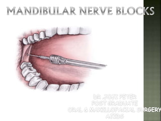
- 7. INTRAORAL NERVE BLOCK TECHNIQUES
- 8. Nerve anesthetized: terminal branches Area anesthetised: area innervated by terminal branches Indication :successful for 6 anterior teeth Techinque: paraperiosteal or interosseous techniques used with 1 5/8 ” needle Symptoms : instrumentation needed BLOCK OF TERMINAL BRANCHES
- 9. MENTAL NERVE BLOCK Nerve anesthetized: Mental nerve Area anesthetised:lower lip Mucolabial fold anterior to the mental foramen Anatomical landmarks: mandibular bicuspids Indication : procedures where manipulation of buccal soft tissue anterior to the mental foramen is necessary. Contraindications : acute inflammation and infection over the injection site.
- 11. Technique: •Mental foramen lies below the apex of the 2nd premolar or between the the two premolars. •It faces posteriorly & thus when making an injection the approach should be from behind •Mental foramen palpated. Symptoms : Tingling & Numbness of lower lip Adv : No loss of lingual sensation –better for children Complication: Hematoma,partial anesthesia of central & lateral incisors
- 14. INCISIVE NERVE BLOCK Nerve anesthetized: Incisive & Mental nerve Area anesthetised:Lower lip,Mandible &Overlying structures anterior to mental foramen,Bicuspids,Cuspids& Incisors Anatomical landmarks: Mandibular bicuspids Indication : Structures anterior to mental foramen, cases where IANB is contraindicated
- 16. Contraindications : acute inflammation & infection over the injection site. Techinque: same as mental nerve block. anaesthetic solution to be penetrated into mental foramen Symptoms : Sub:Numbness & tingling Obj:Anesthesia of anterior teeth & structures on instrumentation Failure: 1.Inadequate volume of anesthetic solution 2.Inadequate duration of pressure. Complication:hematoma
- 17. LONG BUCCAL NERVE BLOCK Nerve anesthetised:Buccal branch of anterior division of V3 Area anesthetised:buccal mucous membrane& mucoperiosteum of mandibular molars Indication : surgery of mandibular buccal mucosa & adjunct to IANB Techinque:Inserted into buccal mucosa distal to 3rd molar. alternative : into retromolar triangle Symptoms : instrumentation
- 18. Infiltration in the buccal sulcus distal to permanent molar tooth Amount deposited-0.2-0.5 ml LONG BUCCAL NERVE BLOCK
- 19. LINGUAL NERVE BLOCK Nerve anesthetised:Lingual nerve Area anesthetised: Anterior 2/3rd of tongue, floor of oral cavity & mucoperiosteum on lingual side of mandible Indication : surgery of anterior 2/3rd of tongue, floor of oral cavity & mucoperiosteum on lingual side of mandible Techinque: same as IANB Symptoms : sub:numbness & tingling obj:anesthesia of anterior teeth & structures on instrumentation
- 20. 3 TECHNIQUES TO BLOCK LINGUAL NERVE Blocking lingual nerve at the same time as an intraoral inferior dental injection by depositing 0.5ml of solution after the needle has been inserted for approx. 1cm By giving submcosal infiltration of 0.5 ml few millimeteres below and behind the region of the lower 3rd molar on its lingual aspect. Infiltration immediately lingual to the gingiva or mucosa to be treated.
- 21. CLASSICAL INFERIOR ALVEOLAR NERVE BLOCK
- 23. Body of mandible Mandibular teeth Mucous membrane and underlying tissue anterior to molar
- 24. Factors affecting the relative position of the mandibular foramen Width of ascending ramus Width of arch of the mandible Obliquity of the angle of the mandible.
- 25. ANATOMIC VARIATIONS Mandible - Mandibular foramen in children 4 years old and less is below the plane of occlusion. - The foramen moves superiorly in the ramus with the eruption of 6’s Adults Children
- 26. Correction for anatomical variation If bone is struck soon after insertion If bone is not reached after insertion of the needle for a reasonable distance.
- 27. Nerve anesthetised:Inferior alveolar nerve ,Mental.N ,Incisive.N, Lingual & Buccinator.N Area anesthetised: body of mandible& inferior portion of ramus, mandibular teeth, mucous membrane &structures anterior to mand 1st molar Anatomical landmarks: Mucobuccal fold Anterior border of ramus of the mandible External oblique ridge Retromolar triangle Internal oblique ridge Pterygomandibular ligament Buccal sucking pad Pteygomandibular space.
- 28. Indication : Analgesia for surgical & operative dentistry on mandibular teeth & supporting structures,Diagnostic & Therapeutic purposes Approximating strucutres when needle is in position: The inferior dental nerve is blocked by the deposition of analgesic solution around it just before it enters the mandibular foramen and when it is in the pterygomandibular space. This is bounded Anteriorly :pterygomandibulr raphe Posteriorly : parotid gland Laterally:ascending ramus of the mandible Medially : medial pterygoid muscle Superiorly: 2 heads of lateral pterygoid Inferiorly : attachment of medial pterygoid
- 31. ADVANTAGES: Wide area of Anesthesia DISADVANTAGES: Wide area of anesthesia Inadequate anesthesia +ve aspiration(10% to 15%) Intra oral landmarks Lingual & lower lip anesthesia Partial anesthesia-bifid mand canals
- 32. For rt IANB 8 o’clk position For lt IANB 10 o’clk position 3 parameters: height of the injection anteroposterior site of injection penetration depth
- 33. Technique: • Mouth open,body of mandible parallel to floor • Operator right side of patient • Thumb palpates mucobuccal fold • Thumb moves posteriorly to contact external oblique ridge on anterior border of ramus • Greatest depth is identified-coronoid notch-height of mand sulcus • Thumb-ligually moved-across retromolar triangle-onto internal oblique ridge. • Thumb moved buccally along with buccal sucking pad-exposure to internal oblique ridge,pterygomandibular raphe & pterygopharygeal depression. • Index finger-extraorally –behind –ramus of mandible. • Syringe-parallel to occlusal plane-opp side of mouth-bisecting finger • Moved until gently bone contacted • Needle withdrawn 1mm,Solution deposited.
- 34. Symptoms : sub:numbness & tingling of lower lip obj:on instrumentation
- 35. FAILURES OF ANESTHESIA: Deposition of anesthetic too low. Deposition of anesthetic too far anteriorly on ramus. Accessory innervation to the mandibular teeth 1o symptom-Incomplete pulpal anesthesia. Accessory sensory innervation (cervical accessory & mylohyoid.N) To correct Technique #1 25 gauge long needle. Retract the tongue toward midline Place the syringe in corner of mouth on opp side & direct the needle tip to apical portion of tooth immediately posterior to tooth of interest Depth of penetration to bone:3-5mm. Aspirate:0.6ml in 20sec Technique #2
- 36. Bifid inferior alveolar nerve to correct-deposit solution inferiorly Incomplete anesthesia to central & lateral incisors Due to innervation of mylohyoid To correct: Supraperiosteal infiltration 27gauge short needle Direction of needle tip Aspirate:0.6ml in 20secs After 2-3mins start dental procedure
- 37. COMPLICATIONS: High injection-Numbness of ear- when auriculotmporl.N is anesthetised. High injection-trismus-injection into lateral pterygoid High injection-toxicity-injection into pterygoid plexus of veins Low injection-Trismus-injection into medial pterygoid Low injection-toxicity-injection into posterior facial vein High & deep injection-paralysis-injection into substances of parotid gland(facial nerve) HEMATOMA TRANSIENT FACIAL PARALYSIS-inj into medial pterygoid
- 38. METHOD OF CLARKE AND HOLMES(1959) This involves depositing the of the solution at a higher level than usual. It’s a modification of indirect technique. Occlusal plane is parallel to the floor. External oblique ridge is palpated Finger is rotated inwards so that the tip lies in the retromolar fossa and the fingernail overlies the internal oblique ridge.
- 39. The needle is advanced from between the premoalrs on the opp.side ,the point passing into the tissue just above the fingernail of the index finger and not at the midpoint as with the standard indirect technique. Bone is encountered Syringe is then gently swung round until it lies over the lower central incisors. The needle is passed another 2cms deeper into the tissue and 1.5ml of the solution is deposited. If lingual analgesia required,then solution is deposited when needle is being withdrawn
- 40. METHOD OF CLARK & HOLMES 1959
- 41. GOW GATES TECHNIQUE 1973 Nerve anesthetised: inferior alveolar,mental,incisive,lingual,mylohyoid,auriculotemporal,bucca l. Area anesthetised:mand teeth to midline, buccal & lingual mucoperiosteum & mucous membrane,ant 2/3rd of tongue, floor of oral cavity, skin over the zygoma, posterior portion of cheek& temporal region. Indication: multiple procedure on mandible, buccal soft procedures from third molar to midline, lingual soft tissue anesthesia, IANB unsuccessful.Diagnosis of facial pain Cotraindication Infection & acute inflammation@ site,young patients, trismus Intraoral approach for mandibular nerve block
- 42. ADV: only 1 inj needed, success rate, aspration rate post injection complication. DISADV : time of onset than IANB
- 43. LANDMARKS Extraoral External ear. Intertragic notch Corner of the mouth Intraoral Tendon of temporal muscle Anterior border of the ramus, Mesiolingual cusp of maxillary 2nd molar
- 44. The patient mouth should be wide open,supine position Needle is inserted at a point lateral to the pterygomandibular depression but medial to the temporal tendon on a plane from the corner of the mouth to intertragic notch. Approximating srtuctures: It should be just inferior to the condyle Inferior to the attacthement of lateral pterygoid muscle.
- 45. TECHNIQUES: Position the patient Locate extra oral landmarks Visualize intraoral landmarks Prepare tissues at site of penetration Direct the syringe Insert the needle Align the needle with the plane Slowly advance the needle Depth of penetration:25mm Withdraw the needle 1mm Aspirate: if –ve slowly deposit 1.8ml in 60-90 secs Withdraw the syringe Request the pt to keep mouth open for 1-2mins Upright position Wait for 3-5mins before starting dental procedure
- 47. METHOD OF ANGELO SARGENTI(1966) Its a modification of the direct technique. The prinicipal difference is that the nerve is approached from a higher level than usual. Index finger placed at retromolar fossa with the nail facing lingually. The needle is then inserted opp.the midpoint of the fingernail and a little beyond its tip. The barrel is placed between and in contact with the upper premolars of opp.side.
- 48. It is kept in this position while the needle is slowly inserted in a downward and backward direction until it touches bone, which is usually at a depth of 1cm.
- 49. METHOD OF ANGELO & SARGENTI 1966
- 50. DR.MENDEL NEVIN Guide finger- Coronoid Notch Pterygotemporal depression in Pterygomandibular space Bone is encountered Syringe is brought in line with the lower teeth on the same side for lingual block DIRECT THRUST TECHNIQUE
- 51. Advantages: One injection provides a wide area of anaesthesia, useful for quadrant surgery. Disadvantages: •Rate of inadequate anesthesia (15 to 20%). •Intraoral landmarks not consistently reliable. •Positive aspiration is high (10 to 15%). •Lingual & lower lip anesthesia is discomfort & possibly dangerous. •Partial anesthesia possible if bifid inferior alveolar nerve or bifid mandibular canals are present.
- 52. Dr.Charles B Hopkins Perpendicular to ramus with barrel in respect to Opposite side 1st & 2nd Molar Height- Above the finger nail Inserted Directly in the mandibular sulcus. DIRECT TECHNIQUE OF CHARLES HOPKINS
- 53. Dr.Brownlee Importance of Posterior border of ramus as landmark Thumb-Coronoid notch Index-Posterior border of ramus Third finger-Angle of mandible Advantages: No unreliable landmarks. Same for all the patients. BROWNLEE’S DIRECT THRUST TECHNIQUE
- 54. Dr.Boris Levitt Modification of Mendel’s technique Midway between the occlusal surface of upper and lower molar teeth Depth not greater than 1 inch Withdraw the needle halfway for Lingual block Deposit solution even without bone contact MODIFIED DIRECT THRUST TECHNIQUE
- 55. ARCHED/CURVED NEEDLE TECHNIQUE In the arched needle technique the flexibility of the fine bore needle is utilized to change the approach angle of the needle tip Inserted at an acute angle, the subsequent arching changes the path & the needle approaches the medial surface of the ramus almost perpendicularly, which is otherwise impossible to achieve with an unarched or unbent needle.
- 56. METHOD OF SUNDER J. VAZIRANI 1960
- 57. VAZIRANI- AKINOSI TECHNIQUE CLOSED MOUTH MANDIBULAR BLOCK Indication: Patient unable to open mouth. Contraindication:Where trismus is present due to infection in the tissues through which a needle would have to passs.
- 59. Landmarks Occlusal plane of occluding teeth mucogingival Junction of the maxillary molar teeth Anterior border of the ramus.
- 60. The needle is placed parallel to the gingival margins of the maxillary teeth or to the alveolar ridge in the edentulous patient. The point of insertion is the mucosa, pterygomandibular fold,buccinator and buccal aponeurosis on the inner aspect of the ascending ramus to reach the pterygomandibular space.
- 62. INDIRECT METHOD-FISCHER 1-2-3 TECHNIQUE Step 1: Barrel of the syringe rests on occlusal surface of the opp. Premolars. Tip of the needle rests on the external oblique ridge at the midpoint of thumbnail. Needle is advanced 6mm and few drops of solution is deposited to block the long buccal nerve.
- 63. Step 2: Barrel of the syringe is withdrawn slightly and shifted to the same side so that ,needle glides over temporalis tendon onto the internal oblique ridge. Needle is further advanced for about 8mm,keeping the barrel of the syringe parallel to mandibular occlusal plane. This is to block the lingual nerve.
- 64. Step3: The barrel is returned to opp.Side , near the 1st premolar and the needle is advanced further for a distance of 12-15mm, until the bony resistance is felt by the tip of the needle. This is to block the inferior dental nerve.
- 65. INDIRECT METHOD
- 68. KURT THOMA TECHNIQUE •Anterior border of masseter is located & its lowest border palpated (A). •This point is marked and line drawn connecting it with tragus of the ear (B). •The mid point of this line is located.(C). •Parallel line drawn with posterior border of ramus (D). •Skin is prepared •Long needle is inserted on the inner aspect of lower border of mandible keeping close to bone throughout & parallel to the line drawn •21 Gauge needle with stopper of 7-8 cms length
- 69. EXTERNAL APPORACH BY – KURT THOMA B C A D
- 71. INDICATION FOR KURT THOMA APPROACH Severe limitation of opening of the jaws TMJ ankylosis
- 72. EXTRAORAL MANDIBULAR NERVE BLOCK TECHNIQUE
- 73. The zygomatic process is palpated and midpoint of the depression on its lower border is marked. Local infiltration should be given A heavy gauge needle min. of 3 1/8 inch(8cm) is used for injection ,incorporating a marker to indicate a depth of 4cm. Needle is inserted little below the zygomatic process and at right angle to the skin, until the lateral pterygoid is reached , which should be at a depth of approx.4cm. The needle is now withdrawn and reinserted a few degrees father distally so as to pass just behind the posterior border of the lateral pterygoid plate. It should never be penetrated more than 5cm. EXTRAORAL MANDIBULAR NERVE BLOCK TECHNIQUE
- 74. Landmarks – - midppoint of zygomatic arch - Zygomatic notch - Coronoid process of ramus - Lateral pterygoid plate Depth – 4.5 cm
- 76. EXTRAORAL MENTAL NERVE BLOCK The mental foramen is palpated externally in the region below the apex of second premolar. This normally equidistant between the upper and lower borders of the mandible when the teeth are present. 25 gauge(2.5 cms) needle is passed downwards at an angle of 30 and forwards at angle of 45 to the body of the mandible until bone is reached.
- 79. ADVANTAGE Avoids the unpleasant loss of lingual sensation, which generally occurs with an inferior dental injection. COMPLICATION & CONTRAINDICATION It should be remembered that the lower central incisors and to a lesser degree the lateral incisors derive some inervation from the incisive nerve of the opposite side. So to achive analgesia a supraperiosteal injection should be used
- 80. •Inferior alveolar nerve block by injection into mandibular foramen in the direction of 1st molar to lateral of pterygomandibular space. •Adv : No danger of injecting the needle and depositing the local anaesthetic contents into the maxillary artery and vein, the middle meningeal artery and vein or the T-M joint capsule, thus avoiding all of the unnecesary complications therein. ANTERIOR RAMUS TECHNIQUE (A.R.T) Dr.Lawrence Gaum &Dr.Allan C Moon Same as that explained by Fischer in 1900
- 84. MASSETERIC & DEEP TEMPORALBLOCKS USEFUL IN MANDIBULAR DISLOCATION ANESTH ESIA PROGRESS. 2009 SPRING; 56(1):
- 85. ANESTHETIC TECHNIQUE FOR INFERIOR ALVEOLAR NERVE BLOCK: A NEW APPROACH. JOURNAL OF APPLIED ORAL SCIENCE. 2011 Jan-Feb; 19(1): 11–15. Potentially effective for inferior alveolar nerve block, especially in pediatric dentistry.
- 86. Insufficient knowledge of local anatomy Individual variations in anatomy Variation due to age Faulty technique injecting too far posteriorly injecting too low down
- 87. AGE CHANGES IN MANDIBLE BIRTH 3 YEARS 6 YEARS ADULT EDENTULOUS OLD AGE Mental foramen Mandibuar foramen
- 88. THANKYOU