
More Related Content
What's hot
What's hot (20)
Viewers also liked
Viewers also liked (18)
Similar to Anatomic landmarks seen in a IOPA
Similar to Anatomic landmarks seen in a IOPA (20)
Recently uploaded
Recently uploaded (20)
Anatomic landmarks seen in a IOPA
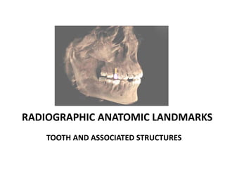
- 1. RADIOGRAPHIC ANATOMIC LANDMARKS TOOTH AND ASSOCIATED STRUCTURES
- 2. REFERENCES White and Pharaoh , Oral Radiology Principles and Interpretation Ongole and Praveen, Clinical Manual for Oral Medicine and Radiology Radiographs given in the material are anatomically correct, not always constant for clinical situations because people are not always the same. ANATOMICAL LANDMARKS, TOOTH AND ASSOCIATED STRUCTURES, SUNDARAM, III BDS, TMDC&H
- 3. R/F OF TOOTH RADIOLUCENT 1. PULP. 2. PERIODONTAL LIGAMENT SPACE. 3. BONE MARROW SPACE. 4. NUTRIENT CANALS. RADIOPAQUE 1. ENAMEL. 2. DENTIN. 3. CEMENTUM. 4. LAMINA DURA. 5. ALVEOLAR CREST. 6. ALVEOLAR BONE. ANATOMICAL LANDMARKS, TOOTH AND ASSOCIATED STRUCTURES, SUNDARAM, III BDS, TMDC&H
- 4. RADIOLUCENT STRUCTURES MAXILLA 1. INCISIVE FORAMEN. 2. INTERMAXILLARY SUTURE. 3. NASAL FOSSA. 4. LATERAL FOSSA. 5. MAXILLARY SINUS. 6. OPENING OF NASOLACRIMAL DUCT. MANDIBLE 1. MANDIBULAR CANAL. 2. MENTAL FORAMEN. 3. LINGUAL FORAMEN. 4. NUTRIENT CANALS. 5. SUBMANDIBULAR GLAND FOSSA. ANATOMICAL LANDMARKS, TOOTH AND ASSOCIATED STRUCTURES, SUNDARAM, III BDS, TMDC&H
- 5. RADIOPAQUE STRUCTURES MAXILLA 1. NASAL SEPTUM. 2. ANTERIOR NASAL SPINE. 3. INVERTED Y OF YENIS. 4. ZYGOMATIC PROCESS OF MAXILLA. 5. MAXILLARY TUBEROSITY. 6. HAMULAR PROCESS. 7. PTERYGOID PLATES. 8. CORONOID PROCESS OF MANDIBLE. 9. TIP OF NOSE. 10. NASOLABIAL FOLD. MANDIBLE 1. EXTERNAL OBLIQUE RIDGE. 2. MYLOHYOID RIDGE. 3. MENTAL RIDGE. 4. LOWER BORDER OF MANDIBLE. 5. GENIAL SPINES. 6. SYMPHYSIS. ANATOMICAL LANDMARKS, TOOTH AND ASSOCIATED STRUCTURES, SUNDARAM, III BDS, TMDC&H
- 6. ANATOMICAL LANDMARKS, TOOTH AND ASSOCIATED STRUCTURES, SUNDARAM, III BDS, TMDC&H
- 7. ROOT CANAL AND APICAL FORAMEN LAMINA DURA ALVEOLAR CREST ANATOMICAL LANDMARKS, TOOTH AND ASSOCIATED STRUCTURES, SUNDARAM, III BDS, TMDC&H
- 8. CANCELLOUS BONE ANATOMICAL LANDMARKS, TOOTH AND ASSOCIATED STRUCTURES, SUNDARAM, III BDS, TMDC&H
- 9. PERIODONTAL LIGAMENT SPACE ANATOMICAL LANDMARKS, TOOTH AND ASSOCIATED STRUCTURES, SUNDARAM, III BDS, TMDC&H
- 10. INTERMAXILLARY SUTURE (A,B) ANTERIOR NASAL SPINE(C) A B C ANATOMICAL LANDMARKS, TOOTH AND ASSOCIATED STRUCTURES, SUNDARAM, III BDS, TMDC&H
- 11. A B (A) NASAL SEPTUM (B) ANTERIOR FLOOR OF NASAL FOSSA ANATOMICAL LANDMARKS, TOOTH AND ASSOCIATED STRUCTURES, SUNDARAM, III BDS, TMDC&H
- 12. ANATOMICAL LANDMARKS, TOOTH AND ASSOCIATED STRUCTURES, SUNDARAM, III BDS, TMDC&H
- 13. LATERAL FOSSA INVERTED ‘Y’ OF YENTIS ANATOMICAL LANDMARKS, TOOTH AND ASSOCIATED STRUCTURES, SUNDARAM, III BDS, TMDC&H
- 14. OPENING OF NASOLACRIMAL CANAL ANATOMICAL LANDMARKS, TOOTH AND ASSOCIATED STRUCTURES, SUNDARAM, III BDS, TMDC&H
- 15. ZYGOMATIC PROCESS OF MAXILLA ANATOMICAL LANDMARKS, TOOTH AND ASSOCIATED STRUCTURES, SUNDARAM, III BDS, TMDC&H
- 16. PTERYGOID PLATES HAMULAR PROCESS MAXILLARY TUBEROSITY ANATOMICAL LANDMARKS, TOOTH AND ASSOCIATED STRUCTURES, SUNDARAM, III BDS, TMDC&H
- 17. FLOOR OF MAXILLARY SINUS ANATOMICAL LANDMARKS, TOOTH AND ASSOCIATED STRUCTURES, SUNDARAM, III BDS, TMDC&H
- 18. ANATOMICAL LANDMARKS, TOOTH AND ASSOCIATED STRUCTURES, SUNDARAM, III BDS, TMDC&H
- 19. ANATOMICAL LANDMARKS, TOOTH AND ASSOCIATED STRUCTURES, SUNDARAM, III BDS, TMDC&H
- 20. ANATOMICAL LANDMARKS, TOOTH AND ASSOCIATED STRUCTURES, SUNDARAM, III BDS, TMDC&H
- 21. ANATOMICAL LANDMARKS, TOOTH AND ASSOCIATED STRUCTURES, SUNDARAM, III BDS, TMDC&H
- 22. ANATOMICAL LANDMARKS, TOOTH AND ASSOCIATED STRUCTURES, SUNDARAM, III BDS, TMDC&H
- 23. (B)(A) (C) (D) (A,B) NUTRIENT CANALS (C,D) MENTAL FORAMEN ANATOMICAL LANDMARKS, TOOTH AND ASSOCIATED STRUCTURES, SUNDARAM, III BDS, TMDC&H
- 24. ANATOMICAL LANDMARKS, TOOTH AND ASSOCIATED STRUCTURES, SUNDARAM, III BDS, TMDC&H
- 25. (A) (B) (C) (A) LINGUAL FORAMEN (B,C) GENIAL TUBERCLES ANATOMICAL LANDMARKS, TOOTH AND ASSOCIATED STRUCTURES, SUNDARAM, III BDS, TMDC&H