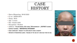
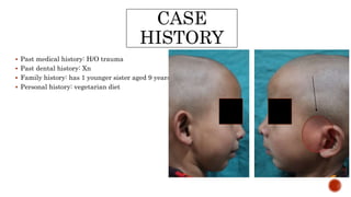
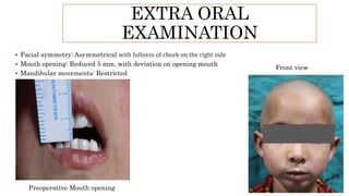
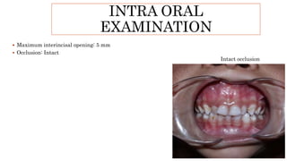
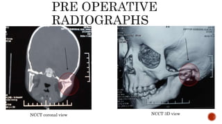
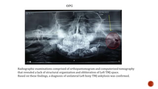
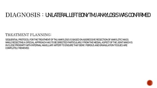
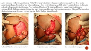
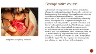
Presented by – Dr. ABHIJEET KAMBLE
PG year 2
CASE
HISTORY
 . A/H/O Trauma 13 months back while the patient was playing with her toys alone and she fell down
and hit her chin on the ground. No bleeding from mouth or face was noted at the time of injury.
Parents recognized the inability to open mouth after sometime and child was then taken to a local
doctor. The child was referred to Department of Maxillofacial and Oral Surgery at Government Dental
College and Hospital, Shimla, for further evaluation and treatment planning
CASE
HISTORY
 Date of Reporting : 06-02-2023
 Cr no : 2022013975
 Name : Kriti
 Age : 5 years
 Sex : female child
 Occupation : Student
 Address : VILLAGE:BANAD ,P.O TUNDA, TEH BHARMOUR –, DISTRICT CHAMBA
 CONTACT NUMBER: - 9805904702,7876582864
 CHIEF COMPLAINT : INABILITY TO OPEN MOUTH SINCE 1 MONTH
 HISTORY OF PRESENTING ILLNESS : TRAUMA AT THE AGE OF 1 AND HALF YEAR OF AGE.
CASE
HISTORY
 Past medical history: H/O trauma
 Past dental history: Xn
 Family history: has 1 younger sister aged 9 years
 Personal history: vegetarian diet
GENERAL
EXAMINATION
 Pallor: Absent
 Icterus: Absent
 Cyanosis: Absent
 Clubbing: Absent
 CNS: Well oriented GCS: 15/51
 CVS: NAD
 Respiratory system: NAD
EXTRA ORAL
EXAMINATION
 Facial symmetry: Asymmetrical with fullness of cheek on the right side
 Mouth opening: Reduced 5 mm, with deviation on opening mouth
 Mandibular movements: Restricted
Preoperative Mouth opening
Front view
INTRA ORAL
EXAMINATION
 Maximum interincisal opening: 5 mm
 Occlusion: Intact
Intact occlusion
NCCT coronal view NCCT 3D view
OPG
Radiographic examinations comprised of orthopantomogram and computerized tomography
that revealed a lack of structural organization and obliteration of Left TMJ space.
Based on these findings, a diagnosis of unilateral Left bony TMJ ankylosis was confirmed.
SEQUENTIAL PROTOCOL FOR THE TREATMENT OF TMJ ANKYLOSIS IS BASED ON AGGRESSIVE RESECTION OF ANKYLOTIC MASS.
WHILE RESECTING A SPECIAL APPROACH HAS TO BE DIRECTED PARTICULARLY FROM THE MEDIAL ASPECT OF THE JOINT WHICH IS
IN CLOSEPROXIMITY WITH INTERNAL MAXILLARY ARTERY TO ENSURE THAT BONY, FIBROUS AND GRANULATION TISSUES ARE
COMPLETELY REMOVED.
UNILATERALLEFTBONYTMJANKYLOSISWASCONFIRMED
After complete evaluation, a unilateral TMJ arthroplasty with interposing temporalis muscle graft was done under
general anesthesia. The patient was intubated using a fiber-optic microscope, which is the recent technique of choice in
patients who present with trismus. A Popowich modification of Al kayat and Bramley preauricular incision was
employed. Full thickness mucoperiosteal flap was reflected, and the ankylotic mass was exposed [FiguresAfter exposing
the joint space, an arthrotomy cut was given at the level of the sigmoid notch.
Reflected full thickness of mucoperiosteal flap Surgical exposure of ankylotic mass One horizontal osteotomy cuts placed
Temporalis sling being harvested
Postoperative course
Active mouth opening exercises are started immediately
after postoperative pain subsides. However, for patients who
have undergone International Monetary Fund, exercises are
started soon after release of fixation. Patients are
encouraged to start gentle, active and gradually increasing
mouth opening exercises using their own fingers as a
monitor to start with, in order to gain self-confidence, and
they are allowed to take a soft diet. Wooden tongue blades
are used thereafter with a gradually increasing number
according to the patient's tolerance, avoiding any passive
force or pain. This is performed under strict supervision for
15 min 5 times a day. Regular weekly visits are arranged
during the 1st month, biweekly for the next 3 months, then
monthly for 1 year .The child regained her beaming smile
and relishing her favorite foods with enthusiasm.
CLINICAL CASE PRESENTATION TMJ ANKYLOSIS.pptx

CLINICAL CASE PRESENTATION TMJ ANKYLOSIS.pptx

  • 1.
    Presented by –Dr. ABHIJEET KAMBLE PG year 2
  • 2.
    CASE HISTORY  . A/H/OTrauma 13 months back while the patient was playing with her toys alone and she fell down and hit her chin on the ground. No bleeding from mouth or face was noted at the time of injury. Parents recognized the inability to open mouth after sometime and child was then taken to a local doctor. The child was referred to Department of Maxillofacial and Oral Surgery at Government Dental College and Hospital, Shimla, for further evaluation and treatment planning
  • 3.
    CASE HISTORY  Date ofReporting : 06-02-2023  Cr no : 2022013975  Name : Kriti  Age : 5 years  Sex : female child  Occupation : Student  Address : VILLAGE:BANAD ,P.O TUNDA, TEH BHARMOUR –, DISTRICT CHAMBA  CONTACT NUMBER: - 9805904702,7876582864  CHIEF COMPLAINT : INABILITY TO OPEN MOUTH SINCE 1 MONTH  HISTORY OF PRESENTING ILLNESS : TRAUMA AT THE AGE OF 1 AND HALF YEAR OF AGE.
  • 4.
    CASE HISTORY  Past medicalhistory: H/O trauma  Past dental history: Xn  Family history: has 1 younger sister aged 9 years  Personal history: vegetarian diet
  • 5.
    GENERAL EXAMINATION  Pallor: Absent Icterus: Absent  Cyanosis: Absent  Clubbing: Absent  CNS: Well oriented GCS: 15/51  CVS: NAD  Respiratory system: NAD
  • 6.
    EXTRA ORAL EXAMINATION  Facialsymmetry: Asymmetrical with fullness of cheek on the right side  Mouth opening: Reduced 5 mm, with deviation on opening mouth  Mandibular movements: Restricted Preoperative Mouth opening Front view
  • 7.
    INTRA ORAL EXAMINATION  Maximuminterincisal opening: 5 mm  Occlusion: Intact Intact occlusion
  • 8.
    NCCT coronal viewNCCT 3D view
  • 9.
    OPG Radiographic examinations comprisedof orthopantomogram and computerized tomography that revealed a lack of structural organization and obliteration of Left TMJ space. Based on these findings, a diagnosis of unilateral Left bony TMJ ankylosis was confirmed.
  • 10.
    SEQUENTIAL PROTOCOL FORTHE TREATMENT OF TMJ ANKYLOSIS IS BASED ON AGGRESSIVE RESECTION OF ANKYLOTIC MASS. WHILE RESECTING A SPECIAL APPROACH HAS TO BE DIRECTED PARTICULARLY FROM THE MEDIAL ASPECT OF THE JOINT WHICH IS IN CLOSEPROXIMITY WITH INTERNAL MAXILLARY ARTERY TO ENSURE THAT BONY, FIBROUS AND GRANULATION TISSUES ARE COMPLETELY REMOVED. UNILATERALLEFTBONYTMJANKYLOSISWASCONFIRMED
  • 11.
    After complete evaluation,a unilateral TMJ arthroplasty with interposing temporalis muscle graft was done under general anesthesia. The patient was intubated using a fiber-optic microscope, which is the recent technique of choice in patients who present with trismus. A Popowich modification of Al kayat and Bramley preauricular incision was employed. Full thickness mucoperiosteal flap was reflected, and the ankylotic mass was exposed [FiguresAfter exposing the joint space, an arthrotomy cut was given at the level of the sigmoid notch. Reflected full thickness of mucoperiosteal flap Surgical exposure of ankylotic mass One horizontal osteotomy cuts placed
  • 12.
    Temporalis sling beingharvested Postoperative course Active mouth opening exercises are started immediately after postoperative pain subsides. However, for patients who have undergone International Monetary Fund, exercises are started soon after release of fixation. Patients are encouraged to start gentle, active and gradually increasing mouth opening exercises using their own fingers as a monitor to start with, in order to gain self-confidence, and they are allowed to take a soft diet. Wooden tongue blades are used thereafter with a gradually increasing number according to the patient's tolerance, avoiding any passive force or pain. This is performed under strict supervision for 15 min 5 times a day. Regular weekly visits are arranged during the 1st month, biweekly for the next 3 months, then monthly for 1 year .The child regained her beaming smile and relishing her favorite foods with enthusiasm.