Download as PDF, PPTX


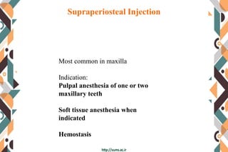


The document provides information on various types of maxillary anesthesia techniques: 1. Supraperiosteal injection is commonly used to anesthetize one or two maxillary teeth and soft tissues. It has a low risk of intravascular administration. 2. Posterior superior alveolar nerve block anesthetizes maxillary molars and buccal tissues through the posterior superior alveolar nerve. It has a high success rate but risks hematoma formation. 3. Nasopalatine nerve block provides wide palatal soft tissue anesthesia using a minimum volume of local anesthetic, minimizing the need for multiple injections.


![Techniques of local anesthesia [autosaved]](https://cdn.slidesharecdn.com/ss_thumbnails/techniquesoflocalanesthesiaautosaved-210618141111-thumbnail.jpg?width=640&height=640&fit=bounds)


