SlideShare a Scribd company logo
The leukemias
Leukaemias
Definition
• Leukaemia is a malignant disease of the
haemopoietic tissue,characterized by the
replacement of normal bone marrow elements
with abnormal blood cells.
• Leukaemic cells are frequently present in the
peripheral blood, commonly invade spleen, liver
and lymph nodes and other tissues of the body,
eg testes, brain, meninges and skin
Classification of Leukaemias
Acute leukaemia
• Acute lymphoblastic leukaemia
• Acute myeloid leukaemia
Chronic leukaemia
• Chronic myeloid leukaemia
• Chronic lymphocytic leukaemia
Classification of leukaemias
• Other chronic lymphoid leukaemias
Hairy cell leukaemias
Prolymphocytic leukaemia
Lymphoma/leukaemia
• ‘Pre - leukaemia’ - myelodysplastic
syndrome
Pathogenesis of Acute leukaemia
• Origin of leukaemia at the genetic level appears
to be related to mutations and altered
expression of oncogenes and tumor suppressor
genes
• Oncogenes regulate cell proliferation and
differentiation
• Abnormal oncogene or tumour suppressor gene
expression induced by translocation and genetic
fusion or mutation results in unregulated cellular
proliferation
Pathogenesis of Acute leukaemia
• Genetic alteration occurs within a single
abnormal stem cell or progenitor cell in the
marrow.
• Successive generation of cells derived by
mitosis from the original abnormal cell
gives rise to a clonal population, which,
when sufficiently large, causes clinically
apparent disease
Pathogenesis of Acute leukaemia
• Acute leukaemia, blasts cells fail to differentiate
normally, further divisions continue, results in
accumulation, replacement of normal cells by
lymphoblasts or myeloblasts – bone marrow
failure
• Clinical condition correlate with total number of
leukaemic cells in the body.
• When 60% or more of total marrow cells –
peripheral blood involvement, liver, spleen and
lymph nodes are infiltrated.
Epidemiologic aspects of
leukaemia
• Host factors
1. Heredity
• Appears not to be inherited but increased
predisposition in some individuals.
Family members of leukaemia pts – possible
shared environment
Identical twin –shared placental circulation
(possible in utero exposures)
Epidemiologic aspects of
leukaemia
2. Congenital chromosomal abnormalities
Disorders with genetic abnormalities predispose
to acute leukemias :
- Down’s syndrome 10 to 20 fold increase of
acute leukaemia. AML 1 gene identified in
chromosome 21
- Fanconi’s anaemia (AML)
- Ataxia telaniectasia (ALL,NHL)
Epidemiologic aspects of
leukaemia
3. Immunodeficiency
• Hereditary immunodeficiency states,
high incidence of lymphoproliferative disorder
4. Chronic marrow dysfunction
MDS, myeloproliferative disorders, aplastic
anaemia,
increased risk of acute leukaemia
transformation
Epidemiologic aspects of
leukaemia
• Environmental factors
1. Ionizing radiation
Exposure associated with develoment of acute and chronic leukaemia
Illustrated in populations exposed to nuclear weapons in Hiroshima and
Nagasaki
2. Chemicals and drugs
Benzene – most frequently documented chemical toxin
Alkylating drugs – pts on combined chemoradiotherapy for Hodgkin’s
disease
Chemotherapy – 10 % - 20% of all AML, secondary AML
Epidemiologic aspects of
leukaemia
3. Viruses
Human T cell leukaemia/lymphoma virus
Implicated causative agent for adult T cell
leukaemia/lymphoma
Epstein Barr virus linked to African Burkitt
lymphoma – high grade B cell lymphoma
Incidence
• Acute leukaemia comprises > 50% of
leukaemia seen in clinical practice
• ALL, common form in children, peak 3-4
yrs falling off by 10 yrs
• AML occurs in all age groups
common type in adults, elderly, only 10
-15% of leukaemia in childhood
Incidence
• Chronic leukaemias
• Generally disease of adults
• CLL extremely rare in children, unusual
before 40 yrs
• CML seen at any age, peak incidence 30-
50 yrs, rare in children
• Distinct juvenile variant (jCML) in children
Comparison of Acute and Chronic
leukaemia
Acute Chronic
• Age All ages Adults
• Clinical onset sudden insidious
• Course <6 months 2-6 yr
• Leukaemic cells immature mature
• Anaemia mild-severe mild
• Thrombocytopenia mild-severe mild
• TWBC variable increase
• Organomegaly mild prominent
Acute leukaemia
• Acute leukaemia is defined as the
presence of >30% blasts in the bone
marrow at clinical presentation.
• Subdivided into two types on the basis of
blasts.
• Acute lymphoblastic leukaemia
• Acute myeloid leukaemia
Clinical features of acute leukaemia
Pathogenesis Clinical features
• Bone marrow failure
• Anaemia Fatigue, malaise,pallor
• Thrombocytopenia Bruising, bleeding
• Neutropenia Fever, infection
Clinical features of acute leukaemia
Organ infiltration
• Marrow expansion Bone or joint pain
• Spleen Splenomegaly
• Liver Hepatomegaly
• Lymph nodes Lymphadenopathy
• CNS CNS symptoms
• Gums, mouth gum hypertropy,
oral lesions
CLASSIFICATION
• Morphology
– French-American-British (FAB) classification
– WHO classification
• Immunophenotype- ALL
Importance of classification
– Determine mode of treatment
– Prognostic value
FRENCH-AMERICAN-BRITISH (FAB)
CLASSIFICATION
ACUTE LEUKEMIA
Acute myeloid leukemia Acute lymphoblastic leukemia
M0 undifferentiated
M1 without maturation
M2 with granulocytic maturation
M3 acute promyelocytic
M4 granulocytic and monocytic maturation
M5 monoblastic (M5a) or monocytic (M5b)
M6 erythroleukemia
M7 megakaryoblastic
L1
L2
L3 (Burkitt)
FAB classification
• Acute lymphoblastic leukaemia subdivided
• Based on morphology and cytochemistry
• L1
• L2
• L3
ALL- L1
• Blasts are small
• Homogenous
• High N/C ratio
• scanty cytoplasm
ALL- L2
• Blasts are larger
• heterogenous
• Low N/C ratio
• More abundant
cytoplasm
ALL- L3
• Blasts are deeply
basophilic
• Vacuolated cytoplasm
FAB classification
AML
• M0 – undifferentiated
• M1 – without maturation
• M2 – with granulocytic maturation
• M3 – acute promyelocytic leukaemia
• M4 – granulocytic and monocytic maturation
• M5 – monoblastic (M5a)
• monocytic (M5b)
• M6 – erythroleukaemia
• M7 - megakaryoblastic
Myeloblast with auer rod
FAB classification
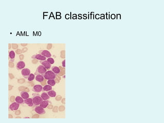
• AML M0
AML-M1
• Blast cells have few
azurophilic granules
• May have auer rods
AML –M2
• Multiple granules
• May have Auer rods
• With granulocytic
differentiation
AML- M3
• Abnormal
promyelocytes
• Prominent granules
• Multiple Auer rods
AML- M4
• Have monocytoid
differentiation
• > 20% or more is
monocytoid cells
AML- M5a
• Monoblastic
• > 80% of blasts are
monoblasts
AML- M5b
• Monocytic
• < 80% of blasts are
monoblasts
AML- M6
• Predominance of
erythroblast
• Dyserythropoiesis
AML- M7
• Megakaryoblast with
cytoplasmic blebs
FAB M3 and M 4 /M 5 features
FAB M3
• Bleeding tendency
• DIVC
• Pancytopenia
• FAB M 4 / M 5
• Gum hypertrophy and infitration
• Skin involvement
• CNS disease
• Granulocytic sarcoma – isolated mass of leukaemia
blasts
Laboratory and Radiographic Work-up:
• CBC with manual differential
• Uric Acid level
• Clotting studies (PT, PTT, D-dimer, fibrinogen)
• Bone marrow aspirate and biopsy
• Chest xray
• Echocardiogram
Diagnosis Of Acute Leukemia
• Demonstrate > 30% blasts in the bone
marrow.
• Diagnosis of acute leukemia is based on
the following:
1.Morphology of the blasts
2.Cytochemistry( MPO,Sudan black- B,NSS,
PAS).
3.Immunophenotyping
4.Cytogenetics
LABORATORY DIAGNOSIS
• Complete blood count
– Normochromic normocytic
anemia
– Thrombocytopenia
– WBC- variable
• Peripheral blood film
– Blasts
• Bone marrow examination
– Hypercellular
– > 30% leukemic blasts
• Cytochemistry
– Myeloperoxidase
– Sudan black
– Non-specific esterase
– Periodic acid-Schiff
– Acid phosphatase
• Flow cytometry
– Myeloid and/or lymphoid
markers
• Genetic analysis
• Others
– Lumbar puncture- leukemic cells
– Uric acid, LDH, Calcium – may be raised
– Renal & liver function test- baseline
– X-ray- lytic bone lesion, enlarge mediastinum
(T-ALL)
Morphology
• Majority of leukemias at most of the
centers world over are diagnosed by
morphology and cytochemistry
• Blasts cells are large cells with:
a)High N:C ratio
b)Nucleus is large with open chromatin
c)Nucleoli 1- 5
d)Thin rim to moderate amount of cytoplsm
Cytochemistry
• Bone marrow smears are stained to
determine specific enzymes or other
proteins produced by cellular organelles.
Cytochemistry
ALL AML
Myeloperoxidase _ +, auer rods
Sudan black _ +, auer rods
Periodic acid schiff + + M6
(coarse granular) (fine blocks)
Nonspecific esterase _ + M4,M5
Acid phosphatase + T ALL +M6
(golgi staining) (diffuse)
Periodic acid- Schiff
Coarse block positivity- ALL Fine blocks – M6
Sudan black
• Black staining in
cytoplasm
Non-specific esterase
• Positive in M4, M5
• Monoblast cytoplasm
– Orange staining
• Myeloblast cytoplasm
– Blue staining
Immunological classification
AML ALL
Marker precursor B T
Myeloid
CD 13 + - -
CD 33 + - -
Glycophorin + M 6 - -
Platelet antigen CD41 +M 7 - -
Myeloperoxidase +M 0 - -
Immunological classification
AML ALL
Marker precursor B T
B lineage ALL
CD 19 - + -
cCD22 - +or- -
CD 10 - + -
Immunological classification
AML ALL
Marker precursor B T
T lineage
CD 7 - - +
cCD3 - - +
TdT - + +
Cytogenetics
• Essential component in newly diagnosed leukaemic
pts
• Major role in diagnosis
• Subclassification
• Selection of appropriate therapy
• Monitering effects of therapy
Cytogenetics
Chromosomal abnormalities associated with distinct
types of leukaemia
• t (15;17) unique to APML (FAB M3)
• Inv (16) AMML with abnormal eosinophilia (M4Eo)
• t (9;22) Ph chromosome in CML , ALL
AML- M2 AML- M4
AML-M3
• blocks differentiation at
the promyelocyte
stage
• All-trans retinoic acid
(ATRA) allow them to
differentiate and to
apoptose
ALL
Prognostic value
good poor
AML t (15;17) – M3
t (8;21) – M2
Inv (16) –M4
Del of chr 5 or 7
t (6;9)
11q23
ALL Ph+
11q23
Molecular genetics
• Primarily used for confirmation of suspected
chromosomal abnormality not detected by
conventional cytogenetics.
• Monitoring minimal residual disease following
therapy
Hand out – molecular correlation of common chromosomal abnormalities in acute
leukaemia
Prognosis in ALL
Good Poor
• WBC Low High > 50,000
• Sex Girls Boys
• Immunophenotype c-ALL B ALL
• Age Child Infants<2 yrs,
adults
• Cytogenetics Normal Ph+, 11q23
or hyperdiploidy
Prognosis in ALL
Good Bad
• Time to clear blasts <1 week >1 week
from blood
• Time to remission <4 weeks >4 weeks
• CNS disease at presentation Absent Present
• Minimal residual disease negative at still positive
1-3 months 3-6 months
Prognosis in AML
Good Bad
• Cytogenetics t(15:17) delection of chromosome
t(8;21) 5or7
inv (16) 11q23
t(6;9)
• Bone marrow response <5% blasts >20% blasts after
to remission induction after first course first course
• Age < 60 yrs >60 yrs
CHRONIC MYELOID LEUKAEMIA
Chronic Myeloid Leukaemia
•Disorder proliferation of haemopoietic stem cells
•Incidence increases with age
– Median age: 40 - 60
– Youngest so far – 12 years old
•Slightly higher incidence in males
– Male-to-female ratio—1.3:1
•Cause
– Unknown
– Slightly increased risk following high dose irradiation: Japanese
atomic bomb survivor
•Median survival: 5 years
Melo. Blood. 1996;88:2375.
Pasternak et al. J Cancer Res Clin Oncol. 1998;124:643.
The Ph Chromosome and the bcr-abl Gene: The t(9;22)
Translocation
FUSION PROTEIN WITH CONSTITUTIVE
TYROSINE KINASE ACTIVITY
bcr-abl
bcr
Philadelphia Chromosome
(or 22q-)
Chromosome 9 q+
abl
Chromosome 9
Chromosome 22
Pathogenesis
•Translocation of genetic material between chromosom 9 and 22
results in fusion gene: bcr – abl (Philadelphia chromososom)
• BCR-ABL fusion gene (M-BCR) encodes a protein of molecular
weight 210 kDa that has greater tyrosine kinase activity than the
normal ABL gene product.
The Ph Chromosome and the bcr-abl Gene: bcr-abl Gene
Structure
p210Bcr-Abl
c-abl1
p185Bcr-Abl2-11
2-11
Chromosome 9
c-bcr
Chromosome 22
2-11
Exon on chromosome 22
Exon on chromosome 9
Introns
CML breakpoints
Melo. Blood. 1996;88:2375.
Pasternak et al. J Cancer Res Clin Oncol. 1998;124:643.
Prevalence of the Philadelphia Chromosome in
Leukemias
Faderl et al. Oncology (Huntingt). 1999;13:169.
%ofPh+patients
0
20
40
60
80
100
CML ALL (adult) ALL (pediatric) AML
95
30
5 2
Clinical Presentation
• Asymptomatic in ~50% of cases
– Incidental finding of leucocytosis
• Common symptoms
– Fatigue
– Weight loss/anorexia
– Abdominal fullness
• Common signs
– Palpable splenomegaly
• Common laboratory findings
– Abnormal differential WCC
– Leukocytosis
– Thrombocytosis
– Anemia
– Basophilia
CML•3 clinical stages
– Chronic phase
– Accelerated phase
– Blastic phase
•40% patient: disease progress from chronic to blastic
phase
•Blast transformation occur: 3 – 5 years
Laboratory investigations in CML
• FBC/FBP – anaemia, leucocytosis, immature
granulocytes, basophilia, thrombocytosis, occasional blast
(chronic phase)
•NAP score – low
•LDH – high
•BMAT – Hypercellular marrow with granulocytic
hyperplasia. Blasts not more than 10%.
•Cytogenetic – Philadelphia chromosome
•Molecular analysis – FISH/RT-PCR
Hematologic Parameters by Phase of CML
Parameter Chronic Accelerated Blastic
WBC count (/L) High High High
Blasts (%) 1-10 ≥10, < 20 ≥30
Basophils (%) ↑ ≥20 Basophilia
Platelets ↑ or normal ↓ or ↑ ↓
Bone marrow Myeloid hyperplasia Blasts+++
Leukemia

More Related Content

What's hot

Benign White blood cell (WBC) Disorders
Benign White blood cell (WBC) DisordersBenign White blood cell (WBC) Disorders
Benign White blood cell (WBC) Disorders
Dr. Varughese George
 
Chronic myeloid leukemia (CML)
Chronic myeloid leukemia (CML)Chronic myeloid leukemia (CML)
Chronic myeloid leukemia (CML)
Dr Shumayla Aslam-Faiz
 
Laboratory diagnosis of anemia
Laboratory diagnosis of anemiaLaboratory diagnosis of anemia
Laboratory diagnosis of anemia
Dr. Varughese George
 
Peripheral blood smear examination
Peripheral blood smear examinationPeripheral blood smear examination
Peripheral blood smear examination
Bahoran Singh Rajput
 
leukemoid reaction and leukemia
leukemoid reaction and leukemialeukemoid reaction and leukemia
leukemoid reaction and leukemia
priya jaswani
 
Chronic myeloid Leukemia
Chronic myeloid LeukemiaChronic myeloid Leukemia
Chronic myeloid Leukemia
MLT LECTURES BY TANVEER TARA
 
Leukemoid and lekoerythroblastic reaction
Leukemoid and lekoerythroblastic reactionLeukemoid and lekoerythroblastic reaction
Leukemoid and lekoerythroblastic reaction
Sindhuja Yella
 
CSF Examination
CSF ExaminationCSF Examination
CSF Examination
Gaurav S
 
Thrombocytopenia
ThrombocytopeniaThrombocytopenia
Thrombocytopenia
Dr Sayan Das
 
Bone marrow examination
Bone marrow examinationBone marrow examination
Bone marrow examination
ariva zhagan
 
Erythrocyte sedimentation rate
Erythrocyte sedimentation rateErythrocyte sedimentation rate
Erythrocyte sedimentation rate
Hajra Mehdi
 
Chronic lymphocytic leukemia
Chronic lymphocytic leukemiaChronic lymphocytic leukemia
Chronic lymphocytic leukemiaJasmine John
 
Peripheral smear
Peripheral smear Peripheral smear
Peripheral smear
Mithila Das Mazumder
 
Cytochemical staining checked
Cytochemical staining checkedCytochemical staining checked
Cytochemical staining checked
BALRAM KRISHAN
 
Chronic lymphocytic leukemia
Chronic lymphocytic leukemiaChronic lymphocytic leukemia
Chronic lymphocytic leukemia
Joyshree Panda
 
Demonstration of le cells
Demonstration of le cellsDemonstration of le cells
Demonstration of le cells
SHRUTHI VASAN
 
Leukemia
LeukemiaLeukemia

What's hot (20)

Benign White blood cell (WBC) Disorders
Benign White blood cell (WBC) DisordersBenign White blood cell (WBC) Disorders
Benign White blood cell (WBC) Disorders
 
Chronic myeloid leukemia (CML)
Chronic myeloid leukemia (CML)Chronic myeloid leukemia (CML)
Chronic myeloid leukemia (CML)
 
Laboratory diagnosis of anemia
Laboratory diagnosis of anemiaLaboratory diagnosis of anemia
Laboratory diagnosis of anemia
 
Peripheral blood smear examination
Peripheral blood smear examinationPeripheral blood smear examination
Peripheral blood smear examination
 
leukemoid reaction and leukemia
leukemoid reaction and leukemialeukemoid reaction and leukemia
leukemoid reaction and leukemia
 
Chronic myeloid Leukemia
Chronic myeloid LeukemiaChronic myeloid Leukemia
Chronic myeloid Leukemia
 
Leukemoid and lekoerythroblastic reaction
Leukemoid and lekoerythroblastic reactionLeukemoid and lekoerythroblastic reaction
Leukemoid and lekoerythroblastic reaction
 
CSF Examination
CSF ExaminationCSF Examination
CSF Examination
 
Thrombocytopenia
ThrombocytopeniaThrombocytopenia
Thrombocytopenia
 
Chronic Myeloid Leukaemia
Chronic Myeloid LeukaemiaChronic Myeloid Leukaemia
Chronic Myeloid Leukaemia
 
Acute leukemia
Acute leukemia Acute leukemia
Acute leukemia
 
Bone marrow examination
Bone marrow examinationBone marrow examination
Bone marrow examination
 
Erythrocyte sedimentation rate
Erythrocyte sedimentation rateErythrocyte sedimentation rate
Erythrocyte sedimentation rate
 
Chronic lymphocytic leukemia
Chronic lymphocytic leukemiaChronic lymphocytic leukemia
Chronic lymphocytic leukemia
 
Peripheral smear
Peripheral smear Peripheral smear
Peripheral smear
 
Cytochemical staining checked
Cytochemical staining checkedCytochemical staining checked
Cytochemical staining checked
 
Haemolytic anemia
Haemolytic anemia Haemolytic anemia
Haemolytic anemia
 
Chronic lymphocytic leukemia
Chronic lymphocytic leukemiaChronic lymphocytic leukemia
Chronic lymphocytic leukemia
 
Demonstration of le cells
Demonstration of le cellsDemonstration of le cells
Demonstration of le cells
 
Leukemia
LeukemiaLeukemia
Leukemia
 

Similar to Leukemia

ACUTE LEUKEMIA
ACUTE LEUKEMIAACUTE LEUKEMIA
ACUTE LEUKEMIA
Suraj Dhara
 
leukemias
leukemiasleukemias
leukemias
drsapnaharsha
 
Ophthalmic manifestations of leukemia
Ophthalmic manifestations of leukemiaOphthalmic manifestations of leukemia
Ophthalmic manifestations of leukemiaDevdutta Nayak
 
LEUKEMIC DISEASES AND THE EYE.pptx. This talks about the ocular manifestation...
LEUKEMIC DISEASES AND THE EYE.pptx. This talks about the ocular manifestation...LEUKEMIC DISEASES AND THE EYE.pptx. This talks about the ocular manifestation...
LEUKEMIC DISEASES AND THE EYE.pptx. This talks about the ocular manifestation...
BARNABASMUGABI
 
Leukemia slides pathology 4th year mbbs
Leukemia slides pathology 4th year mbbsLeukemia slides pathology 4th year mbbs
Leukemia slides pathology 4th year mbbs
IhtishamHaq9
 
acuteleukemiacomplt-161017163342 (1).pptx
acuteleukemiacomplt-161017163342 (1).pptxacuteleukemiacomplt-161017163342 (1).pptx
acuteleukemiacomplt-161017163342 (1).pptx
vandana thakur
 
acuteleukemiacomplt-161017163342.pdf
acuteleukemiacomplt-161017163342.pdfacuteleukemiacomplt-161017163342.pdf
acuteleukemiacomplt-161017163342.pdf
VandanaChandan1
 
Acute leukemia
Acute leukemia Acute leukemia
Acute leukemia
anil kumar g
 
Acute lymphoblastic leukemia dr narmada
Acute lymphoblastic leukemia dr narmadaAcute lymphoblastic leukemia dr narmada
Acute lymphoblastic leukemia dr narmada
Narmada Tiwari
 
acute myeloid leukemia - neoplastic disease.pptx
acute myeloid leukemia - neoplastic disease.pptxacute myeloid leukemia - neoplastic disease.pptx
acute myeloid leukemia - neoplastic disease.pptx
MeethuRappai1
 
Acute Lymphoblastic Leukemia
Acute Lymphoblastic LeukemiaAcute Lymphoblastic Leukemia
Acute Lymphoblastic Leukemia
MLT LECTURES BY TANVEER TARA
 
Leukaemia for bds
Leukaemia for bdsLeukaemia for bds
Leukaemia for bds
Sunil Rao
 
2leukemia
2leukemia2leukemia
2leukemia
Engidaw Ambelu
 
Childhood leukemia long vr
Childhood leukemia  long vrChildhood leukemia  long vr
Childhood leukemia long vr
Nahar Kamrun
 
Acute leukemias
Acute leukemiasAcute leukemias
Acute leukemias
Syed Muhammad Ali Shah
 
Acute myeloid leukemia
Acute myeloid leukemiaAcute myeloid leukemia
Acute myeloid leukemia
Pradip Katwal
 
Multiple Myeloma
Multiple MyelomaMultiple Myeloma
Multiple Myeloma
OscarKwan6
 
Acute leukaemia
Acute leukaemia Acute leukaemia
Acute leukaemia
NITISH SHAH
 
ACUTE LEUKAEMIA Anika.ppt
ACUTE LEUKAEMIA Anika.pptACUTE LEUKAEMIA Anika.ppt
ACUTE LEUKAEMIA Anika.ppt
AngaiAnika
 
leukemia.pptx
leukemia.pptxleukemia.pptx
leukemia.pptx
Lawrenceshamboko
 

Similar to Leukemia (20)

ACUTE LEUKEMIA
ACUTE LEUKEMIAACUTE LEUKEMIA
ACUTE LEUKEMIA
 
leukemias
leukemiasleukemias
leukemias
 
Ophthalmic manifestations of leukemia
Ophthalmic manifestations of leukemiaOphthalmic manifestations of leukemia
Ophthalmic manifestations of leukemia
 
LEUKEMIC DISEASES AND THE EYE.pptx. This talks about the ocular manifestation...
LEUKEMIC DISEASES AND THE EYE.pptx. This talks about the ocular manifestation...LEUKEMIC DISEASES AND THE EYE.pptx. This talks about the ocular manifestation...
LEUKEMIC DISEASES AND THE EYE.pptx. This talks about the ocular manifestation...
 
Leukemia slides pathology 4th year mbbs
Leukemia slides pathology 4th year mbbsLeukemia slides pathology 4th year mbbs
Leukemia slides pathology 4th year mbbs
 
acuteleukemiacomplt-161017163342 (1).pptx
acuteleukemiacomplt-161017163342 (1).pptxacuteleukemiacomplt-161017163342 (1).pptx
acuteleukemiacomplt-161017163342 (1).pptx
 
acuteleukemiacomplt-161017163342.pdf
acuteleukemiacomplt-161017163342.pdfacuteleukemiacomplt-161017163342.pdf
acuteleukemiacomplt-161017163342.pdf
 
Acute leukemia
Acute leukemia Acute leukemia
Acute leukemia
 
Acute lymphoblastic leukemia dr narmada
Acute lymphoblastic leukemia dr narmadaAcute lymphoblastic leukemia dr narmada
Acute lymphoblastic leukemia dr narmada
 
acute myeloid leukemia - neoplastic disease.pptx
acute myeloid leukemia - neoplastic disease.pptxacute myeloid leukemia - neoplastic disease.pptx
acute myeloid leukemia - neoplastic disease.pptx
 
Acute Lymphoblastic Leukemia
Acute Lymphoblastic LeukemiaAcute Lymphoblastic Leukemia
Acute Lymphoblastic Leukemia
 
Leukaemia for bds
Leukaemia for bdsLeukaemia for bds
Leukaemia for bds
 
2leukemia
2leukemia2leukemia
2leukemia
 
Childhood leukemia long vr
Childhood leukemia  long vrChildhood leukemia  long vr
Childhood leukemia long vr
 
Acute leukemias
Acute leukemiasAcute leukemias
Acute leukemias
 
Acute myeloid leukemia
Acute myeloid leukemiaAcute myeloid leukemia
Acute myeloid leukemia
 
Multiple Myeloma
Multiple MyelomaMultiple Myeloma
Multiple Myeloma
 
Acute leukaemia
Acute leukaemia Acute leukaemia
Acute leukaemia
 
ACUTE LEUKAEMIA Anika.ppt
ACUTE LEUKAEMIA Anika.pptACUTE LEUKAEMIA Anika.ppt
ACUTE LEUKAEMIA Anika.ppt
 
leukemia.pptx
leukemia.pptxleukemia.pptx
leukemia.pptx
 

More from Badheeb

Molnupiravir
MolnupiravirMolnupiravir
Molnupiravir
Badheeb
 
Arfid
ArfidArfid
Arfid
Badheeb
 
Neoadjuvant NSAIDs in colon cancer
Neoadjuvant NSAIDs in  colon cancerNeoadjuvant NSAIDs in  colon cancer
Neoadjuvant NSAIDs in colon cancer
Badheeb
 
Hepatitis c guidance 2018 update aasld idsa recommendations for testing, mana...
Hepatitis c guidance 2018 update aasld idsa recommendations for testing, mana...Hepatitis c guidance 2018 update aasld idsa recommendations for testing, mana...
Hepatitis c guidance 2018 update aasld idsa recommendations for testing, mana...
Badheeb
 
Antibiotics guide
Antibiotics guide Antibiotics guide
Antibiotics guide
Badheeb
 
Fluids and electrolyte pediatrics
Fluids and electrolyte pediatrics Fluids and electrolyte pediatrics
Fluids and electrolyte pediatrics
Badheeb
 
Abdomen xray signs
Abdomen xray signsAbdomen xray signs
Abdomen xray signs
Badheeb
 
Equivalent doses
Equivalent dosesEquivalent doses
Equivalent doses
Badheeb
 
DNR
DNRDNR
DNR
Badheeb
 
Taxanes induced Neuropathy
Taxanes induced Neuropathy Taxanes induced Neuropathy
Taxanes induced Neuropathy
Badheeb
 
Taxanes induced Neuropathy
Taxanes induced Neuropathy Taxanes induced Neuropathy
Taxanes induced Neuropathy
Badheeb
 
Taxanes induced Neuropathy
Taxanes induced Neuropathy Taxanes induced Neuropathy
Taxanes induced Neuropathy
Badheeb
 
Sepsis 2017
Sepsis 2017Sepsis 2017
Sepsis 2017
Badheeb
 
Flowcytometry
FlowcytometryFlowcytometry
Flowcytometry
Badheeb
 
Hypertension in elderly
Hypertension in elderlyHypertension in elderly
Hypertension in elderly
Badheeb
 
التميز في خدمة العملاء
التميز في خدمة العملاءالتميز في خدمة العملاء
التميز في خدمة العملاء
Badheeb
 
Introduction to hematology lab
Introduction to hematology labIntroduction to hematology lab
Introduction to hematology lab
Badheeb
 
MERS CORONA
MERS CORONAMERS CORONA
MERS CORONA
Badheeb
 
Diabetes in Ramadan guidelines
Diabetes in Ramadan guidelinesDiabetes in Ramadan guidelines
Diabetes in Ramadan guidelines
Badheeb
 
Carboplatin desensitization
Carboplatin desensitizationCarboplatin desensitization
Carboplatin desensitization
Badheeb
 

More from Badheeb (20)

Molnupiravir
MolnupiravirMolnupiravir
Molnupiravir
 
Arfid
ArfidArfid
Arfid
 
Neoadjuvant NSAIDs in colon cancer
Neoadjuvant NSAIDs in  colon cancerNeoadjuvant NSAIDs in  colon cancer
Neoadjuvant NSAIDs in colon cancer
 
Hepatitis c guidance 2018 update aasld idsa recommendations for testing, mana...
Hepatitis c guidance 2018 update aasld idsa recommendations for testing, mana...Hepatitis c guidance 2018 update aasld idsa recommendations for testing, mana...
Hepatitis c guidance 2018 update aasld idsa recommendations for testing, mana...
 
Antibiotics guide
Antibiotics guide Antibiotics guide
Antibiotics guide
 
Fluids and electrolyte pediatrics
Fluids and electrolyte pediatrics Fluids and electrolyte pediatrics
Fluids and electrolyte pediatrics
 
Abdomen xray signs
Abdomen xray signsAbdomen xray signs
Abdomen xray signs
 
Equivalent doses
Equivalent dosesEquivalent doses
Equivalent doses
 
DNR
DNRDNR
DNR
 
Taxanes induced Neuropathy
Taxanes induced Neuropathy Taxanes induced Neuropathy
Taxanes induced Neuropathy
 
Taxanes induced Neuropathy
Taxanes induced Neuropathy Taxanes induced Neuropathy
Taxanes induced Neuropathy
 
Taxanes induced Neuropathy
Taxanes induced Neuropathy Taxanes induced Neuropathy
Taxanes induced Neuropathy
 
Sepsis 2017
Sepsis 2017Sepsis 2017
Sepsis 2017
 
Flowcytometry
FlowcytometryFlowcytometry
Flowcytometry
 
Hypertension in elderly
Hypertension in elderlyHypertension in elderly
Hypertension in elderly
 
التميز في خدمة العملاء
التميز في خدمة العملاءالتميز في خدمة العملاء
التميز في خدمة العملاء
 
Introduction to hematology lab
Introduction to hematology labIntroduction to hematology lab
Introduction to hematology lab
 
MERS CORONA
MERS CORONAMERS CORONA
MERS CORONA
 
Diabetes in Ramadan guidelines
Diabetes in Ramadan guidelinesDiabetes in Ramadan guidelines
Diabetes in Ramadan guidelines
 
Carboplatin desensitization
Carboplatin desensitizationCarboplatin desensitization
Carboplatin desensitization
 

Recently uploaded

Flu Vaccine Alert in Bangalore Karnataka
Flu Vaccine Alert in Bangalore KarnatakaFlu Vaccine Alert in Bangalore Karnataka
Flu Vaccine Alert in Bangalore Karnataka
addon Scans
 
Lung Cancer: Artificial Intelligence, Synergetics, Complex System Analysis, S...
Lung Cancer: Artificial Intelligence, Synergetics, Complex System Analysis, S...Lung Cancer: Artificial Intelligence, Synergetics, Complex System Analysis, S...
Lung Cancer: Artificial Intelligence, Synergetics, Complex System Analysis, S...
Oleg Kshivets
 
Are There Any Natural Remedies To Treat Syphilis.pdf
Are There Any Natural Remedies To Treat Syphilis.pdfAre There Any Natural Remedies To Treat Syphilis.pdf
Are There Any Natural Remedies To Treat Syphilis.pdf
Little Cross Family Clinic
 
Factory Supply Best Quality Pmk Oil CAS 28578–16–7 PMK Powder in Stock
Factory Supply Best Quality Pmk Oil CAS 28578–16–7 PMK Powder in StockFactory Supply Best Quality Pmk Oil CAS 28578–16–7 PMK Powder in Stock
Factory Supply Best Quality Pmk Oil CAS 28578–16–7 PMK Powder in Stock
rebeccabio
 
Ocular injury ppt Upendra pal optometrist upums saifai etawah
Ocular injury  ppt  Upendra pal  optometrist upums saifai etawahOcular injury  ppt  Upendra pal  optometrist upums saifai etawah
Ocular injury ppt Upendra pal optometrist upums saifai etawah
pal078100
 
ARTIFICIAL INTELLIGENCE IN HEALTHCARE.pdf
ARTIFICIAL INTELLIGENCE IN  HEALTHCARE.pdfARTIFICIAL INTELLIGENCE IN  HEALTHCARE.pdf
ARTIFICIAL INTELLIGENCE IN HEALTHCARE.pdf
Anujkumaranit
 
Cervical & Brachial Plexus By Dr. RIG.pptx
Cervical & Brachial Plexus By Dr. RIG.pptxCervical & Brachial Plexus By Dr. RIG.pptx
Cervical & Brachial Plexus By Dr. RIG.pptx
Dr. Rabia Inam Gandapore
 
Hemodialysis: Chapter 3, Dialysis Water Unit - Dr.Gawad
Hemodialysis: Chapter 3, Dialysis Water Unit - Dr.GawadHemodialysis: Chapter 3, Dialysis Water Unit - Dr.Gawad
Hemodialysis: Chapter 3, Dialysis Water Unit - Dr.Gawad
NephroTube - Dr.Gawad
 
Report Back from SGO 2024: What’s the Latest in Cervical Cancer?
Report Back from SGO 2024: What’s the Latest in Cervical Cancer?Report Back from SGO 2024: What’s the Latest in Cervical Cancer?
Report Back from SGO 2024: What’s the Latest in Cervical Cancer?
bkling
 
heat stroke and heat exhaustion in children
heat stroke and heat exhaustion in childrenheat stroke and heat exhaustion in children
heat stroke and heat exhaustion in children
SumeraAhmad5
 
Superficial & Deep Fascia of the NECK.pptx
Superficial & Deep Fascia of the NECK.pptxSuperficial & Deep Fascia of the NECK.pptx
Superficial & Deep Fascia of the NECK.pptx
Dr. Rabia Inam Gandapore
 
Antiulcer drugs Advance Pharmacology .pptx
Antiulcer drugs Advance Pharmacology .pptxAntiulcer drugs Advance Pharmacology .pptx
Antiulcer drugs Advance Pharmacology .pptx
Rohit chaurpagar
 
Charaka Samhita Sutra sthana Chapter 15 Upakalpaniyaadhyaya
Charaka Samhita Sutra sthana Chapter 15 UpakalpaniyaadhyayaCharaka Samhita Sutra sthana Chapter 15 Upakalpaniyaadhyaya
Charaka Samhita Sutra sthana Chapter 15 Upakalpaniyaadhyaya
Dr KHALID B.M
 
POST OPERATIVE OLIGURIA and its management
POST OPERATIVE OLIGURIA and its managementPOST OPERATIVE OLIGURIA and its management
POST OPERATIVE OLIGURIA and its management
touseefaziz1
 
Couples presenting to the infertility clinic- Do they really have infertility...
Couples presenting to the infertility clinic- Do they really have infertility...Couples presenting to the infertility clinic- Do they really have infertility...
Couples presenting to the infertility clinic- Do they really have infertility...
Sujoy Dasgupta
 
Ozempic: Preoperative Management of Patients on GLP-1 Receptor Agonists
Ozempic: Preoperative Management of Patients on GLP-1 Receptor Agonists  Ozempic: Preoperative Management of Patients on GLP-1 Receptor Agonists
Ozempic: Preoperative Management of Patients on GLP-1 Receptor Agonists
Saeid Safari
 
New Directions in Targeted Therapeutic Approaches for Older Adults With Mantl...
New Directions in Targeted Therapeutic Approaches for Older Adults With Mantl...New Directions in Targeted Therapeutic Approaches for Older Adults With Mantl...
New Directions in Targeted Therapeutic Approaches for Older Adults With Mantl...
i3 Health
 
basicmodesofventilation2022-220313203758.pdf
basicmodesofventilation2022-220313203758.pdfbasicmodesofventilation2022-220313203758.pdf
basicmodesofventilation2022-220313203758.pdf
aljamhori teaching hospital
 
TEST BANK for Operations Management, 14th Edition by William J. Stevenson, Ve...
TEST BANK for Operations Management, 14th Edition by William J. Stevenson, Ve...TEST BANK for Operations Management, 14th Edition by William J. Stevenson, Ve...
TEST BANK for Operations Management, 14th Edition by William J. Stevenson, Ve...
kevinkariuki227
 
24 Upakrama.pptx class ppt useful in all
24 Upakrama.pptx class ppt useful in all24 Upakrama.pptx class ppt useful in all
24 Upakrama.pptx class ppt useful in all
DrSathishMS1
 

Recently uploaded (20)

Flu Vaccine Alert in Bangalore Karnataka
Flu Vaccine Alert in Bangalore KarnatakaFlu Vaccine Alert in Bangalore Karnataka
Flu Vaccine Alert in Bangalore Karnataka
 
Lung Cancer: Artificial Intelligence, Synergetics, Complex System Analysis, S...
Lung Cancer: Artificial Intelligence, Synergetics, Complex System Analysis, S...Lung Cancer: Artificial Intelligence, Synergetics, Complex System Analysis, S...
Lung Cancer: Artificial Intelligence, Synergetics, Complex System Analysis, S...
 
Are There Any Natural Remedies To Treat Syphilis.pdf
Are There Any Natural Remedies To Treat Syphilis.pdfAre There Any Natural Remedies To Treat Syphilis.pdf
Are There Any Natural Remedies To Treat Syphilis.pdf
 
Factory Supply Best Quality Pmk Oil CAS 28578–16–7 PMK Powder in Stock
Factory Supply Best Quality Pmk Oil CAS 28578–16–7 PMK Powder in StockFactory Supply Best Quality Pmk Oil CAS 28578–16–7 PMK Powder in Stock
Factory Supply Best Quality Pmk Oil CAS 28578–16–7 PMK Powder in Stock
 
Ocular injury ppt Upendra pal optometrist upums saifai etawah
Ocular injury  ppt  Upendra pal  optometrist upums saifai etawahOcular injury  ppt  Upendra pal  optometrist upums saifai etawah
Ocular injury ppt Upendra pal optometrist upums saifai etawah
 
ARTIFICIAL INTELLIGENCE IN HEALTHCARE.pdf
ARTIFICIAL INTELLIGENCE IN  HEALTHCARE.pdfARTIFICIAL INTELLIGENCE IN  HEALTHCARE.pdf
ARTIFICIAL INTELLIGENCE IN HEALTHCARE.pdf
 
Cervical & Brachial Plexus By Dr. RIG.pptx
Cervical & Brachial Plexus By Dr. RIG.pptxCervical & Brachial Plexus By Dr. RIG.pptx
Cervical & Brachial Plexus By Dr. RIG.pptx
 
Hemodialysis: Chapter 3, Dialysis Water Unit - Dr.Gawad
Hemodialysis: Chapter 3, Dialysis Water Unit - Dr.GawadHemodialysis: Chapter 3, Dialysis Water Unit - Dr.Gawad
Hemodialysis: Chapter 3, Dialysis Water Unit - Dr.Gawad
 
Report Back from SGO 2024: What’s the Latest in Cervical Cancer?
Report Back from SGO 2024: What’s the Latest in Cervical Cancer?Report Back from SGO 2024: What’s the Latest in Cervical Cancer?
Report Back from SGO 2024: What’s the Latest in Cervical Cancer?
 
heat stroke and heat exhaustion in children
heat stroke and heat exhaustion in childrenheat stroke and heat exhaustion in children
heat stroke and heat exhaustion in children
 
Superficial & Deep Fascia of the NECK.pptx
Superficial & Deep Fascia of the NECK.pptxSuperficial & Deep Fascia of the NECK.pptx
Superficial & Deep Fascia of the NECK.pptx
 
Antiulcer drugs Advance Pharmacology .pptx
Antiulcer drugs Advance Pharmacology .pptxAntiulcer drugs Advance Pharmacology .pptx
Antiulcer drugs Advance Pharmacology .pptx
 
Charaka Samhita Sutra sthana Chapter 15 Upakalpaniyaadhyaya
Charaka Samhita Sutra sthana Chapter 15 UpakalpaniyaadhyayaCharaka Samhita Sutra sthana Chapter 15 Upakalpaniyaadhyaya
Charaka Samhita Sutra sthana Chapter 15 Upakalpaniyaadhyaya
 
POST OPERATIVE OLIGURIA and its management
POST OPERATIVE OLIGURIA and its managementPOST OPERATIVE OLIGURIA and its management
POST OPERATIVE OLIGURIA and its management
 
Couples presenting to the infertility clinic- Do they really have infertility...
Couples presenting to the infertility clinic- Do they really have infertility...Couples presenting to the infertility clinic- Do they really have infertility...
Couples presenting to the infertility clinic- Do they really have infertility...
 
Ozempic: Preoperative Management of Patients on GLP-1 Receptor Agonists
Ozempic: Preoperative Management of Patients on GLP-1 Receptor Agonists  Ozempic: Preoperative Management of Patients on GLP-1 Receptor Agonists
Ozempic: Preoperative Management of Patients on GLP-1 Receptor Agonists
 
New Directions in Targeted Therapeutic Approaches for Older Adults With Mantl...
New Directions in Targeted Therapeutic Approaches for Older Adults With Mantl...New Directions in Targeted Therapeutic Approaches for Older Adults With Mantl...
New Directions in Targeted Therapeutic Approaches for Older Adults With Mantl...
 
basicmodesofventilation2022-220313203758.pdf
basicmodesofventilation2022-220313203758.pdfbasicmodesofventilation2022-220313203758.pdf
basicmodesofventilation2022-220313203758.pdf
 
TEST BANK for Operations Management, 14th Edition by William J. Stevenson, Ve...
TEST BANK for Operations Management, 14th Edition by William J. Stevenson, Ve...TEST BANK for Operations Management, 14th Edition by William J. Stevenson, Ve...
TEST BANK for Operations Management, 14th Edition by William J. Stevenson, Ve...
 
24 Upakrama.pptx class ppt useful in all
24 Upakrama.pptx class ppt useful in all24 Upakrama.pptx class ppt useful in all
24 Upakrama.pptx class ppt useful in all
 

Leukemia

  • 2. Leukaemias Definition • Leukaemia is a malignant disease of the haemopoietic tissue,characterized by the replacement of normal bone marrow elements with abnormal blood cells. • Leukaemic cells are frequently present in the peripheral blood, commonly invade spleen, liver and lymph nodes and other tissues of the body, eg testes, brain, meninges and skin
  • 3. Classification of Leukaemias Acute leukaemia • Acute lymphoblastic leukaemia • Acute myeloid leukaemia Chronic leukaemia • Chronic myeloid leukaemia • Chronic lymphocytic leukaemia
  • 4. Classification of leukaemias • Other chronic lymphoid leukaemias Hairy cell leukaemias Prolymphocytic leukaemia Lymphoma/leukaemia • ‘Pre - leukaemia’ - myelodysplastic syndrome
  • 5. Pathogenesis of Acute leukaemia • Origin of leukaemia at the genetic level appears to be related to mutations and altered expression of oncogenes and tumor suppressor genes • Oncogenes regulate cell proliferation and differentiation • Abnormal oncogene or tumour suppressor gene expression induced by translocation and genetic fusion or mutation results in unregulated cellular proliferation
  • 6. Pathogenesis of Acute leukaemia • Genetic alteration occurs within a single abnormal stem cell or progenitor cell in the marrow. • Successive generation of cells derived by mitosis from the original abnormal cell gives rise to a clonal population, which, when sufficiently large, causes clinically apparent disease
  • 7. Pathogenesis of Acute leukaemia • Acute leukaemia, blasts cells fail to differentiate normally, further divisions continue, results in accumulation, replacement of normal cells by lymphoblasts or myeloblasts – bone marrow failure • Clinical condition correlate with total number of leukaemic cells in the body. • When 60% or more of total marrow cells – peripheral blood involvement, liver, spleen and lymph nodes are infiltrated.
  • 8. Epidemiologic aspects of leukaemia • Host factors 1. Heredity • Appears not to be inherited but increased predisposition in some individuals. Family members of leukaemia pts – possible shared environment Identical twin –shared placental circulation (possible in utero exposures)
  • 9. Epidemiologic aspects of leukaemia 2. Congenital chromosomal abnormalities Disorders with genetic abnormalities predispose to acute leukemias : - Down’s syndrome 10 to 20 fold increase of acute leukaemia. AML 1 gene identified in chromosome 21 - Fanconi’s anaemia (AML) - Ataxia telaniectasia (ALL,NHL)
  • 10. Epidemiologic aspects of leukaemia 3. Immunodeficiency • Hereditary immunodeficiency states, high incidence of lymphoproliferative disorder 4. Chronic marrow dysfunction MDS, myeloproliferative disorders, aplastic anaemia, increased risk of acute leukaemia transformation
  • 11. Epidemiologic aspects of leukaemia • Environmental factors 1. Ionizing radiation Exposure associated with develoment of acute and chronic leukaemia Illustrated in populations exposed to nuclear weapons in Hiroshima and Nagasaki 2. Chemicals and drugs Benzene – most frequently documented chemical toxin Alkylating drugs – pts on combined chemoradiotherapy for Hodgkin’s disease Chemotherapy – 10 % - 20% of all AML, secondary AML
  • 12. Epidemiologic aspects of leukaemia 3. Viruses Human T cell leukaemia/lymphoma virus Implicated causative agent for adult T cell leukaemia/lymphoma Epstein Barr virus linked to African Burkitt lymphoma – high grade B cell lymphoma
  • 13. Incidence • Acute leukaemia comprises > 50% of leukaemia seen in clinical practice • ALL, common form in children, peak 3-4 yrs falling off by 10 yrs • AML occurs in all age groups common type in adults, elderly, only 10 -15% of leukaemia in childhood
  • 14. Incidence • Chronic leukaemias • Generally disease of adults • CLL extremely rare in children, unusual before 40 yrs • CML seen at any age, peak incidence 30- 50 yrs, rare in children • Distinct juvenile variant (jCML) in children
  • 15. Comparison of Acute and Chronic leukaemia Acute Chronic • Age All ages Adults • Clinical onset sudden insidious • Course <6 months 2-6 yr • Leukaemic cells immature mature • Anaemia mild-severe mild • Thrombocytopenia mild-severe mild • TWBC variable increase • Organomegaly mild prominent
  • 16. Acute leukaemia • Acute leukaemia is defined as the presence of >30% blasts in the bone marrow at clinical presentation. • Subdivided into two types on the basis of blasts. • Acute lymphoblastic leukaemia • Acute myeloid leukaemia
  • 17. Clinical features of acute leukaemia Pathogenesis Clinical features • Bone marrow failure • Anaemia Fatigue, malaise,pallor • Thrombocytopenia Bruising, bleeding • Neutropenia Fever, infection
  • 18. Clinical features of acute leukaemia Organ infiltration • Marrow expansion Bone or joint pain • Spleen Splenomegaly • Liver Hepatomegaly • Lymph nodes Lymphadenopathy • CNS CNS symptoms • Gums, mouth gum hypertropy, oral lesions
  • 19. CLASSIFICATION • Morphology – French-American-British (FAB) classification – WHO classification • Immunophenotype- ALL Importance of classification – Determine mode of treatment – Prognostic value
  • 20. FRENCH-AMERICAN-BRITISH (FAB) CLASSIFICATION ACUTE LEUKEMIA Acute myeloid leukemia Acute lymphoblastic leukemia M0 undifferentiated M1 without maturation M2 with granulocytic maturation M3 acute promyelocytic M4 granulocytic and monocytic maturation M5 monoblastic (M5a) or monocytic (M5b) M6 erythroleukemia M7 megakaryoblastic L1 L2 L3 (Burkitt)
  • 21. FAB classification • Acute lymphoblastic leukaemia subdivided • Based on morphology and cytochemistry • L1 • L2 • L3
  • 22. ALL- L1 • Blasts are small • Homogenous • High N/C ratio • scanty cytoplasm
  • 23. ALL- L2 • Blasts are larger • heterogenous • Low N/C ratio • More abundant cytoplasm
  • 24. ALL- L3 • Blasts are deeply basophilic • Vacuolated cytoplasm
  • 25. FAB classification AML • M0 – undifferentiated • M1 – without maturation • M2 – with granulocytic maturation • M3 – acute promyelocytic leukaemia • M4 – granulocytic and monocytic maturation • M5 – monoblastic (M5a) • monocytic (M5b) • M6 – erythroleukaemia • M7 - megakaryoblastic
  • 28. AML-M1 • Blast cells have few azurophilic granules • May have auer rods
  • 29. AML –M2 • Multiple granules • May have Auer rods • With granulocytic differentiation
  • 30. AML- M3 • Abnormal promyelocytes • Prominent granules • Multiple Auer rods
  • 31. AML- M4 • Have monocytoid differentiation • > 20% or more is monocytoid cells
  • 32. AML- M5a • Monoblastic • > 80% of blasts are monoblasts
  • 33. AML- M5b • Monocytic • < 80% of blasts are monoblasts
  • 34. AML- M6 • Predominance of erythroblast • Dyserythropoiesis
  • 35. AML- M7 • Megakaryoblast with cytoplasmic blebs
  • 36. FAB M3 and M 4 /M 5 features FAB M3 • Bleeding tendency • DIVC • Pancytopenia • FAB M 4 / M 5 • Gum hypertrophy and infitration • Skin involvement • CNS disease • Granulocytic sarcoma – isolated mass of leukaemia blasts
  • 37. Laboratory and Radiographic Work-up: • CBC with manual differential • Uric Acid level • Clotting studies (PT, PTT, D-dimer, fibrinogen) • Bone marrow aspirate and biopsy • Chest xray • Echocardiogram
  • 38. Diagnosis Of Acute Leukemia • Demonstrate > 30% blasts in the bone marrow. • Diagnosis of acute leukemia is based on the following: 1.Morphology of the blasts 2.Cytochemistry( MPO,Sudan black- B,NSS, PAS). 3.Immunophenotyping 4.Cytogenetics
  • 39. LABORATORY DIAGNOSIS • Complete blood count – Normochromic normocytic anemia – Thrombocytopenia – WBC- variable • Peripheral blood film – Blasts • Bone marrow examination – Hypercellular – > 30% leukemic blasts • Cytochemistry – Myeloperoxidase – Sudan black – Non-specific esterase – Periodic acid-Schiff – Acid phosphatase • Flow cytometry – Myeloid and/or lymphoid markers • Genetic analysis
  • 40. • Others – Lumbar puncture- leukemic cells – Uric acid, LDH, Calcium – may be raised – Renal & liver function test- baseline – X-ray- lytic bone lesion, enlarge mediastinum (T-ALL)
  • 41. Morphology • Majority of leukemias at most of the centers world over are diagnosed by morphology and cytochemistry • Blasts cells are large cells with: a)High N:C ratio b)Nucleus is large with open chromatin c)Nucleoli 1- 5 d)Thin rim to moderate amount of cytoplsm
  • 42. Cytochemistry • Bone marrow smears are stained to determine specific enzymes or other proteins produced by cellular organelles.
  • 43. Cytochemistry ALL AML Myeloperoxidase _ +, auer rods Sudan black _ +, auer rods Periodic acid schiff + + M6 (coarse granular) (fine blocks) Nonspecific esterase _ + M4,M5 Acid phosphatase + T ALL +M6 (golgi staining) (diffuse)
  • 44. Periodic acid- Schiff Coarse block positivity- ALL Fine blocks – M6
  • 45. Sudan black • Black staining in cytoplasm
  • 46. Non-specific esterase • Positive in M4, M5 • Monoblast cytoplasm – Orange staining • Myeloblast cytoplasm – Blue staining
  • 47. Immunological classification AML ALL Marker precursor B T Myeloid CD 13 + - - CD 33 + - - Glycophorin + M 6 - - Platelet antigen CD41 +M 7 - - Myeloperoxidase +M 0 - -
  • 48. Immunological classification AML ALL Marker precursor B T B lineage ALL CD 19 - + - cCD22 - +or- - CD 10 - + -
  • 49. Immunological classification AML ALL Marker precursor B T T lineage CD 7 - - + cCD3 - - + TdT - + +
  • 50. Cytogenetics • Essential component in newly diagnosed leukaemic pts • Major role in diagnosis • Subclassification • Selection of appropriate therapy • Monitering effects of therapy
  • 51. Cytogenetics Chromosomal abnormalities associated with distinct types of leukaemia • t (15;17) unique to APML (FAB M3) • Inv (16) AMML with abnormal eosinophilia (M4Eo) • t (9;22) Ph chromosome in CML , ALL
  • 53. AML-M3 • blocks differentiation at the promyelocyte stage • All-trans retinoic acid (ATRA) allow them to differentiate and to apoptose
  • 54. ALL
  • 55. Prognostic value good poor AML t (15;17) – M3 t (8;21) – M2 Inv (16) –M4 Del of chr 5 or 7 t (6;9) 11q23 ALL Ph+ 11q23
  • 56. Molecular genetics • Primarily used for confirmation of suspected chromosomal abnormality not detected by conventional cytogenetics. • Monitoring minimal residual disease following therapy Hand out – molecular correlation of common chromosomal abnormalities in acute leukaemia
  • 57. Prognosis in ALL Good Poor • WBC Low High > 50,000 • Sex Girls Boys • Immunophenotype c-ALL B ALL • Age Child Infants<2 yrs, adults • Cytogenetics Normal Ph+, 11q23 or hyperdiploidy
  • 58. Prognosis in ALL Good Bad • Time to clear blasts <1 week >1 week from blood • Time to remission <4 weeks >4 weeks • CNS disease at presentation Absent Present • Minimal residual disease negative at still positive 1-3 months 3-6 months
  • 59. Prognosis in AML Good Bad • Cytogenetics t(15:17) delection of chromosome t(8;21) 5or7 inv (16) 11q23 t(6;9) • Bone marrow response <5% blasts >20% blasts after to remission induction after first course first course • Age < 60 yrs >60 yrs
  • 61. Chronic Myeloid Leukaemia •Disorder proliferation of haemopoietic stem cells •Incidence increases with age – Median age: 40 - 60 – Youngest so far – 12 years old •Slightly higher incidence in males – Male-to-female ratio—1.3:1 •Cause – Unknown – Slightly increased risk following high dose irradiation: Japanese atomic bomb survivor •Median survival: 5 years
  • 62. Melo. Blood. 1996;88:2375. Pasternak et al. J Cancer Res Clin Oncol. 1998;124:643. The Ph Chromosome and the bcr-abl Gene: The t(9;22) Translocation FUSION PROTEIN WITH CONSTITUTIVE TYROSINE KINASE ACTIVITY bcr-abl bcr Philadelphia Chromosome (or 22q-) Chromosome 9 q+ abl Chromosome 9 Chromosome 22
  • 63. Pathogenesis •Translocation of genetic material between chromosom 9 and 22 results in fusion gene: bcr – abl (Philadelphia chromososom) • BCR-ABL fusion gene (M-BCR) encodes a protein of molecular weight 210 kDa that has greater tyrosine kinase activity than the normal ABL gene product.
  • 64. The Ph Chromosome and the bcr-abl Gene: bcr-abl Gene Structure p210Bcr-Abl c-abl1 p185Bcr-Abl2-11 2-11 Chromosome 9 c-bcr Chromosome 22 2-11 Exon on chromosome 22 Exon on chromosome 9 Introns CML breakpoints Melo. Blood. 1996;88:2375. Pasternak et al. J Cancer Res Clin Oncol. 1998;124:643.
  • 65. Prevalence of the Philadelphia Chromosome in Leukemias Faderl et al. Oncology (Huntingt). 1999;13:169. %ofPh+patients 0 20 40 60 80 100 CML ALL (adult) ALL (pediatric) AML 95 30 5 2
  • 66. Clinical Presentation • Asymptomatic in ~50% of cases – Incidental finding of leucocytosis • Common symptoms – Fatigue – Weight loss/anorexia – Abdominal fullness • Common signs – Palpable splenomegaly • Common laboratory findings – Abnormal differential WCC – Leukocytosis – Thrombocytosis – Anemia – Basophilia
  • 67. CML•3 clinical stages – Chronic phase – Accelerated phase – Blastic phase •40% patient: disease progress from chronic to blastic phase •Blast transformation occur: 3 – 5 years
  • 68. Laboratory investigations in CML • FBC/FBP – anaemia, leucocytosis, immature granulocytes, basophilia, thrombocytosis, occasional blast (chronic phase) •NAP score – low •LDH – high •BMAT – Hypercellular marrow with granulocytic hyperplasia. Blasts not more than 10%. •Cytogenetic – Philadelphia chromosome •Molecular analysis – FISH/RT-PCR
  • 69.
  • 70.
  • 71. Hematologic Parameters by Phase of CML Parameter Chronic Accelerated Blastic WBC count (/L) High High High Blasts (%) 1-10 ≥10, < 20 ≥30 Basophils (%) ↑ ≥20 Basophilia Platelets ↑ or normal ↓ or ↑ ↓ Bone marrow Myeloid hyperplasia Blasts+++

Editor's Notes

  1. A gene that contributes to the production of a cancer. Oncogenes are generally mutated forms of normal cellular genes (proto-oncogenes). A gene capable, when activated, of transforming a cell. Oncogenes are found in the oncogenically activated state in retroviruses and transformed cells and in their normal non-oncogenically activated state in non-transformed cells in which they are called proto-oncogenes.
  2. the fact or condition of being genetically identical, as to a parent, sibling, or other biological source
  3. MRD stands for Minimal Residual Disease. It is a term that simply put means that they detect leukemia cells with a much greater sensitivity than they did in the early days of childhood leukemia treatment. Instead of looking at 100 cells and finding 1 leukemia cell, they can now look at 10,000 cells and find 1 leukemia cell
  4. Globally, CML has an incidence of 1 to 2 cases per 100,000 population and is responsible for 15% to 20% of all adult leukemia. The median age at presentation is 53 years, with a median range of 45 to 55 years. The incidence of CML increases with age; up to 30% of patients are 60 years of age or older at presentation, which may influence the selection of treatment options in this population. CML is less common in children, with approximately 10% of patients less than 20 years of age. CML occurs somewhat more frequently in males, with a male-to-female ratio of 1.3:1. Approximately 50% of patients are asymptomatic at diagnosis, with CML discovered through routine laboratory blood tests. Eighty-five percent of patients are diagnosed during the chronic phase of disease.
  5. The Ph chromosome is the result of a reciprocal translocation, t(9;22)(q34;q11), between the long arms of chromosomes 9 and 22. A segment of the abl gene (Abelson mouse leukemia proto-oncogene) on chromosome 9q34 coding for a nonreceptor tyrosine kinase is translocated to the bcr gene (breakpoint cluster region) on chromosome 22q11 to form an abnormal hybrid bcr-abl gene. The bcr-abl gene is transcribed into a hybrid messenger RNA; the translation product of this RNA is an abnormal fusion protein tyrosine kinase. The Ph chromosome was first described in 1960 as a shortened chromosome 22 present in myeloid cells from patients with CML. This was the first report of a human cancer associated with a specific genetic abnormality. Ninety-five percent of patients with CML have the Ph chromosome—hence, this chromosome is the hallmark of CML. The Ph chromosome can be detected in BM cells in metaphase by standard cytogenetic techniques. The Ph chromosome is present in all myeloid cell lineages, including erythrocytes, granulocytes, monocytes, and megakaryocytes, as well as some cells of lymphocytic lineage, indicating that malignant transformation to CML originates at the stem cell level. Irradiated mice that received BM infected with a retrovirus carrying the p210 Bcr-Abl kinase encoded by the Ph chromosome developed hematologic malignancies, including a myoproliferative disease similar to chronic phase CML.
  6. Globally, CML has an incidence of 1 to 2 cases per 100,000 population and is responsible for 15% to 20% of all adult leukemia. The median age at presentation is 53 years, with a median range of 45 to 55 years. The incidence of CML increases with age; up to 30% of patients are 60 years of age or older at presentation, which may influence the selection of treatment options in this population. CML is less common in children, with approximately 10% of patients less than 20 years of age. CML occurs somewhat more frequently in males, with a male-to-female ratio of 1.3:1. Approximately 50% of patients are asymptomatic at diagnosis, with CML discovered through routine laboratory blood tests. Eighty-five percent of patients are diagnosed during the chronic phase of disease.
  7. The abl gene, which spans exons 2 through 11 on chromosome 9, encodes native tyrosine kinase (145 kDa). Wild-type Abl (or c-Abl) kinase has normal signal transduction activity in a nonmalignant cell. During the translocation between chromosomes 9 and 22, the breakpoint in the c-abl gene fragment usually occurs 5&amp;apos; to exon 2. In contrast, the breakpoint site on the bcr gene may vary (eg, between exons b1 and b2, b3, or b4), resulting in fusion gene products of varying lengths, which in turn code for fusion protein tyrosine kinases of different molecular masses: 185/190 kDa, 210 kDa, and 230 kDa (not shown), all with tyrosine kinase activity. The Bcr portion of the protein interferes with the regulatory component of the c-Abl kinase activity, resulting in kinase activity that is constitutively activated. The p210Bcr-Abl fusion protein tyrosine kinase is expressed primarily in CML, the p190Bcr-Abl primarily in Ph+ ALL, and the p230Bcr-Abl in a subset of patients with CML.
  8. The Ph chromosome is also seen in leukemias other than CML. About 5% of childhood ALL, 15% to 30% of adult ALL, and 2% of cases of AML are Ph+. In some patients with CML, the Ph chromosome may not be detectable by cytogenetic analysis. For those cases, molecular techniques such as Southern blotting, fluorescence in situ hybridization (FISH), and reverse transcriptase polymerase chain reaction (RT-PCR) can be used to detect the presence of the bcr-abl gene.
  9. The most common symptoms at presentation in the chronic phase of CML include fatigue, weight loss, abdominal fullness, and night sweats. In 50% of cases, the clinical presentation is asymptomatic. Physical examination shows palpable splenomegaly in more than 50% of patients. Typical laboratory results include leukocytosis with an abnormal differential, anemia, basophilia, and thrombocytosis.
  10. CML is a clonal proliferation of a malignant hematopoietic progenitor cell. The disease has been studied extensively and is characterized by defined clinical stages and prognostic factors. The efficacy and safety outcomes from a number of treatment regimens are well documented. In 1961, an aberrant chromosome that is unique to this disease was identified in bone marrow (BM) cells from patients with CML. Termed the Philadelphia (Ph) chromosome, it is a shortened chromosome 22 resulting from a reciprocal translocation, t(9;22)(q34;q11), between the long arms of chromosomes 9 and 22. The Ph chromosome, which is present in 95% of patients with CML, produces an abnormal, constitutively activated Bcr-Abl tyrosine kinase that is linked to malignant transformation. Thus, the Bcr-Abl kinase is an attractive target for rational drug design.
  11. CML is a clonal proliferation of a malignant hematopoietic progenitor cell. The disease has been studied extensively and is characterized by defined clinical stages and prognostic factors. The efficacy and safety outcomes from a number of treatment regimens are well documented. In 1961, an aberrant chromosome that is unique to this disease was identified in bone marrow (BM) cells from patients with CML. Termed the Philadelphia (Ph) chromosome, it is a shortened chromosome 22 resulting from a reciprocal translocation, t(9;22)(q34;q11), between the long arms of chromosomes 9 and 22. The Ph chromosome, which is present in 95% of patients with CML, produces an abnormal, constitutively activated Bcr-Abl tyrosine kinase that is linked to malignant transformation. Thus, the Bcr-Abl kinase is an attractive target for rational drug design.
  12. Diagnostic tests are critical for confirming the presence of CML and staging the disease to determine potential treatment options. In a peripheral complete blood count with differential, the white blood cell (WBC) count is typically 20×109/L and may increase as the disease progresses. Increased numbers of granulocytes at all stages of maturation account for many of the elevated WBC counts. The increase in peripheral blast counts is characteristic as the disease enters the accelerated phase and blastic phase, at which time the disease resembles acute leukemia. Although elevated platelets are frequently seen in the chronic phase, thrombocytopenia may signal the onset of advanced disease. Progressive anemia and basophilia are also characteristic of the advanced stages of CML, although lymphocyte and monocyte counts may remain relatively normal. In BM aspirates and biopsy samples, the marrow is hypercellular with excessive myeloid hyperplasia and a shift to more immature myeloid forms and blasts; the myeloid-to-erythroid ratio is elevated to 10:1 to 30:1 (normal, 2:1 to 5:1). Eosinophils, basophils, and megakaryocytes are increased in number and may appear dysplastic. Cytogenetic analysis shows the presence of the Ph chromosome in 95% of patients; however, with advanced disease, additional karyotypic abnormalities may develop.The detection of bcr-abl RNA transcripts by the sensitive polymerase chain reaction is also used.