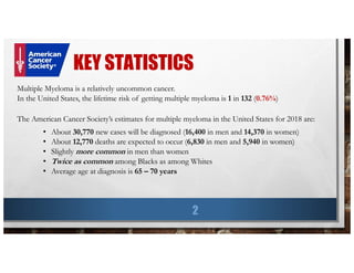
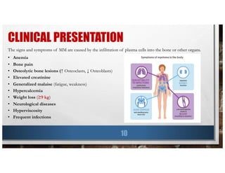
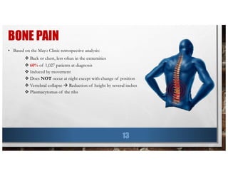
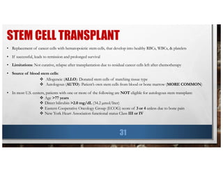
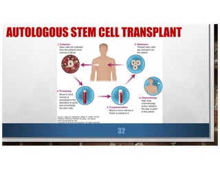
This document provides information about multiple myeloma, including key statistics, risk factors, symptoms, diagnosis, and tests. It summarizes findings from a retrospective analysis of over 1,000 multiple myeloma patients at the Mayo Clinic from 1985 to 1988. The analysis found that the median age at diagnosis was 66 years old, common symptoms at presentation included anemia (73% of patients), bone pain (60% of patients), and renal disease (20% of patients had elevated creatinine). The median survival time was 33 months and did not improve from 1985 to 1988.
Multiple myeloma is a cancer of plasma cells that is characterized by malignant proliferation of a single clone of plasma cells in the bone marrow. It most commonly affects bones and can cause bone pain, fractures, anemia, renal failure and recurrent infections. Diagnosis involves blood and urine tests showing monoclonal proteins and bone marrow biopsy with over 30% plasma cells. Treatment involves chemotherapy, stem cell transplantation, radiation, steroids and supportive care. Newer targeted therapies are improving outcomes for patients.
Multiple myeloma is a cancer of plasma cells that produce abnormal proteins and can cause issues like kidney damage, anemia, and bone lesions. It is diagnosed based on blood tests, bone marrow biopsy, and imaging showing signs of bone damage. Prognosis depends on factors like age, kidney function, and chromosome abnormalities. Treatment may include chemotherapy, steroids, stem cell transplant, and newer drugs like bortezomib, lenalidomide, and thalidomide. Patients are monitored after treatment for signs of recurrence.
Multiple myeloma is a malignant proliferation of plasma cells that commonly affects bone. It causes bone pain, fractures, renal failure, anemia, and susceptibility to infection. The cause is unknown but genetic factors may be involved. Myeloma cells interact with bone marrow to increase osteoclast activity, causing lytic bone lesions. Diagnosis requires bone marrow plasmocytosis, serum/urine monoclonal protein, and end-organ damage. Risk is stratified using beta-2 microglobulin and FISH. Treatment involves induction, consolidation, and maintenance therapies such as bortezomib, lenalidomide, and dexamethasone combinations. Supportive care focuses on complications like hypercalcemia, fractures, and anemia
This document provides an overview of multiple myeloma, including its definition, etiology, incidence, pathogenesis, clinical features, diagnosis, and staging. Some key points:
- Multiple myeloma is a malignant proliferation of plasma cells from a single clone that results in organ dysfunction. Unknown etiology but some associations with occupational exposures.
- Chromosomal alterations like deletions and translocations are significant for prognosis. IL-6 plays a role in driving proliferation.
- Presenting symptoms often include bone pain, renal failure, anemia, and bone lesions. Laboratory findings include M protein, increased calcium, and bone marrow plasmacytosis.
- Staging includes distinguishing between monoclonal gammopathy of un
Multiple myeloma is a cancer of plasma cells that produce abnormal antibodies. It causes bone destruction and can damage the kidneys and suppress the bone marrow. While the cause is unknown, risk factors include age, family history, and exposure to radiation. Symptoms include bone pain, fatigue, recurrent infection, and kidney problems. Diagnosis involves blood and urine tests and a bone marrow biopsy. Staging uses tests such as MRI, blood tests, and bone surveys. Treatment may include chemotherapy, steroids, radiation, stem cell transplants, and newer drugs that target specific pathways in myeloma cells. While not yet curable, novel agents have improved survival rates and quality of life compared to conventional chemotherapy alone.
Multiple myeloma is a group of diseases characterized by proliferation of a single plasma cell clone that overproduces monoclonal immunoglobulin molecules known as paraprotein. Myeloma cells are located in the bone marrow and blood, while paraprotein is found in the blood and urine. Diagnostic features include more than 10% plasma cells in bone marrow, presence of an M-band on serum electrophoresis, hypercalcemia, and Bence-Jones proteins in urine that precipitate with heating. Clinical manifestations include bone disease, impaired renal function, anemia, hypercalcemia, and infections.
This document discusses multiple myeloma, a plasma cell neoplasm. It covers the epidemiology, etiology, clinical presentation, diagnostic workup, staging, prognostic factors, and treatment approaches for multiple myeloma. Key points include that multiple myeloma accounts for 22% of all mature B-cell neoplasms and presents with hematologic dysfunction, bone disease, infections, and organ impairment. Diagnosis involves blood and bone marrow tests to detect monoclonal proteins and clonal plasma cells. Treatment may include chemotherapy, stem cell transplantation, radiation therapy, immunotherapy, and management of symptoms.
Multiple myeloma is a cancer of plasma cells that produces abnormal proteins. It most commonly affects people over age 65 and is more common in African populations. Symptoms include bone pain, infections, anemia, kidney problems, and neurological issues. Diagnosis requires the presence of clonal plasma cells in bone marrow, monoclonal proteins in blood or urine, or biomarkers of malignancy. Initial tests evaluate for paraproteins, organ damage, tumor burden, and prognosis.
A 55-year-old male presented with new symptoms of exertional fatigue. Bloodwork found low hemoglobin and a serum protein electrophoresis demonstrated a monoclonal IgA protein. A skeletal survey showed lytic bone lesions in the skull and humeri. A bone marrow biopsy showed 30% involvement by abnormal plasma cells positive for CD138. This suggests a diagnosis of multiple myeloma based on the presence of a monoclonal protein, bone lesions, and bone marrow involvement by plasma cells.
The passage summarizes key changes in the 2016 revision of the World Health Organization classification of lymphoid neoplasms compared to the 2008 classification. Some notable changes include new entities recognized based on next-generation sequencing and other molecular data, as well as splits or merges of some subtypes. The revised classification includes updated categories for mature B-cell, mature T-cell and NK-cell neoplasms, Hodgkin lymphoma, post-transplant lymphoproliferative disorders, and histiocytic and dendritic cell neoplasms. New provisional entities are also included to reflect evolving clinical and biological understanding.
This document provides information on multiple myeloma. Key points include:
- Multiple myeloma is a neoplastic proliferation of plasma cells producing monoclonal immunoglobulins that can cause symptoms.
- It occurs more commonly in older adults, blacks, and those with radiation/chemical exposure. Common signs are anemia, bone pain, elevated creatinine, and hypercalcemia.
- Diagnosis requires clonal plasma cells ≥10% on bone marrow biopsy or related end organ damage like kidney impairment. Staging involves tests like SPEP, UPEP, and imaging to identify bone lesions.
Multiple myeloma is a cancer of plasma cells that results in excessive numbers of abnormal plasma cells in the bone marrow. It is characterized by overproduction of monoclonal proteins, known as M proteins, and can cause symptoms such as anemia, kidney damage, and bone fractures. Diagnosis involves blood and urine tests to detect M proteins, bone marrow biopsy to identify plasma cell percentage, and imaging tests to check for bone lesions. Treatment may include chemotherapy, steroids, stem cell transplantation, and other newer drugs depending on the stage of disease and patient health. The goals of treatment are to eliminate the myeloma, control disease activity, and relieve symptoms in order to prolong survival and quality of life.
This document provides an overview of the approach to cystic bone lesions. It begins with definitions and classifications of benign and malignant cystic bone lesions. Key aspects to consider in the approach include the age of the patient, location of the lesion, characteristics of the transitional zone, presence of a matrix, status of the bone cortex, periosteal reaction, and soft tissue swelling. Specific cystic lesions discussed individually include aneurysmal bone cyst, solitary bone cyst, fibrous dysplasia, and enchondroma. Treatment approaches are also summarized for some of the lesions.
The document summarizes key topics related to plasma cell dyscrasias and multiple myeloma, including definitions, investigations, classifications, and treatment approaches. It describes the typical features of plasma cells, abnormalities like Russell bodies and Mott cells. It outlines criteria for monoclonal gammopathy of undetermined significance (MGUS), smoldering myeloma, solitary plasmacytoma, extramedullary plasmacytoma, and multiple myeloma. It discusses workup, staging, cytogenetics, and management options for multiple myeloma including stem cell transplantation and novel agents.
- A 55-year-old male presented with peripheral and central lymphadenopathy and splenomegaly but was asymptomatic. A lymph node biopsy was performed.
- Microscopic examination revealed features consistent with follicular lymphoma (FL), a neoplasm composed of follicle center B-cells which usually has at least a partially follicular pattern. FL is characterized by t(14;18) translocation and involves bone marrow in 40-70% of cases.
- FL was further classified based on the number of centroblasts per high power field according to the Mann and Berard grading system into Grade I (0-5 centroblasts/HPF), Grade II (6-15 centroblasts/
Plasma cell disorders are a group of lymphoid neoplasms involving the expansion of a single plasma cell clone secreting monoclonal immunoglobulins. Multiple myeloma is a malignant plasma cell disorder characterized by proliferation of plasma cells in the bone marrow, resulting in anemia, bone lesions, hypercalcemia and renal failure. Treatment involves alkylating agents, glucocorticoids, immunomodulatory drugs and proteasome inhibitors. New targeted therapies and personalized treatment approaches based on disease risk factors are improving outcomes.
Multiple myeloma is a malignant proliferation of plasma cells in the bone marrow that produces monoclonal immunoglobulins. It causes organ dysfunction through tumor infiltration, paraprotein production, and impaired immunity. Common signs include bone pain, fractures, infections, anemia, renal failure, and hypercalcemia. Diagnosis is based on bone marrow plasmacytosis, monoclonal proteins, and organ or tissue impairment. Treatment aims to relieve symptoms, control disease activity, extend survival, and preserve quality of life. Novel agents like bortezomib provide additional options for patients with relapsed or refractory disease.
Multiple myeloma is a cancer of plasma cells that produce abnormal antibodies. When myeloma cells spread throughout the bone marrow, it is called multiple myeloma. Renal impairment is a common feature of symptomatic multiple myeloma and can provide a clue to the diagnosis. The International Myeloma Working Group diagnostic criteria require either 10% or more plasma cells in the bone marrow or a biopsy-proven plasmacytoma plus evidence of end organ damage like hypercalcemia, renal insufficiency, anemia, or bone lesions.
This document discusses various types of liver lesions including regenerative nodules, dysplastic nodules, hepatocellular adenoma, focal nodular hyperplasia, and hepatocellular carcinoma. It provides details on the histological and immunohistochemical features that can help differentiate these lesions. Key points include that dysplastic nodules are believed to be HCC precursors, hepatocellular adenomas can be single or multifocal and classified based on molecular features, and the distinction between well-differentiated HCC and hepatocellular adenoma can be challenging based on overlapping histological features alone.
This document summarizes acute myeloid leukemia (AML), a cancer of the myeloid line of blood cells. It discusses key genetic abnormalities in AML, including those involving RUNX1, CBFB, and KIT genes. The RUNX1 and CBFB genes are involved in chromosomal translocations that result in fusion proteins and interfere with normal hematopoiesis. KIT mutations also occur in some cases and activate proliferation and survival pathways. Diagnosis involves blood tests, bone marrow biopsy, and genetic testing to identify specific abnormalities.
Multiple myeloma is a cancer of plasma cells that results in excessive proliferation of monoclonal proteins. It commonly causes lytic bone lesions, anemia, hypercalcemia and renal failure. Diagnosis involves blood and bone marrow tests detecting the monoclonal protein and plasma cell percentage. Staging systems include Durie-Salmon and International Staging System. Treatment involves disease-specific therapies like chemotherapy, steroids, stem cell transplantation and supportive care for complications. Bisphosphonates are the standard treatment for bone disease and fractures may require surgical stabilization.
This document provides an overview of acute myeloid leukemia (AML). It discusses the historical background, classification, clinical features, risk stratification, diagnostic evaluation, and treatment regimens for AML. Key points include that AML is characterized by infiltration of blood and bone marrow by proliferative myeloid cells, the WHO classification system is based on clinical features, morphology, cytogenetics and molecular abnormalities, risk is stratified by cytogenetics and molecular markers, and treatment involves supportive care, induction chemotherapy, and consideration of novel targeted therapies or stem cell transplant depending on risk factors.
Acute lymphoblastic leukemia (ALL) is the most common malignancy in children. It is characterized by the overproduction of immature white blood cells called lymphoblasts. The disease is classified based on immunophenotyping and cytogenetics. Prognostic factors include age, white blood cell count, cytogenetics, and immunophenotype. Diagnosis involves examination of peripheral blood, bone marrow aspirate, immunophenotyping, cytogenetics, and molecular testing. Treatment and monitoring of minimal residual disease is important. Genetic conditions and environmental exposures can predispose children to developing ALL.
Imaging gives a clue on pattern recognition for differential diagnosis. Therefore imaging alone may not be sufficient for the diagnosis of MM.
Hematopathologic and Histopathologic correlation are important key for differentiation and definitive diagnosis.
This document discusses the classification of acute myeloid leukemia (AML). It provides an overview of classifications proposed over time including the French-American-British (FAB) classification from 1976 based on morphology and cytochemistry. The 2008 World Health Organization classification expanded genetic subtypes and incorporated cytogenetic and molecular information along with morphology. Accurate classification involves evaluating blast percentage, cell morphology, dysplasia, immunophenotyping, cytogenetics and molecular genetics to identify distinct biological entities of AML.
This document provides an overview of multiple myeloma, including its definition, clinical presentation, workup, staging, management, and treatment regimens. Key points include:
- Multiple myeloma is a malignant neoplasm of plasma cells that accumulate in the bone marrow, leading to bone destruction.
- Workup involves blood and urine tests, bone marrow biopsy, skeletal survey, and imaging to determine disease severity and stage according to the Durie-Salmon or ISS staging systems.
- Treatment may include chemotherapy, steroids, immunomodulators, stem cell transplantation, and supportive care. The goal of initial therapy is to achieve remission prior to stem cell transplantation in eligible patients.
Hodgkin Lymphoma - Diagnosis to ManagementSubhash Thakur
Presentation is about Hodgkin lymphoma, its incidence and epidemiology, diagnosis, molecular and immunophenotype, work up, staging, treatment and follow up
This case presentation describes a 55-year-old male referred for management of severe anemia. He has a history of recurrent kidney stones, hypertension, diabetes, and back pain. Laboratory tests reveal severe anemia with rouleaux formation. Further workup shows evidence of a gammopathy. A bone marrow biopsy is needed to confirm a diagnosis of multiple myeloma, which can cause anemia and kidney damage through paraprotein production and bone lesions. Treatment involves supportive care, chemotherapy, and stem cell transplantation in eligible patients.
This document discusses leukocyte disorders and provides information about acute leukemia. It defines leukocytosis and leukopenia as increases or decreases in white blood cell count. Non-neoplastic causes of changes in white blood cells include neutrophilia, lymphocytosis, eosinophilia, monocytosis, and basophilia. Neoplastic disorders include acute myeloid leukemia and acute lymphoblastic leukemia. The document outlines the classification, pathogenesis, clinical features, diagnosis and treatment of acute leukemias. Diagnosis involves examination of blood and bone marrow smears, cytochemistry, immunophenotyping, cytogenetics and molecular analysis to determine the leukemia subtype and guide treatment.
LEUKEMIC DISEASES AND THE EYE.pptx. This talks about the ocular manifestation...BARNABASMUGABI
This document provides an overview of leukemic diseases and their ocular manifestations. It discusses the different types of leukemia including acute myeloid leukemia, acute lymphoblastic leukemia, chronic myeloid leukemia, and chronic lymphocytic leukemia. It covers the pathogenesis, classification, signs and symptoms, diagnostic testing including blood counts, bone marrow examination, and cytochemical staining. It also describes the various ocular changes that can occur due to anemia, thrombocytopenia, hyperviscosity, thrombosis, infiltration of tissues, and metabolic abnormalities associated with leukemias.
Acute leukemias are malignant disorders of hematopoietic tissues characterized by increased white blood cells in the bone marrow and blood. They are classified as either acute lymphoblastic leukemia (ALL) or acute myeloid leukemia (AML) based on the affected cell lineage. Treatment involves chemotherapy to induce remission through combination regimens, with the goals of restoring normal hematopoiesis and preventing relapse through additional consolidation therapy and long-term maintenance treatment. Prognosis depends on several risk factors like age, subtype, initial response to treatment, and specific genetic abnormalities.
MDS and myeloproliferative disorders are heterogeneous groups of clonal stem cell disorders characterized by ineffective or excessive blood cell production. MDS involves dysplastic and ineffective hematopoiesis that can progress to acute leukemia, while myeloproliferative disorders feature overgrowth of one or more myeloid cell lines. Key disorders discussed include MDS, polycythemia vera involving excessive red blood cell production, essential thrombocytosis with marked thrombocytosis, myelofibrosis with bone marrow fibrosis, and chronic myeloid leukemia characterized by the Philadelphia chromosome translocation. Diagnosis and classification of these disorders has evolved with new genetic and molecular testing.
Plasma cell disorders are characterized by the disproportionate proliferation of a single clone of plasma cells resulting in monoclonal immunoglobulins. The most common types are monoclonal gammopathy of undetermined significance (MGUS), multiple myeloma, and solitary plasmacytoma. Diagnosis involves detecting the monoclonal protein by serum and urine protein electrophoresis and bone marrow biopsy. Risk stratification uses tools like ISS staging and FISH. Treatment depends on disease stage and eligibility for stem cell transplant, and may include chemotherapy, corticosteroids, immunomodulatory drugs, proteasome inhibitors, and autologous stem cell transplant.
Multiple myeloma is a cancer of plasma cells that results in overproduction of abnormal antibodies in the bone marrow. It commonly causes bone pain, fractures, anemia, and kidney problems. Risk factors include age over 60 and exposure to chemicals like pesticides, radiation, or certain industrial chemicals. Treatment may include chemotherapy, steroids, stem cell transplantation, radiation, surgery, and newer drugs like thalidomide, lenalidomide, and bortezomib to improve outcomes. Despite recent advances, multiple myeloma remains incurable and patients often relapse, highlighting the need for additional therapeutic options.
acute leukemia
For More Medicine Free PPT - http://playnever.blogspot.com/
For Health benefits and medicine videos Subscribe youtube channel - https://www.youtube.com/playlist?list=PLKg-H-sMh9G01zEg4YpndngXODW2bq92w
The document discusses various myeloproliferative neoplasms (MPNs) including classic MPNs such as polycythemia vera, essential thrombocythemia, and primary myelofibrosis as well as atypical MPNs. It provides details on the diagnostic criteria, signs and symptoms, genetic mutations involved, and treatment approaches for these different MPNs. Chronic myelogenous leukemia is also examined as it represents a distinct MPN characterized by the Philadelphia chromosome genetic abnormality.
Leukemia is the most common pediatric malignancy, accounting for 1/3 of all childhood cancers. It is defined as the malignant clonal proliferation of lymphoid or myeloid precursor cells in the bone marrow and infiltration of other organs. The presentation includes general symptoms like fever, fatigue, and pallor as well as hematological effects from bone marrow invasion including anemia, neutropenia, and thrombocytopenia. Diagnosis involves blood tests, bone marrow examination, and other investigations. Treatment consists of induction chemotherapy followed by consolidation and maintenance therapy to achieve remission and prevent relapse.
This document discusses multiple myeloma, a plasma cell disorder. It begins with an overview of plasma cell disorders and defines multiple myeloma. It then covers the epidemiology, etiology, pathophysiology, clinical features, diagnostic tests including serum protein electrophoresis and immunofixation, bone marrow examination, skeletal survey, staging, prognostic factors. It also discusses related conditions like monoclonal gammopathy of undetermined significance, smoldering myeloma, non-secretory myeloma, solitary plasmacytoma, extramedullary plasmacytoma. It concludes with discussing criteria for diagnosing multiple myeloma and initial treatment approaches.
Leukemias are malignant diseases of the bone marrow characterized by the abnormal proliferation of white blood cells. The document discusses the classification, pathogenesis, epidemiology, clinical features, diagnosis and prognosis of various types of leukemias. It describes the key subtypes of acute and chronic leukemias, focusing on chronic myeloid leukemia, which results from a genetic translocation forming the Philadelphia chromosome and the constitutively active BCR-ABL fusion oncogene that drives uncontrolled myeloid cell growth.
This document provides an overview of acute myeloid leukemia (AML). It discusses the etiology, classification, clinical features, laboratory findings, treatment including induction chemotherapy, post-remission therapy such as stem cell transplantation, and prognostic factors of AML. The key points are that AML is a cancer of the myeloid line of blood cells, its incidence increases with age, and treatment involves induction chemotherapy to achieve remission followed by post-remission therapy to prevent relapse.
Multiple myeloma is characterized by neoplastic proliferation of plasma cells producing monoclonal immunoglobulin. It commonly affects elderly individuals and presents with bone disease, anemia, infections and renal failure. Diagnosis involves finding monoclonal proteins in serum or urine and plasma cell infiltration in bone marrow. Treatment aims to improve quality of life and may include chemotherapy, stem cell transplant, supportive care and management of complications.
This document provides information about multiple myeloma. It begins by describing multiple myeloma as a malignant proliferation of plasma cells from a single clone that most commonly affects bone. It then discusses the incidence, risk factors, clinical features, recurrent infections, renal failure, anemia, classifications, diagnostic features, laboratory findings, radiology, and management of multiple myeloma. In summary, it provides an overview of multiple myeloma including its causes, symptoms, diagnostic criteria, staging systems, and treatment options.
This document discusses aplastic anemia, including its definition, causes, pathophysiology, symptoms, diagnosis, treatment, and differential diagnosis. Aplastic anemia is a condition where the bone marrow fails to produce sufficient new blood cells, leading to pancytopenia. It can be caused by radiation, chemicals, drugs, infections, autoimmune or genetic disorders. Diagnosis involves blood tests showing low blood cell counts and a bone marrow biopsy appearing hypocellular. Treatment options include stem cell transplant or immunosuppression with antithymocyte globulin and cyclosporine to recover bone marrow function.
This document discusses leukemias and provides information about leukemoid reactions. It defines a leukemoid reaction as a high white blood cell count with neutrophilia usually in response to infection, which can mimic chronic myelogenous leukemia or acute myeloid leukemia. The document notes that serum leukocyte alkaline phosphatase is normally elevated in leukemoid reactions, distinguishing it from CML where it is depressed. Features suggesting a leukemoid reaction rather than leukemia include toxic granulation, a high LAP score, and an obvious cause of the neutrophilia such as infection. The document provides several potential causes of leukemoid reactions and discusses methods for distinguishing leukemoid reactions from leukemia.
This document discusses multiple myeloma (MM) and monoclonal gammopathy of undetermined significance (MGUS). It defines MGUS and describes its three distinct types. It outlines the workup, disease course, and monitoring for MGUS. Complications of MGUS discussed include anemia, fractures, and thrombosis. MM is then reviewed, including typical presentations and complications like renal disease. Renal disease in MM is further explained, focusing on light chain cast nephropathy.
Multiple myeloma - Etiopathogenesis, Clinical features, Advances in ManagementChetan Ganteppanavar
Multiple myeloma is a cancer of plasma cells that accumulate in the bone marrow. It causes anemia, bone lesions, high calcium levels, and kidney damage. The disease arises from post-germinal center plasma cells and is characterized by genetic abnormalities. Myeloma cells interact with the bone marrow microenvironment to gain growth and drug resistance advantages. Treatment involves systemic therapy to control disease progression and supportive care for complications. High-dose chemotherapy with autologous stem cell transplantation is the standard initial treatment for eligible patients.
Aplastic anemia is a bone marrow failure syndrome characterized by pancytopenia and a hypocellular bone marrow. It can be caused by radiation, chemicals, drugs, infections, autoimmune or genetic conditions. Patients present with bleeding, anemia symptoms, or infection. Diagnosis involves blood tests showing pancytopenia and low reticulocytes, along with a bone marrow biopsy demonstrating a hypocellular marrow. Treatment involves stem cell transplant or immunosuppression with antithymocyte globulin and cyclosporine.
Dr Andrew Stewart, Consultant Haematologist, The Royal Gwent Hospital
Dr Chris Jenkins, Consultant Haematologist, University Hospital of the North Midlands
Oscar Kwan is an experienced pharmacist with over 3 years of retail pharmacy experience. He has interned at Pfizer where he analyzed safety reports and regulatory documents. Kwan received his PharmD from St. John's University with a GPA of 3.77. He is proficient in medical writing, data analysis, and using technology to communicate health information.
Rapid sequence intubation (RSI) is an emergency method used to safely and rapidly intubate a patient's airway while minimizing the risks of regurgitation and aspiration. The steps of RSI include preoxygenation, positioning the patient, administering premedication, applying cricoid pressure, rapidly inducing unconsciousness with drugs like propofol or ketamine, paralyzing the patient with succinylcholine or rocuronium, intubating the trachea, and providing post-intubation care and ventilation. RSI aims to protect patients from complications of aspiration that can occur if the airway is not properly protected during emergency intubation.
Diabetic ketoacidosis (DKA) is typically associated with type 1 diabetes but can also occur in type 2 diabetes under extreme stress. DKA results from a lack of insulin and excess glucagon and catecholamines, leading to fat breakdown, ketone production, and high blood sugar. Symptoms include nausea, vomiting, fruity breath, and signs of dehydration. DKA is diagnosed based on hyperglycemia, ketonemia, and metabolic acidosis.
Aminoglycosides like gentamicin are effective against many gram-negative bacteria but can cause nephrotoxicity and ototoxicity. Penicillins such as amoxicillin are effective against streptococci and have good oral availability but are susceptible to beta-lactamases. Cephalosporins have expanding spectra of activity against gram-positive and gram-negative organisms but can cause diarrhea and interact with warfarin. Carbapenems like imipenem are very broad-spectrum but can induce seizures if renal dosing is not adjusted.
Estrogen Trimestegone Effect on Breast CarcinomaOscarKwan6
Breast cancer is the most commonly diagnosed cancer and the second leading cause of cancer death among women in the United States. Risk factors for breast cancer include older age, family history, certain genetic mutations, high breast density, radiation exposure, late menopause, early menstruation, late or no pregnancy. Studies have found that oral contraceptive use is associated with a slight increased risk of breast cancer, though this risk decreases 10 years after stopping use. Certain combined hormone replacement therapies of estrogen and progestin are also associated with increased breast cancer risk apparent after 3 years of use, but risk returns to baseline within 5 years of stopping treatment.
Inotuzumab ozogamicin is an antibody-drug conjugate being studied for the treatment of acute lymphoblastic leukemia (ALL) in adults. The INO-VATE ALL study compared inotuzumab ozogamicin to standard chemotherapy for adults with relapsed or refractory ALL. The primary objectives were to demonstrate higher rates of complete remission and longer overall survival with inotuzumab ozogamicin. Complete remission, safety, and overall survival were evaluated. If shown to be effective, inotuzumab ozogamicin could provide a new treatment option for adults with relapsed/refractory ALL.
Sunitinib Malate and its Important Identified Risk of Fistula FormationOscarKwan6
Sunitinib (Sutent) is associated with an important identified risk of fistula formation. Fistulas are abnormal connections between organs that can develop spontaneously or after surgery and radiation. The mechanism may involve disturbed vascular integrity from VEGF inhibition. Incidence of fistula with sunitinib is highest in gastrointestinal stromal tumor and pancreatic neuroendocrine tumor patients. Fistulas can be life-threatening and require hospitalization but are sometimes preventable through monitoring of high-risk patients. More information is still needed on pediatric use and in patients with cardiac or severe liver impairment.
The survey assessed pharmacy students' preferences for the structure of advanced pharmacy practice experiences (APPEs). It found that the majority of students preferred shorter 4-week APPE rotations so they could experience a greater number and more diverse range of practice settings. While some felt 5 weeks would allow sufficient time to meet objectives, most did not want longer 6-week rotations if it meant fewer total rotations. Almost half of students spent over 1 day completing orientation activities at sites, indicating a need for more time at each APPE. The survey provided insights into optimizing the APPE curriculum, but had limitations as not all students responded.
This document provides information about stroke, including:
1. Stroke is caused by a lack of oxygen to the brain from blocked or ruptured arteries, and is a leading cause of death and disability in the US and worldwide.
2. The two main types of stroke are ischemic (87% of cases) and hemorrhagic (13% of cases).
3. Signs of stroke include sudden weakness, confusion, trouble speaking, vision issues, loss of balance, and severe headaches with no known cause. Early treatment leads to better outcomes.
This document summarizes a case of a 24-year-old female diagnosed with type 1 diabetes mellitus. She presented with symptoms of polydipsia, polyuria, weight loss, and chest pain. Her blood glucose was elevated at 520 mg/dL and HbA1c was 10.4%. She was initially diagnosed with type 2 diabetes due to family history but further testing revealed positive antibodies for type 1 diabetes. She was started on insulin therapy and educated on diabetes self-management.
TEST BANK For Community Health Nursing A Canadian Perspective, 5th Edition by...Donc Test
TEST BANK For Community Health Nursing A Canadian Perspective, 5th Edition by Stamler, Verified Chapters 1 - 33, Complete Newest Version Community Health Nursing A Canadian Perspective, 5th Edition by Stamler, Verified Chapters 1 - 33, Complete Newest Version Community Health Nursing A Canadian Perspective, 5th Edition by Stamler Community Health Nursing A Canadian Perspective, 5th Edition TEST BANK by Stamler Test Bank For Community Health Nursing A Canadian Perspective, 5th Edition Pdf Chapters Download Test Bank For Community Health Nursing A Canadian Perspective, 5th Edition Pdf Download Stuvia Test Bank For Community Health Nursing A Canadian Perspective, 5th Edition Study Guide Test Bank For Community Health Nursing A Canadian Perspective, 5th Edition Ebook Download Stuvia Test Bank For Community Health Nursing A Canadian Perspective, 5th Edition Questions and Answers Quizlet Test Bank For Community Health Nursing A Canadian Perspective, 5th Edition Studocu Test Bank For Community Health Nursing A Canadian Perspective, 5th Edition Quizlet Test Bank For Community Health Nursing A Canadian Perspective, 5th Edition Stuvia Community Health Nursing A Canadian Perspective, 5th Edition Pdf Chapters Download Community Health Nursing A Canadian Perspective, 5th Edition Pdf Download Course Hero Community Health Nursing A Canadian Perspective, 5th Edition Answers Quizlet Community Health Nursing A Canadian Perspective, 5th Edition Ebook Download Course hero Community Health Nursing A Canadian Perspective, 5th Edition Questions and Answers Community Health Nursing A Canadian Perspective, 5th Edition Studocu Community Health Nursing A Canadian Perspective, 5th Edition Quizlet Community Health Nursing A Canadian Perspective, 5th Edition Stuvia Community Health Nursing A Canadian Perspective, 5th Edition Test Bank Pdf Chapters Download Community Health Nursing A Canadian Perspective, 5th Edition Test Bank Pdf Download Stuvia Community Health Nursing A Canadian Perspective, 5th Edition Test Bank Study Guide Questions and Answers Community Health Nursing A Canadian Perspective, 5th Edition Test Bank Ebook Download Stuvia Community Health Nursing A Canadian Perspective, 5th Edition Test Bank Questions Quizlet Community Health Nursing A Canadian Perspective, 5th Edition Test Bank Studocu Community Health Nursing A Canadian Perspective, 5th Edition Test Bank Quizlet Community Health Nursing A Canadian Perspective, 5th Edition Test Bank Stuvia
These lecture slides, by Dr Sidra Arshad, offer a quick overview of the physiological basis of a normal electrocardiogram.
Learning objectives:
1. Define an electrocardiogram (ECG) and electrocardiography
2. Describe how dipoles generated by the heart produce the waveforms of the ECG
3. Describe the components of a normal electrocardiogram of a typical bipolar lead (limb II)
4. Differentiate between intervals and segments
5. Enlist some common indications for obtaining an ECG
6. Describe the flow of current around the heart during the cardiac cycle
7. Discuss the placement and polarity of the leads of electrocardiograph
8. Describe the normal electrocardiograms recorded from the limb leads and explain the physiological basis of the different records that are obtained
9. Define mean electrical vector (axis) of the heart and give the normal range
10. Define the mean QRS vector
11. Describe the axes of leads (hexagonal reference system)
12. Comprehend the vectorial analysis of the normal ECG
13. Determine the mean electrical axis of the ventricular QRS and appreciate the mean axis deviation
14. Explain the concepts of current of injury, J point, and their significance
Study Resources:
1. Chapter 11, Guyton and Hall Textbook of Medical Physiology, 14th edition
2. Chapter 9, Human Physiology - From Cells to Systems, Lauralee Sherwood, 9th edition
3. Chapter 29, Ganong’s Review of Medical Physiology, 26th edition
4. Electrocardiogram, StatPearls - https://www.ncbi.nlm.nih.gov/books/NBK549803/
5. ECG in Medical Practice by ABM Abdullah, 4th edition
6. Chapter 3, Cardiology Explained, https://www.ncbi.nlm.nih.gov/books/NBK2214/
7. ECG Basics, http://www.nataliescasebook.com/tag/e-c-g-basics
Recomendações da OMS sobre cuidados maternos e neonatais para uma experiência pós-natal positiva.
Em consonância com os ODS – Objetivos do Desenvolvimento Sustentável e a Estratégia Global para a Saúde das Mulheres, Crianças e Adolescentes, e aplicando uma abordagem baseada nos direitos humanos, os esforços de cuidados pós-natais devem expandir-se para além da cobertura e da simples sobrevivência, de modo a incluir cuidados de qualidade.
Estas diretrizes visam melhorar a qualidade dos cuidados pós-natais essenciais e de rotina prestados às mulheres e aos recém-nascidos, com o objetivo final de melhorar a saúde e o bem-estar materno e neonatal.
Uma “experiência pós-natal positiva” é um resultado importante para todas as mulheres que dão à luz e para os seus recém-nascidos, estabelecendo as bases para a melhoria da saúde e do bem-estar a curto e longo prazo. Uma experiência pós-natal positiva é definida como aquela em que as mulheres, pessoas que gestam, os recém-nascidos, os casais, os pais, os cuidadores e as famílias recebem informação consistente, garantia e apoio de profissionais de saúde motivados; e onde um sistema de saúde flexível e com recursos reconheça as necessidades das mulheres e dos bebês e respeite o seu contexto cultural.
Estas diretrizes consolidadas apresentam algumas recomendações novas e já bem fundamentadas sobre cuidados pós-natais de rotina para mulheres e neonatos que recebem cuidados no pós-parto em unidades de saúde ou na comunidade, independentemente dos recursos disponíveis.
É fornecido um conjunto abrangente de recomendações para cuidados durante o período puerperal, com ênfase nos cuidados essenciais que todas as mulheres e recém-nascidos devem receber, e com a devida atenção à qualidade dos cuidados; isto é, a entrega e a experiência do cuidado recebido. Estas diretrizes atualizam e ampliam as recomendações da OMS de 2014 sobre cuidados pós-natais da mãe e do recém-nascido e complementam as atuais diretrizes da OMS sobre a gestão de complicações pós-natais.
O estabelecimento da amamentação e o manejo das principais intercorrências é contemplada.
Recomendamos muito.
Vamos discutir essas recomendações no nosso curso de pós-graduação em Aleitamento no Instituto Ciclos.
Esta publicação só está disponível em inglês até o momento.
Prof. Marcus Renato de Carvalho
www.agostodourado.com
Does Over-Masturbation Contribute to Chronic Prostatitis.pptxwalterHu5
In some case, your chronic prostatitis may be related to over-masturbation. Generally, natural medicine Diuretic and Anti-inflammatory Pill can help mee get a cure.
Histololgy of Female Reproductive System.pptxAyeshaZaid1
Dive into an in-depth exploration of the histological structure of female reproductive system with this comprehensive lecture. Presented by Dr. Ayesha Irfan, Assistant Professor of Anatomy, this presentation covers the Gross anatomy and functional histology of the female reproductive organs. Ideal for students, educators, and anyone interested in medical science, this lecture provides clear explanations, detailed diagrams, and valuable insights into female reproductive system. Enhance your knowledge and understanding of this essential aspect of human biology.
Basavarajeeyam is an important text for ayurvedic physician belonging to andhra pradehs. It is a popular compendium in various parts of our country as well as in andhra pradesh. The content of the text was presented in sanskrit and telugu language (Bilingual). One of the most famous book in ayurvedic pharmaceutics and therapeutics. This book contains 25 chapters called as prakaranas. Many rasaoushadis were explained, pioneer of dhatu druti, nadi pareeksha, mutra pareeksha etc. Belongs to the period of 15-16 century. New diseases like upadamsha, phiranga rogas are explained.
Integrating Ayurveda into Parkinson’s Management: A Holistic ApproachAyurveda ForAll
Explore the benefits of combining Ayurveda with conventional Parkinson's treatments. Learn how a holistic approach can manage symptoms, enhance well-being, and balance body energies. Discover the steps to safely integrate Ayurvedic practices into your Parkinson’s care plan, including expert guidance on diet, herbal remedies, and lifestyle modifications.
Here is the updated list of Top Best Ayurvedic medicine for Gas and Indigestion and those are Gas-O-Go Syp for Dyspepsia | Lavizyme Syrup for Acidity | Yumzyme Hepatoprotective Capsules etc
Cell Therapy Expansion and Challenges in Autoimmune DiseaseHealth Advances
There is increasing confidence that cell therapies will soon play a role in the treatment of autoimmune disorders, but the extent of this impact remains to be seen. Early readouts on autologous CAR-Ts in lupus are encouraging, but manufacturing and cost limitations are likely to restrict access to highly refractory patients. Allogeneic CAR-Ts have the potential to broaden access to earlier lines of treatment due to their inherent cost benefits, however they will need to demonstrate comparable or improved efficacy to established modalities.
In addition to infrastructure and capacity constraints, CAR-Ts face a very different risk-benefit dynamic in autoimmune compared to oncology, highlighting the need for tolerable therapies with low adverse event risk. CAR-NK and Treg-based therapies are also being developed in certain autoimmune disorders and may demonstrate favorable safety profiles. Several novel non-cell therapies such as bispecific antibodies, nanobodies, and RNAi drugs, may also offer future alternative competitive solutions with variable value propositions.
Widespread adoption of cell therapies will not only require strong efficacy and safety data, but also adapted pricing and access strategies. At oncology-based price points, CAR-Ts are unlikely to achieve broad market access in autoimmune disorders, with eligible patient populations that are potentially orders of magnitude greater than the number of currently addressable cancer patients. Developers have made strides towards reducing cell therapy COGS while improving manufacturing efficiency, but payors will inevitably restrict access until more sustainable pricing is achieved.
Despite these headwinds, industry leaders and investors remain confident that cell therapies are poised to address significant unmet need in patients suffering from autoimmune disorders. However, the extent of this impact on the treatment landscape remains to be seen, as the industry rapidly approaches an inflection point.
Osteoporosis - Definition , Evaluation and Management .pdfJim Jacob Roy
Osteoporosis is an increasing cause of morbidity among the elderly.
In this document , a brief outline of osteoporosis is given , including the risk factors of osteoporosis fractures , the indications for testing bone mineral density and the management of osteoporosis
2. KEY STATISTICS
2
Multiple Myeloma is a relatively uncommon cancer.
In the United States, the lifetime risk of getting multiple myeloma is 1 in 132 (0.76%)
The American Cancer Society’s estimates for multiple myeloma in the United States for 2018 are:
• About 30,770 new cases will be diagnosed (16,400 in men and 14,370 in women)
• About 12,770 deaths are expected to occur (6,830 in men and 5,940 in women)
• Slightly more common in men than women
• Twice as common among Blacks as among Whites
• Average age at diagnosis is 65 – 70 years
3. WHAT IS MULTIPLE MYELOMA?
• A CANCER of plasma cells (fully mature B cell)
• Lymphocytes: Main types of WBCs (B cells, T cells, NK cells)
• Overproduction of monoclonal Antibodies (immunoglobulins)
• Adaptive Immunity vs. Innate Immunity
3
4. PATHOLOGY
• Bone Marrow: Home site for hematopoiesis (Disrupted balance in Multiple Myeloma)
• Neoplastic proliferation in the bone marrow of a SINGLE CLONE of malignant plasma cells
• Immortal cancerous plasma cells accumulate and deplete the production of normal healthy blood cells
• Production of abnormal antibodies known by monoclonal immunoglobulin, M-protein, monoclonal protein,
M-spike, or paraprotein
4
5. RISK FACTORS
5
• Genetic predisposition (Familial clusters of Multiple myeloma has been reported)
• Environmental factors:
q Radiation exposure (Hiroshima and Nagasaki atomic bomb survivors, Radiologists, Nuclear plant workers)
q Organic chemicals (Aromatic hydrocarbons; benzene)
q Herbicides
q Insecticides
• Old age ≥65 years old
• Gender: Male > Female
• Race: African Americans >2x more common than White Americans
• Weight: Overweight or Obesity (↑ BMI 5 kg/m2 = ↑ 11% Risk)
• Plasma cell diseases: MGUS (Monoclonal Gammopathy of Undetermined Significance)
Solitary plasmacytoma
SMM (Smoldering Multiple Myeloma)
6. RISK STRATIFICATION
6
High-Risk Cytogenetics: 15% of MM patients
1. Deletion of a piece (p13) of Chromosome 17
2. Translocation between Chromosomes 14 and 20
3. Translocation between Chromosomes 14 and 16
4. Elevated Lactate dehydrogenase levels 2x ULN (200 units/L)
• Associated with poor prognosis and shorten survival
• Requires more aggressive treatment
7. RISK STRATIFICATION
7
Intermediate-Risk Cytogenetics: 10% of MM patients
1. Translocation between Chromosomes 4 and 14
Standard-Risk Cytogenetics: 85% of MM patients
1. All patients that lack High-risk and Intermediate-risk
genetic abnormalities
2. Estimated median survival of 8 – 10 years
8. ETIOLOGY
8
The cause of Multiple Myeloma remains unknown.
Genetic mutations: Recent studies have shown primary cytogenetic abnormalities play a major role in MM development
1. MYC oncogene – Plasma cell tumors
2. RAS oncogene – Myeloma cells in the bone marrow after treatment
3. p53 tumor suppressor gene – Metastasis to other organs
4. Immunoglobulin Heavy Chain Translocations = “Class Switching” or “Switch Recombination”
v 11q13 – cyclin D1 gene
v 6p21 – cyclin D3 gene
v 4p16.3 – multiple myeloma set domain (MMSET)
v 16q23 – musculoaponeurotic fibrosarcoma (c-maf)
v 20q11 – musculoaponeurotic fibrosarcoma oncology family, protein B (mafB)
9. TRIGGERS OF GENETIC MUTATIONS
9
Abnormal overproduction of Toll-like receptors and IL-6 receptors
Antigenic Stimulation
Abnormal and sustained growth signal
Increased proliferative rate of plasma cells
Increased risk of chromosomal changes and damage
Creation of a plasma cell clone
Overproduction of a single type of immunoglobulin
Toll-like receptors
10. CLINICAL PRESENTATION
10
The signs and symptoms of MM are caused by the infiltration of plasma cells into the bone or other organs.
• Anemia
• Bone pain
• Osteolytic bone lesions (↑ Osteoclasts, ↓ Osteoblasts)
• Elevated creatinine
• Generalized malaise (fatigue, weakness)
• Hypercalcemia
• Weight loss (≥9 kg)
• Neurological diseases
• Hyperviscosity
• Frequent infections
11. RETROSPECTIVE ANALYSIS
11
Institution: Mayo Clinic
Location: Rochester, Minn
Objective: To determine the clinical and laboratory features of newly diagnosed multiple myeloma.
Methods: Records of 1,027 patients in whom Multiple Myeloma was initially diagnosed from January 1, 1985 to
December 31, 1988 were reviewed.
Patients:
• 2% were younger than 40 years old
• 38% were 70 years old or older
• Median Age: 66 years old
Conclusion: The median duration of survival was 33 months and did not improve from 1985 through 1998.
12. ANEMIA
12
• Based on the Mayo Clinic retrospective analysis:
v Normocytic, Normochromic
v Hgb ≤12 g/dl
v 73% of 1,027 patients at diagnosis
v 97% of 1,027 patients during the course of the disease
• Precipitating Causes:
v Bone marrow replacement by malignant plasma cells
v Kidney damage (↓ EPO hormone)
v Dilution due to large M-protein
• Symptoms:
v Fatigue
v Pallor
v Weakness
v Heart palpitations
13. BONE PAIN
13
• Based on the Mayo Clinic retrospective analysis:
v Back or chest, less often in the extremities
v 60% of 1,027 patients at diagnosis
v Induced by movement
v Does NOT occur at night except with change of position
v Vertebral collapse à Reduction of height by several inches
v Plasmacytomas of the ribs
14. RENAL DISEASE
14
• Based on the Mayo Clinic retrospective analysis:
v 20% of 1,027 patients’ SCr was >2 mg/dL
v Kidney failure à requires emergent treatment
v Symptoms: Weakness, shortness of breath, Itching, Leg swelling
• Precipitating Causes:
v Light chain cast nephropathy “Myeloma Kidney”: >1,500 mg/dL of free light chain levels
v Hypercalcemia
• Other causes:
v Light chain amyloidosis (AL)
v Light chain deposition disease
v Drug-induced renal damage
15. HYPERCALCEMIA
15
• Based on the Mayo Clinic retrospective analysis:
v 13% of 1,027 patients’ serum calcium was ≥11 mg/dL
• Precipitating Causes:
v Break down of bone marrow tissue
v Binding of monoclonal protein with calcium
• Symptoms:
v Excessive thirst/Dehydration
v Frequent urination
v Stomach upset
v Nausea and vomiting
v Severe constipation
v Bone weakness
v Lethargy/fatigue
v Loss of appetite
16. NEUROLOGICAL DISEASES
16
• Radiculopathy:
v Most common neurological complication of Multiple Myeloma
v Thoracic/lumbosacral area
v Compression of nerve by paravertebral plasmacytoma or collapsed bone
• Cord Compression:
v 5% of patients
v Compression by extramedullary plasmacytoma or bone fragment of vertebral bone fracture
v Symptoms: Severe back pain with weakness, lower extremity paresthesias, bladder/bowel dysfunction
• Peripheral Neuropathy “Pins and Needles”:
v Uncommon in Multiple Myeloma
v If present, due to amyloidosis
• CNS Involvement:
v Intracranial plasmacytomas are rare
v Poor prognosis: Leptomeningeal myelomatosis (Lactate dehydrogenase [LDH] levels ↑)
v Rare: Encephalopathy due to hyperviscosity or high blood levels of ammonia
17. HYPERVISCOSITY
17
• Thickening of the blood:
v Large amounts of M-protein
v Slow blood flow to the brain
• Symptoms:
v Confusion
v Dizziness
v One-sided body weakness
v Slurred speech
• Patients with these symptoms should call their doctor
• Plasmapheresis:
v Rapid reversal
v Removal of protein from blood
• CANNOT treat with “BLOOD THINNERS”
18. INFECTION
18
• Patients with Multiple Myeloma are at increased risk of infection:
1. Immunocompromised:
v Leukopenia
v Impaired lymphocyte function
v Suppression of normal plasma cell function
v Hypogammaglobulinemia
2. Physical factors:
v Hypoventilation due to bone fracture and pain of the spine or rib cage
• Streptococcus pneumoniae and Gram-negative organisms are the most frequent pathogens
19. DIAGNOSIS
19
Criteria:
• Presence of ≥10% plasma cells in the bone marrow
OR
• Biopsy proven plasmacytoma
AND at least one of the following:
v ≥ 60% plasma cells in the bone marrow
v Calcium (Serum Ca2+>11 mg/dL)
v Renal insufficiency (CrCl <40 ml/min or SCr >2 mg/dl)
v Anemia (Hgb <10 g/dL or <2 g/dL below normal)
v Bone lesions: (≥1 osteolytic lesions ≥5 mm in size on skeletal radiography, CT, or PET scan)
It is important to rule out other plasma cell dyscrasias (MGUS, SMM) for appropriate prognosis and treatment.
Treatment of MM is time-dependent, a major delay in diagnosis has been associated with a negative impact on disease course.
20. DIAGNOSTIC TESTS
20
• Complete Blood Count:
v Red cells – Anemia
v White cells – Leukopenia
v Platelets – Thrombocytopenia
• Blood Chemistry Tests:
v Creatinine – Kidney function
v Albumin – Low levels in MM
v Calcium – Hypercalcemia
v Lactic dehydrogenase – High levels in more advanced MM with worse prognosis
v Beta-2 microglobulin – Prognosis indicator
• Blood Protein Tests:
v Serum Protein Electrophoresis (SPEP) – measures blood levels of intact whole monoclonal antibodies
v Immunofixation – determines the exact type of abnormal antibody
21. MONOCLONAL ANTIBODY
21
Light chain ( λ, κ)
Normally, present in
equal amounts in
the blood; (1:1) ratio Bence Jones protein:
Light chains of M protein.
If found in the urine,
suggestive of Multiple Myeloma.
22. DIAGNOSTIC TESTS
22
• Serum Free Light Chain (FLC) Analysis:
v Measures blood levels of light chain fragments
v Helpful when no whole M protein is found in SPEP
v Sign of Myeloma: Ratio of lambda to kappa light chains is NOT 1:1
• Urine Tests:
v Urine Protein Electrophoresis (UPEP): Detects proteinuria over a 24-hour time period
v Urine Immunofixation: Characterizes urine proteins
• Bone Marrow Aspiration and Biopsy:
v Immunohistochemistry: Special proteins identifies myeloma cells through color changes
v Flow cytometry: Bone marrow is treated with special proteins that stick only to cancer cells
v Fluorescent In Situ Hybridization (FISH): Locates translocations and deletions using fluorescent dyes
v Cytogenetics: Evaluates chromosomes for abnormalities to predict prognosis
• Fine Needle Aspiration Biopsy:
v Withdraw tissue from tumor or enlarged lymph node using a very thin needle and syringe
v Advantage: Does NOT require surgery
v Disadvantage: Insufficient amount of tissue removed due to thin needle
23. WHEN SHOULD TREATMENT START?
23
• Treatment depends on many factors:
v Age
v Comorbidities
v Presence of symptoms
v Stage of multiple myeloma
v Genetic risk level
• Weigh the risks against the benefits of treatment with your doctor
Risk
Benefit
25. CHEMOTHERAPY
25
• Stops or slows cancer cells’ ability to grow and divide
• Enters the bloodstream and reaches almost all areas of the body (NON-TARGETED THERAPY)
• Routes of Administration:
v PO
v IV
• Chemo drugs used to treat MM include:
v Melphalan (AlkeranTM)
v Vincristine (Oncovin®)
v Cyclophosphamide (Cytoxan®)
v Etoposide (Toposar®)
v Doxorubicin (Adriamycin®)
v Liposomal doxorubicin (Doxil®)
v Bendamustin (Treanda®)
26. CHEMOTHERAPY
26
• To increase effectiveness, combined with other types of treatment, such as:
v Targeted therapies
v Corticosteroids
• If a stem cell transplant is planned, avoid using bone-marrow damaging chemo drugs (e.g. Mephalan)
• Chemo drugs kill cancer cells, but also can damage normal cells
• Dose carefully to avoid or reduce side effects of chemotherapy
• Common side effects of chemotherapy:
v Alopecia
v Mouth sores
v Loss of appetite
v Nausea, vomiting, and diarrhea
v Pancytopenia
v Peripheral neuropathy
v Hemophilia
27. TARGETED THERAPY
27
• Immuno-modulating agents: Suppression of angiogenesis, strengthen immune attack on cancer cells
v Thalidomide (Thalomid®, Synovir®): severe constipation, permanent neuropathy, blood clots (DVT, PE)
v Lenalidomide (Revlimid®): thrombocytopenia, leukopenia, neuropathy, lower blood clot risk
v Pomalidomide (Pomalyst®): anemia, leukopenia, less severe neuropathy, thromboembolism
• Proteasome inhibitors: Activates the apoptosis of cancer cells by inhibiting the breakdown of proteins that control it
v Bortezomib (Velcade®): IV/SQ once or twice a week
SQ preferred, due to less peripheral neuropathy
May cause shingles (prevent with acyclovir)
v Carfilzomib (Kyprolis®): IV every 4 weeks
Allergic reactions (prevent with dexamethasone before each dose in 1st cycle)
Serious side effects (pneumonia, heart, kidney/liver failure)
Alternative if other drugs failed
v Ixazomib (Ninlaro®): PO once weekly for 3 weeks, then 1 week off
Alternative if other drugs failed
28. TARGETED THERAPY
28
• Histone deacetylase (HDAC) inhibitors: Uncoils DNA & activates genes for apoptosis, blocks aggresome (escape route)
v Panobinostat (Farydak®): PO three times a week for 2 weeks, then 1 week off
Common side effects (NVD, fatigue, arm/leg swelling, fever, weakness)
Severe side effects (electrolyte imbalance, internal bleeding, liver damage, arrhythmias)
• Monoclonal antibodies: Bind to antigens of myeloma cells and label them for immune attack and removal
v Daratumumab (Darzalex®): Attaches on the CD38 protein
IV infusion
Acute allergic reactions (coughing, wheezing, SOB, chest tightness, rhinitis, rash)
Side effects (back pain, nausea, fatigue, fever, cough, bruising, bleeding)
Alternative if other drugs failed
v Elotuzumab (Empliciti®): Attaches on the SLAMF7 protein
IV infusion
Acute allergic reactions (coughing, wheezing, SOB, chest tightness, rhinitis, rash)
Side effects (peripheral neuropathy, upper respiratory tract infections, loss of appetite)
Alternative if other drugs failed
29. OTHER DRUG THERAPIES
29
• Corticosteroids: Decrease nausea and emetogenic potential of chemotherapy
v Dexamethasone (Decadron®)
v Prednisone (Intensol®)
v Given alone or in combination with chemotherapy
v Side effects: Hyperglycemia
Weight gain
Increased appetite
Insomnia
Agitation
Weak bones
Immunosuppression
Diaphoresis
Hiccups
v Most of these side effects are temporary, and go away after stopping treatment
30. OTHER DRUG THERAPIES
30
• Bone-modifying Agents: Strengthen bones, reduce bone pain, and decrease the risk of bone fractures
v Bisphosphonates: Decreases bone resorption by inhibiting osteoclastic activity
1. Zoledronic acid (Zometa®): 4 mg IV over 15 minutes every 3 – 4 weeks
NOT recommended in renal impairment
2. Pamidronate (Aredia®): 60 – 90 mg IV over at least 2 hours every 3 – 4 weeks
Dose decreased over longer time in renal impairment
3. Common side effects: Flu-like symptoms, join pain, anemia, muscle pain
4. Osteonecrosis of the Jaw (ONJ): Infection and death of jaw bone à Open sores, tooth loss, jaw pain
Triggered by tooth removal or jaw surgery, AVOID them!
Maintain good oral hygiene (flossing, brushing, properly-fitted dentures)
Have regular dental checkups
Treat tooth or gum infections right away
If ONJ occurs, stop bisphosphonate immediately
v Denosumab (Xgeva®): 120 mg SQ every 4 weeks
Monoclonal antibody to RANK (Receptor Activator of Nuclear Factor Kappa B) ligand
Better option for patients with severe kidney disease
More expensive, but equally efficacious to bisphosphonates
31. STEM CELL TRANSPLANT
31
• Replacement of cancer cells with hematopoietic stem cells, that develop into healthy RBCs, WBCs, & platelets
• If successful, leads to remission and prolonged survival
• Limitations: Not curative, relapse after transplantation due to residual cancer cells left after chemotherapy
• Source of blood stem cells:
v Allogeneic (ALLO): Donated stem cells of matching tissue type
v Autologous (AUTO): Patient’s own stem cells from blood or bone marrow (MORE COMMON)
• In most U.S. centers, patients with one or more of the following are NOT eligible for autologous stem transplant:
v Age >77 years
v Direct bilirubin >2.0 mg/dL (34.2 µmol/liter)
v Eastern Cooperative Oncology Group (ECOG) score of 3 or 4 unless due to bone pain
v New York Heart Association functional status Class III or IV
33. LOCAL THERAPY
33
• Treats cancer tumor without affecting the rest of the body
• More useful for earlier stage (less advanced) cancers
• Types:
v Surgery: Used to relief symptoms of MM, does NOT treat the disease
Spinal cord compressions à paralysis, severe muscle weakness, numbness
Remove single plasmacytomas
Prevent or treat bone fractures with metal plates and rods
v Radiation: Use of high-energy rays or particles to kill cancer cells
Eases bone pain unresponsive to other drugs
Prevents paralysis in spinal cord collapse
External beam radiation therapy: machine outside the body
Side effects: skin changes in treated area (redness, peeling, blistering), fatigue,
nausea, diarrhea, low blood counts [SEs go away once treatment is over]
34. SUPPORTIVE CARE
34
• Antibiotics: To treat or prevent infections due to leukopenia
• Pain medications (Tylenol, Opiates, AVOID NSAIDs): To alleviate bone pain
• Antivirals: To prevent viral infections such as herpes zoster, particularly treatment with proteasome inhibitors
• Blood thinners: To prevent blood clots in patients treated with immuno-modulating agents
• Antidepressants: To help cope emotionally with cancer battle and/or treat neuropathy
• Exercise: To maintain bone strength, reduce loss of calcium, and prevent fatigue
• Drinking water: Hydrate with adequate amount of fluids to flush out impurities from the blood and kidneys
• High-calorie protein diet: Prevent fatigue and infection
• Rest: Reduce stress and fatigue, increase overall health
35. REFERENCES
35
1. American Cancer Society. Cancer Facts & Figures 2018. Atlanta, Ga: American Cancer Society; 2018.
2. Dipiro, J. T. (2017). Pharmacotherapy: a pathophysiologic approach. Retrieved from https://accesspharmacy-mhmedical-
com.jerome.stjohns.edu/content.aspx?bookid=1861§ionid=146028752
3. https://www.uptodate.com/contents/multiple-myeloma-symptoms-diagnosis-and-staging-beyond-the-basics
4. Landgren O, Kristinsson SY, Goldin LR, et al. Risk of plasma cell and lymphoproliferative disorders among 14,621 first-degree relatives of 4,458
patients with monoclonal gammopathy of undetermined significance in Sweden. Blood 2009;114(4):791–795.
5. Riedel, DA, Pottern LM. The epidemiology of multiple myeloma. Hematol Oncol Clin North Am. 1992 Apr;6(2):225-47. Retrieved from
https://www-ncbi-nlm-nih-gov.jerome.stjohns.edu/pubmed?term=1582971
6. Ichimaru M, Ishimaru T, Mikami M, Matsunaga M. Multiple myeloma among atomic bomb survivors in Hiroshima and Nagasaki, 1950-76:
relationship to radiation dose absorbed by marrow. J Natl Cancer Inst. 1982;69(2):323.
7. Iwanaga M, Tagawa M, Tsukasaki K. Relationship between monoclonal gammopathy of undetermined significance and radiation exposure in Nagasaki
atomic bomb survivors. Blood. 2009;113(8):1639.
8. Preston DL, Kusumi S, Tomonaga M. Cancer incidence in atomic bomb survivors. Part III. Leukemia, lymphoma and multiple myeloma, 1950-
1987. Radiat Res. 1994;137(2 Suppl):S68.
9. LEWIS EB. Leukemia, multiple myeloma, and aplastic anemia in american radiologists. Science. 1963;142(3598):1492.
10. Marshall A. Lichtman. Obesity and the Risk for a Hematological Malignancy: Leukemia, Lymphoma, or Myeloma. Oncologist. 2010
Oct; 15(10): 1083–1101.
36. REFERENCES
36
11. VanValkenburg ME, Pruitt GI, Brill IK, et al. Family history of hematologic malignancies and risk of multiple
myeloma: differences by race and clinical features. Cancer Causes Control. 2016 Jan;27(1):81-91.
12. Rajkumar SV, Dispenzieri A. Multiple myeloma and related disorders. In: Niederhuber JE, Armitage JO, Doroshow JH, Kastan MB,
Tepper JE. Abeloff’sClinical Oncology. 5th edition. Philadelphia, PA. Elsevier: 2014:1991-2017.
13. Munshi NC, Anderson KC. Ch. 112 Plasma cell neoplasms. In: DeVita VT, Hellman S, Rosenberg SA, eds. Cancer: Principles and Practice
of Oncology. 10th edition. Philadelphia, PA: Lippincott Williams & Wilkins; 2015.
14. Roberts, DL; Dive, C; Renehan, AG (2010). "Biological mechanisms linking obesity and cancer risk: new perspectives". Annual Review
of Medicine. 61: 301–16. doi:10.1146/annurev.med.080708.082713. PMID 19824817.
15. Kariyawasan CC, Hughes DA, Jayatillake MM, Mehta AB. Multiple myeloma: causes and consequences of delay in diagnosis. QJM
2007; 100:635.
16. Kyle RA, Gertz MA, Witzig TE, et al. Review of 1027 patients with newly diagnosed multiple myeloma. Mayo Clin Proc 2003; 78:21.
17. https://www-uptodate-com.jerome.stjohns.edu/contents/overview-of-the-management-of-multiple-
myeloma?sectionName=DETERMINING%20TRANSPLANT%20ELIGIBILITY&topicRef=6647&anchor=H7&source=see_link
#H55653075