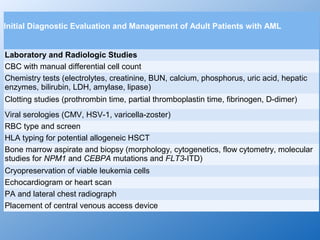
Downloaded 1,337 times


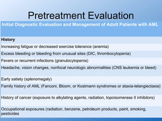
![Physical Examination
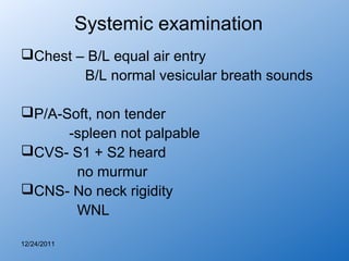
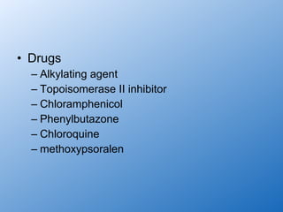
Performance status (prognostic factor)
Ecchymosis and oozing from IV sites (DIC, possible acute promyelocytic leukemia)
Fever and tachycardia (signs of infection)
Papilledema, retinal infiltrates, cranial nerve abnormalities (CNS leukemia)
Poor dentition, dental abscesses
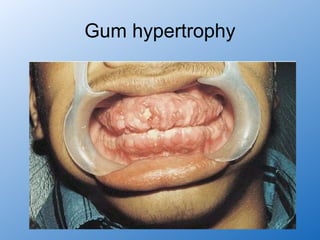
Gum hypertrophy (leukemic infiltration, most common in monocytic leukemia)
Skin infiltration or nodules (leukemia infiltration, most common in monocytic leukemia)
Lymphadenopathy, splenomegaly, hepatomegaly
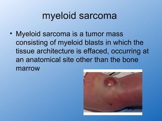
Back pain, lower extremity weakness [spinal granulocytic sarcoma, most likely in t(8;21)
patients]](https://image.slidesharecdn.com/aml-130116015226-phpapp01/85/Acute-myeloid-leukemia-41-320.jpg)


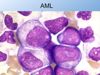
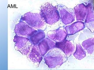
This document discusses the case of a 35-year-old female patient presenting with fever, fatigue, and shortness of breath. Her medical history includes a hysterectomy for menorrhagia and treatment for genitourinary tuberculosis. On examination, she has pallor and tachycardia. Laboratory tests show pancytopenia and blasts in her peripheral blood smear. A bone marrow biopsy confirms the diagnosis of acute myeloid leukemia. The discussion reviews the epidemiology, etiology, classification, clinical presentation, diagnostic workup, and initial treatment evaluation for AML.


