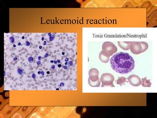
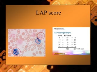
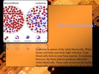
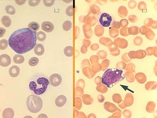
LEUKEMIAS
Dr SAPNA M
Leukemoid Reaction
• A leukemoid reaction describes a high
WBC count with neutrophilia,usually in
response to infection.
• The WBC count may be as high as 50,000
/microL and can easily mimic CML or
AML.
• Serum leukocyte alkaline phosphatase is
normal or elevated in leukemoid reaction,
but is depressed in chronic myelogenous
leukemia.
• The bone marrow in a leukemoid reaction,
if examined, may be hypercellular but is
otherwise typically unremarkable.
Features Suggesting Leukemoid
Reaction
• Toxic granulation.
• High LAP score.
• Presence of an obvious cause for the
neutrophilia.
• As noted above, a leukemoid reaction is typically a response to an underlying medical issue. Causes of leukemoid reactions include:
• Hemorrhage
• Drugs
– Use of Sulfa drugs
– Use of Dapsone
– Use of glucocorticoids
– Use of G-CSF or related growth factors
– All-trans retinoic acid (ATRA)
• Infections
– Clostridium difficile
– Tuberculosis
– Pertussis
– Infectious mononucleosis (lymphocyte predominant)
– Visceral Larva Migrans (eosinophil predominant)
• Asplenia
• Diabetic ketoacidosis
• Organ necrosis
– Hepatic necrosis
– Ischemic colitis
• As a feature of Trisomy 21 in infancy (incidence of ~10%)
• As a paraneoplastic phenomenon (rare)
Leukemoid reaction
LAP score
What is leukemia?
Leukemia is cancer of the white blood cells. White
blood cells help your body fight infection. Your
blood cells form in your bone marrow. In leukemia,
however, the bone marrow produces abnormal
white blood cells. These cells crowd out the healthy
blood cells, making it hard for blood to do its work.
Hematopoietic
stem cell
Neutrophils
Eosinophils
Basophils
Monocytes
Platelets
Red cells
Myeloid
progenitor
Lymphoid
progenitor
B-lymphocytes
T-lymphocytes
Plasma
cells
naïve
ALL
AML
Myeloid maturation
myelobl
ast
promyelocytemyelocytemetamyelocyteband neutrophil
MATURATION
Adapted and modified from U Va website
Definition
• Leukemia: is a cancer of the blood or bone marrow
characterized by abnormal proliferation of blood
cells,usually WBCs(Leukocytes).
• Acute leukemia: rapid increase of immature blood
cells.
• Chronic leukemia: excessive build up of relatively
mature,but still abnormal blood cells.
About the Disease
• Leukemia, lymphoma and myeloma are
cancers that originate in the bone marrow
(leukemia & myeloma) or in lymphatic
tissues (lymphoma).
Different Types of Blood
Cancers
• Leukemia
• Non-Hodgkin Lymphoma
• Hodgkin Lymphoma
• Myeloma
• Myelodysplastic Syndromes
What is Leukemia
• Greek word which means “white blood”
• Leukemia is when cells spread rapidly and destroy living
tissue.
• It grows/invades the bone marrow which is the factory of
blood and replaces normal blood elements with cancer
cells.
• Cancer cells replace all bone marrow cells which causes
infection and bleeding problems.
• Leukemia is basically white blood cells that don‟t work
well and cause trouble.
Signs and Symptoms
• Most of the signs and symptoms are due to:
1-Anemia.
2-Leukopenia.
3-Thrombocytopenia.
• Bicytopenia,Pancytopenia.
• All symptoms associated with leukemia
can be attributed to other diseases,
consequently,leukemia is always
diagnosed by laboratory investigations.
Causes
• Leukemia,like other malignancies, results
from somatic mutations in the DNA.
• Certain mutations produce leukemia by
activating oncogenes or deactivating
tumor suppressor genes.
• These mutations may occur spontaneously
or as a result of exposure to radiation
or carcinogenic substances,and likely
to be influenced by genetic factors.
Causes-cont‟d
• Ionizing radiation
• Viruses: Human T-lymphotropic virus (HTLV-1)
• Chemicals: Benzene,chemotherapy.
• Smoking: slight increase in leukemia
incidence.
• Genetic predisposition toward developing
leukemia: Down syn.,Fanconi anemia
Classification
• Multiple classification systems.
• FAB classification:
French-American-British Classification.
• FAB Classification relies on morphologic,
cytochemical,and immunophenotyping
criteria to define 8 major subtypes
(M0-M7)
FAB vs WHO Classification
• French-American-British (FAB) Cx
– Cellular morphology and cytochemical stain
– Acute leukemia as > 30% bone marrow blasts
– Widely used
• World Health Organization Cx
– Cellular morphology and cytochemical stain
– Immunologic probes of cell markers, cytogenetics,
molecular abnormalities & clinical syndrome
– Acute leukemia as > 20% bone marrow blasts
– Standard for diagnosis
Acute myeloid leukemias (AML)
Classification - FAB
1. M0: minimally differentiated
2. M1: myeloblastic leukemia without maturation
3. M2: myeloblastic leukemia with maturation
4. M3: hypergranular promyelocytic leukemia
5. M4: myelomonocytic leukemia
6. M4Eo: variant, increase in marrow eosinophils
7. M5: monocytic leukemia
8. M6: erythroleukemia (DiGuglielmo's disease)
9. M7: megakaryoblastic leukemia
AML classification - WHO
AML not otherwise categorized
1. AML minimally differentiated
2. AML without maturation
3. AML with maturation
4. Acute myelomonocytic leukemia
5. Acute monocytic leukemia
6. Acute erythroid leukemia
7. Acute megakaryocytic leukemia
8. Acute basophilic leukemia
9. Acute panmyelosis with myelofibrosis
Acute vs Chronic Leukemia
Acute Chronic
Age Children & young
adults
Middle age and
elderly
Onset Sudden insidious
Duration weeks to months years
WBC count Variable High
Acute vs Chronic Leukemia
Acute Chronic
Platelets Low Early: Normal/ High
Late: Low
Anemia High (>90%) None/ mild
Predomi-
nant cells
Blast cells Mature cells
AML = myeloblast
ALL= lymphoblast
CML=granulocytes
CLL=lymphocytes
Acute vs Chronic Leukemia
Acute Chronic
Marrow
cellularity
>20% marrow
blasts (WHO)
> 30% marrow
blasts (FAB)
>70% marrow
cellularity
(hypercellular);
No dysplasia
Diagnosis PBS, BM exam,
cytochemical
stains, surface
markers,
EM,chromosome
PBS (peripheral
blood smear)
Acute Myeloid Leukemia
Definition
• Acute myeloid leukemia (AML): acute
myelogenous leukemia,acute non-
lymphocytic leukemia.
• AML consists of a group of relatively well-
defined hematopoietic neoplasms
involving precursor cells commited
to the myeloid line(WBCs,RBCs,PLTs)
Chracteristics
• AML is characterized by a clonal proli-
feration of myeloid precursors with a
reduced capacity to differentiate into
mature cellular elements.
• As a result,there is an accumulation of
leukemic blasts or other immature
forms in the BM,peripheral blood,and
other tissues with a variable reduction in
the production of normal RBCs,platelets,
and mature granulocytes.
Specific:
• M2 : Chloroma:-presents as a mass lesion „tumor
of leukemic cells‟
• M3 : DIC
• M4/M5 : Infiltration of soft tissues,
gum infiltration, skin deposits ,Meningeal
involvement-headache, vomiting, eye symptoms
A
B
C
Chloromas
NEJM 1998
Gum hypertrophy
Leukostasis
• accumulation of blasts in microcirculation
with impaired perfusion
• lungs: hypoxemia, pulmonary infiltrates
• CNS: stroke
• only seen with WBC >> 50 x 109/L
Pathological Features
• CBC and differential.
• Blood film (smear).
• Bone marrow examination: BM aspirate
and trephine biopsy.
1-Morphology.
2-Immunephenotyping.
3-Cytogenetics and molecular biology.
Jemshidi trephine &
Salah aspiration needle
Bone marrow in acute leukemia
• necessary for diagnosis
• useful for determining type
• useful for prognosis
• Acute leukemias are defined by the
presence of > 20% blasts in bone marrow
(% of nucleated marrow cells)
• Bone marrow aspirate & trephine:
Hypercellular,
– blast cells ( > 20%),
– presence of Auer rods - AML type
• Cytochemistry :
Special stains to differentiate AML from
ALL ;
Positivity with Sudan black &
Myeloperoxidase (MPO) in AML
WBC Count in AML
• WBC count in AML can be high,normal,or
low.
• Median WBC count in AML is 15 000/uL.
• 20% of patients have > 100 000/uL
• 25-40% of patients have <5000/uL
• 95% of patients have blast cells on blood
film.
Distinguishing AML from ALL
• light microscopy
– AML: Auer rods, cytoplasmic granules
– ALL: no Auer rods or granules.
• flow cytometry
• special stains (cytochemistry)
Cytochemical Stains
• Since the early 20th century, cytochemical staining of
cells has been a useful tool in differentiating
hematopoietic diseases.
• Smears and imprints made from bone marrow, lymph
nodes, spleen, or peripheral blood are preferred.
– In enzymatic techniques, fresh smears are used to
ensure optimal enzyme activity
• Certain elements may be inhibited during the fixation
of smears and imprints
Myeloperoxidase (MPX/MPO)• The proxidase enzyme reacts with H2O2 & release O2,
which oxidizes the indicator dye and produce orange-
brown granules in the cells (3-amino-9-erythrocarbazol)
• Enzyme MPX is found in the 1o granules of
granulocytes, neutrophils and precursors (from the
promyelocyte stage on) & eosinophils
• Monocytes may be weakly pos
• Leukemic myeloblasts are usually pos and Auer rods
stain very strongly
• Used for differentiating AML (+) from ALL (-)
• Normal bone marrow smear <5 days old used for control
slides (promyelocyte - neutrophils)
MPO (right) & Sudan black (left)
showing intense localised positivity in
blasts
Myeloperoxidase
(MPO)
p-Phenylene diamine + Catecol + H2O2
MPO > Brown black deposits
Chloracetate (Specific) Esterase
Myeloid Cell Line
Naphthol-ASD-chloracetate
CAE > Free naphthol compounds
+ Stable diazonium salt (eg, Fast Corinth)
> Red deposit
Non-Specific Esterase (NSE)
• Nonspecific esterase liberates alpha-naphthyl from
the substrate alpha-naphthyl acetate. Alpha-
naphthyl is couples with the dye molecule to form
dark reddish-brown granules
• Monocytes, monblasts, macrophages, histiocytes,
megakaryocytes and some carcinomas are NSE pos
• Abnormal erythroblasts are strongly pos
• Lymphocytes are neg or may show dot positivity
Non-Specific Esterase
Monocytic Line
Naphthyl acetate
ANAE > Free naphthyl compounds
+Stable diazonium salt (eg, Fast blue RR)
> Brown deposits
NSE continued
• Used for differentiating myelomonocytic and
monocytic leukemia (+) from granulocytic leukemia (-
)
• Monocyte NSE are fluoride sensitive
• Peripheral smear with appreciable # of monocytes or a
normal BM smear used for control slides
http://www.healthsystem.virginia.edu/internet/hematology/hessedd/malignanthematologicdisorders/leukemias/aml-m4.cfm
Double Esterase in M4
NSE with Fl inhibition
Histiocyte
Periodic Acid Schiff (PAS)
• Periodic acid oxidizes glycogen, mucoproteins, and
other high-molecular weight carbohydrates to
aldehydes.
• Aldehydes react with colorless Schiff reagent,
staining them a bright red-pink
• Megakaryocytes and platelets stain strongly pos
• Normoblasts will stain Pos
• Lymphoblasts in ALL show course and granular
(block) positivity
PAS Continued
• Myeloblasts are Neg
• Aids in diagnosis of ALL, erythroleukemia, and
megakaryoblastic leukemia
• Normal bone marrow smear used for control slides
http://www.pathologyoutlines.com/leukemia.html
Periodic Acid Schiff
Periodic acid + Glycogen
oxidation > Aldehyde + Schiff reagent
(para-rosaniline, Na metabisulfite)
> Red deposit
AML
AML
Auer rods in AML
ALL
P. Smear AML
MO: Minimally differentiated
• Undifferentiated Blasts
(No maturation)
• Myeloid phenotype -
CD13, CD33, CD34
• (-) SBB, MPO
• Negative: Auer rods,
Esterase
M1 AML without maturation
 > 30% myeloblasts
 Large cells, round nucleus
 Nucleoli (+)
 scanty cytoplasm
 >3% MPO, SBB (+)
 <20% NSE (+)
 CD 13, 33, 117
M1 AML without maturation
M2 AML with maturation
• Common type
• >30% myeloblasts
• >10% granulocyte
• Kidney shape nucleus
• Nucleoli (+)
• (+) Auer rods
• Eosinophilic granules
• >50% MPO, SBB (+)
• CD 13, 33
M2 AML with maturation
Auer Rods
M3 (hypergranular promyelocytic)
• Promyelocyte-predominant
• Large, kidney shape
• (+) Auer rods (faggot cells)
• basophilic, bilobed nuclei
• CD 13,33
• High incidence of DIC
Acute myeloid leukemia with very
abnormal cells (AML M3/ t15;17)
M4 Acute myelomonocytic
• >30% myeloblast (FAB)
• >20% granulocyte
• >20% promonocytes and
monocytes
• CD 11, 13, 33,14
• (+) Auer rods common
• High serum lysozyme level
– M4Eo = w/ eosinophilia
M3
M5
M4
M5: acute monocytic leukemia
1. M5a – without maturation
– Monoblasts , few promonocytes
2. M5b – with maturation
– Blast, Promonocytes (BM), Monocytes
(Blood)
M5a
• Monoblast ameboid with
round to oval nuclei,
• prominent nucleoli,
• <20% promonocytes/mono
• Vacuolated cytoplasm
AML M5a
M5b
• > 20% promonocytes,
monocytes
• Promonocytes folded,
convulated nucleus
• Azurophilic granules
AML M5b
M6 - erythroleukemia
 Large, bizarre,
round-to-oval cells
 (+) nucleoli
 > 50% Erythroblasts
 > 30 % Myeloblasts
 CD 45,71 Glycophorin A
 CD 13, 15,33 myeloblast
 PAS (+)
M6 (erythroblast)
M6 (erythroblast)
M7 – acute megakaryoblastic
• >30% megakaryoblasts
• platelet like granules on
PAS stain
• NSE (but not BE) (+)
• Myeloid blasts may show
SBB or MPO (+)
• CD 41,42,61
M7 (Megakaryoblast)
Megakaryoblast
Acute Nonlymphocytic Leukemias
• Acute lymphoblastic leukemias
FAB Classification of ALL
L1: Small homogeneous blasts; mostly in
children
L2: Large heterogeneous blasts; mostly in
adults
L3: “Burkitt” large basophilic B-cell blasts
with vacuoles
WHO Classification of
Lymphoproliferative Syndromes
 Precursor B Lymphoblastic Leukemia/Lymphoma
(ALL/LBL) -- ALL in children (80-85% of
childhood ALL); LBL in young adults and rare;
FAB L1 or L2 blast morphology
 Precursor T ALL/LBL -- 15% of childhood ALL
and 25% of adult ALL
 Burkitt Leukemia/Lymphoma (FAB L3)
• Confirmation:
– Immunophenotyping
– Molecular genetics
– Cytogenetics: Chromosomal abnormalities
ALL L1
L3L2
Burkits / ALL L3
Prognostic factors
• High WBC – relapse in testis /cns
• Infants <1 yr, children >10 yr poor outcome
• L1- good prognosis
• L2,3- bad prognosis
Prognosis
• The response to treatment and overall
survival of patients with AML are
heterogenous.
• Prognostic factors are related to patient
and tumor characteristics:
1-Age
2-Performance status
3- Karyotype
Adverse Clinical Predictors
• Advanced age.
• Poor performance status.
• History of exposure to cytostatic agents or
radiotherapy.(Therapy-related AML).
• History of MDS or other hematological
diseases
THANK YOU

leukemias

  • 1.
  • 2.
    Leukemoid Reaction • Aleukemoid reaction describes a high WBC count with neutrophilia,usually in response to infection. • The WBC count may be as high as 50,000 /microL and can easily mimic CML or AML.
  • 3.
    • Serum leukocytealkaline phosphatase is normal or elevated in leukemoid reaction, but is depressed in chronic myelogenous leukemia. • The bone marrow in a leukemoid reaction, if examined, may be hypercellular but is otherwise typically unremarkable.
  • 4.
    Features Suggesting Leukemoid Reaction •Toxic granulation. • High LAP score. • Presence of an obvious cause for the neutrophilia.
  • 5.
    • As notedabove, a leukemoid reaction is typically a response to an underlying medical issue. Causes of leukemoid reactions include: • Hemorrhage • Drugs – Use of Sulfa drugs – Use of Dapsone – Use of glucocorticoids – Use of G-CSF or related growth factors – All-trans retinoic acid (ATRA) • Infections – Clostridium difficile – Tuberculosis – Pertussis – Infectious mononucleosis (lymphocyte predominant) – Visceral Larva Migrans (eosinophil predominant) • Asplenia • Diabetic ketoacidosis • Organ necrosis – Hepatic necrosis – Ischemic colitis • As a feature of Trisomy 21 in infancy (incidence of ~10%) • As a paraneoplastic phenomenon (rare)
  • 6.
  • 7.
  • 9.
    What is leukemia? Leukemiais cancer of the white blood cells. White blood cells help your body fight infection. Your blood cells form in your bone marrow. In leukemia, however, the bone marrow produces abnormal white blood cells. These cells crowd out the healthy blood cells, making it hard for blood to do its work.
  • 10.
  • 11.
  • 12.
    Definition • Leukemia: isa cancer of the blood or bone marrow characterized by abnormal proliferation of blood cells,usually WBCs(Leukocytes). • Acute leukemia: rapid increase of immature blood cells. • Chronic leukemia: excessive build up of relatively mature,but still abnormal blood cells.
  • 13.
    About the Disease •Leukemia, lymphoma and myeloma are cancers that originate in the bone marrow (leukemia & myeloma) or in lymphatic tissues (lymphoma).
  • 14.
    Different Types ofBlood Cancers • Leukemia • Non-Hodgkin Lymphoma • Hodgkin Lymphoma • Myeloma • Myelodysplastic Syndromes
  • 15.
    What is Leukemia •Greek word which means “white blood” • Leukemia is when cells spread rapidly and destroy living tissue. • It grows/invades the bone marrow which is the factory of blood and replaces normal blood elements with cancer cells. • Cancer cells replace all bone marrow cells which causes infection and bleeding problems. • Leukemia is basically white blood cells that don‟t work well and cause trouble.
  • 16.
    Signs and Symptoms •Most of the signs and symptoms are due to: 1-Anemia. 2-Leukopenia. 3-Thrombocytopenia. • Bicytopenia,Pancytopenia. • All symptoms associated with leukemia can be attributed to other diseases, consequently,leukemia is always diagnosed by laboratory investigations.
  • 18.
    Causes • Leukemia,like othermalignancies, results from somatic mutations in the DNA. • Certain mutations produce leukemia by activating oncogenes or deactivating tumor suppressor genes. • These mutations may occur spontaneously or as a result of exposure to radiation or carcinogenic substances,and likely to be influenced by genetic factors.
  • 19.
    Causes-cont‟d • Ionizing radiation •Viruses: Human T-lymphotropic virus (HTLV-1) • Chemicals: Benzene,chemotherapy. • Smoking: slight increase in leukemia incidence. • Genetic predisposition toward developing leukemia: Down syn.,Fanconi anemia
  • 21.
    Classification • Multiple classificationsystems. • FAB classification: French-American-British Classification. • FAB Classification relies on morphologic, cytochemical,and immunophenotyping criteria to define 8 major subtypes (M0-M7)
  • 22.
    FAB vs WHOClassification • French-American-British (FAB) Cx – Cellular morphology and cytochemical stain – Acute leukemia as > 30% bone marrow blasts – Widely used • World Health Organization Cx – Cellular morphology and cytochemical stain – Immunologic probes of cell markers, cytogenetics, molecular abnormalities & clinical syndrome – Acute leukemia as > 20% bone marrow blasts – Standard for diagnosis
  • 23.
    Acute myeloid leukemias(AML) Classification - FAB 1. M0: minimally differentiated 2. M1: myeloblastic leukemia without maturation 3. M2: myeloblastic leukemia with maturation 4. M3: hypergranular promyelocytic leukemia 5. M4: myelomonocytic leukemia 6. M4Eo: variant, increase in marrow eosinophils 7. M5: monocytic leukemia 8. M6: erythroleukemia (DiGuglielmo's disease) 9. M7: megakaryoblastic leukemia
  • 24.
    AML classification -WHO AML not otherwise categorized 1. AML minimally differentiated 2. AML without maturation 3. AML with maturation 4. Acute myelomonocytic leukemia 5. Acute monocytic leukemia 6. Acute erythroid leukemia 7. Acute megakaryocytic leukemia 8. Acute basophilic leukemia 9. Acute panmyelosis with myelofibrosis
  • 27.
    Acute vs ChronicLeukemia Acute Chronic Age Children & young adults Middle age and elderly Onset Sudden insidious Duration weeks to months years WBC count Variable High
  • 28.
    Acute vs ChronicLeukemia Acute Chronic Platelets Low Early: Normal/ High Late: Low Anemia High (>90%) None/ mild Predomi- nant cells Blast cells Mature cells AML = myeloblast ALL= lymphoblast CML=granulocytes CLL=lymphocytes
  • 29.
    Acute vs ChronicLeukemia Acute Chronic Marrow cellularity >20% marrow blasts (WHO) > 30% marrow blasts (FAB) >70% marrow cellularity (hypercellular); No dysplasia Diagnosis PBS, BM exam, cytochemical stains, surface markers, EM,chromosome PBS (peripheral blood smear)
  • 30.
  • 31.
    Definition • Acute myeloidleukemia (AML): acute myelogenous leukemia,acute non- lymphocytic leukemia. • AML consists of a group of relatively well- defined hematopoietic neoplasms involving precursor cells commited to the myeloid line(WBCs,RBCs,PLTs)
  • 32.
    Chracteristics • AML ischaracterized by a clonal proli- feration of myeloid precursors with a reduced capacity to differentiate into mature cellular elements. • As a result,there is an accumulation of leukemic blasts or other immature forms in the BM,peripheral blood,and other tissues with a variable reduction in the production of normal RBCs,platelets, and mature granulocytes.
  • 33.
    Specific: • M2 :Chloroma:-presents as a mass lesion „tumor of leukemic cells‟ • M3 : DIC • M4/M5 : Infiltration of soft tissues, gum infiltration, skin deposits ,Meningeal involvement-headache, vomiting, eye symptoms
  • 34.
  • 35.
  • 36.
    Leukostasis • accumulation ofblasts in microcirculation with impaired perfusion • lungs: hypoxemia, pulmonary infiltrates • CNS: stroke • only seen with WBC >> 50 x 109/L
  • 37.
    Pathological Features • CBCand differential. • Blood film (smear). • Bone marrow examination: BM aspirate and trephine biopsy. 1-Morphology. 2-Immunephenotyping. 3-Cytogenetics and molecular biology.
  • 38.
    Jemshidi trephine & Salahaspiration needle
  • 40.
    Bone marrow inacute leukemia • necessary for diagnosis • useful for determining type • useful for prognosis • Acute leukemias are defined by the presence of > 20% blasts in bone marrow (% of nucleated marrow cells)
  • 41.
    • Bone marrowaspirate & trephine: Hypercellular, – blast cells ( > 20%), – presence of Auer rods - AML type • Cytochemistry : Special stains to differentiate AML from ALL ; Positivity with Sudan black & Myeloperoxidase (MPO) in AML
  • 42.
    WBC Count inAML • WBC count in AML can be high,normal,or low. • Median WBC count in AML is 15 000/uL. • 20% of patients have > 100 000/uL • 25-40% of patients have <5000/uL • 95% of patients have blast cells on blood film.
  • 43.
    Distinguishing AML fromALL • light microscopy – AML: Auer rods, cytoplasmic granules – ALL: no Auer rods or granules. • flow cytometry • special stains (cytochemistry)
  • 44.
    Cytochemical Stains • Sincethe early 20th century, cytochemical staining of cells has been a useful tool in differentiating hematopoietic diseases. • Smears and imprints made from bone marrow, lymph nodes, spleen, or peripheral blood are preferred. – In enzymatic techniques, fresh smears are used to ensure optimal enzyme activity • Certain elements may be inhibited during the fixation of smears and imprints
  • 46.
    Myeloperoxidase (MPX/MPO)• Theproxidase enzyme reacts with H2O2 & release O2, which oxidizes the indicator dye and produce orange- brown granules in the cells (3-amino-9-erythrocarbazol) • Enzyme MPX is found in the 1o granules of granulocytes, neutrophils and precursors (from the promyelocyte stage on) & eosinophils • Monocytes may be weakly pos • Leukemic myeloblasts are usually pos and Auer rods stain very strongly • Used for differentiating AML (+) from ALL (-) • Normal bone marrow smear <5 days old used for control slides (promyelocyte - neutrophils)
  • 47.
    MPO (right) &Sudan black (left) showing intense localised positivity in blasts
  • 48.
    Myeloperoxidase (MPO) p-Phenylene diamine +Catecol + H2O2 MPO > Brown black deposits
  • 49.
    Chloracetate (Specific) Esterase MyeloidCell Line Naphthol-ASD-chloracetate CAE > Free naphthol compounds + Stable diazonium salt (eg, Fast Corinth) > Red deposit
  • 50.
    Non-Specific Esterase (NSE) •Nonspecific esterase liberates alpha-naphthyl from the substrate alpha-naphthyl acetate. Alpha- naphthyl is couples with the dye molecule to form dark reddish-brown granules • Monocytes, monblasts, macrophages, histiocytes, megakaryocytes and some carcinomas are NSE pos • Abnormal erythroblasts are strongly pos • Lymphocytes are neg or may show dot positivity
  • 51.
    Non-Specific Esterase Monocytic Line Naphthylacetate ANAE > Free naphthyl compounds +Stable diazonium salt (eg, Fast blue RR) > Brown deposits
  • 52.
    NSE continued • Usedfor differentiating myelomonocytic and monocytic leukemia (+) from granulocytic leukemia (- ) • Monocyte NSE are fluoride sensitive • Peripheral smear with appreciable # of monocytes or a normal BM smear used for control slides http://www.healthsystem.virginia.edu/internet/hematology/hessedd/malignanthematologicdisorders/leukemias/aml-m4.cfm
  • 53.
    Double Esterase inM4 NSE with Fl inhibition Histiocyte
  • 54.
    Periodic Acid Schiff(PAS) • Periodic acid oxidizes glycogen, mucoproteins, and other high-molecular weight carbohydrates to aldehydes. • Aldehydes react with colorless Schiff reagent, staining them a bright red-pink • Megakaryocytes and platelets stain strongly pos • Normoblasts will stain Pos • Lymphoblasts in ALL show course and granular (block) positivity
  • 55.
    PAS Continued • Myeloblastsare Neg • Aids in diagnosis of ALL, erythroleukemia, and megakaryoblastic leukemia • Normal bone marrow smear used for control slides http://www.pathologyoutlines.com/leukemia.html
  • 56.
    Periodic Acid Schiff Periodicacid + Glycogen oxidation > Aldehyde + Schiff reagent (para-rosaniline, Na metabisulfite) > Red deposit
  • 57.
  • 58.
  • 59.
  • 60.
  • 61.
  • 62.
    MO: Minimally differentiated •Undifferentiated Blasts (No maturation) • Myeloid phenotype - CD13, CD33, CD34 • (-) SBB, MPO • Negative: Auer rods, Esterase
  • 63.
    M1 AML withoutmaturation  > 30% myeloblasts  Large cells, round nucleus  Nucleoli (+)  scanty cytoplasm  >3% MPO, SBB (+)  <20% NSE (+)  CD 13, 33, 117
  • 64.
    M1 AML withoutmaturation
  • 65.
    M2 AML withmaturation • Common type • >30% myeloblasts • >10% granulocyte • Kidney shape nucleus • Nucleoli (+) • (+) Auer rods • Eosinophilic granules • >50% MPO, SBB (+) • CD 13, 33
  • 66.
    M2 AML withmaturation
  • 67.
  • 68.
    M3 (hypergranular promyelocytic) •Promyelocyte-predominant • Large, kidney shape • (+) Auer rods (faggot cells) • basophilic, bilobed nuclei • CD 13,33 • High incidence of DIC
  • 69.
    Acute myeloid leukemiawith very abnormal cells (AML M3/ t15;17)
  • 70.
    M4 Acute myelomonocytic •>30% myeloblast (FAB) • >20% granulocyte • >20% promonocytes and monocytes • CD 11, 13, 33,14 • (+) Auer rods common • High serum lysozyme level – M4Eo = w/ eosinophilia
  • 71.
  • 72.
    M5: acute monocyticleukemia 1. M5a – without maturation – Monoblasts , few promonocytes 2. M5b – with maturation – Blast, Promonocytes (BM), Monocytes (Blood)
  • 74.
    M5a • Monoblast ameboidwith round to oval nuclei, • prominent nucleoli, • <20% promonocytes/mono • Vacuolated cytoplasm
  • 75.
  • 76.
    M5b • > 20%promonocytes, monocytes • Promonocytes folded, convulated nucleus • Azurophilic granules
  • 77.
  • 78.
    M6 - erythroleukemia Large, bizarre, round-to-oval cells  (+) nucleoli  > 50% Erythroblasts  > 30 % Myeloblasts  CD 45,71 Glycophorin A  CD 13, 15,33 myeloblast  PAS (+)
  • 79.
  • 80.
  • 81.
    M7 – acutemegakaryoblastic • >30% megakaryoblasts • platelet like granules on PAS stain • NSE (but not BE) (+) • Myeloid blasts may show SBB or MPO (+) • CD 41,42,61
  • 82.
  • 83.
  • 84.
  • 85.
  • 86.
    FAB Classification ofALL L1: Small homogeneous blasts; mostly in children L2: Large heterogeneous blasts; mostly in adults L3: “Burkitt” large basophilic B-cell blasts with vacuoles
  • 87.
    WHO Classification of LymphoproliferativeSyndromes  Precursor B Lymphoblastic Leukemia/Lymphoma (ALL/LBL) -- ALL in children (80-85% of childhood ALL); LBL in young adults and rare; FAB L1 or L2 blast morphology  Precursor T ALL/LBL -- 15% of childhood ALL and 25% of adult ALL  Burkitt Leukemia/Lymphoma (FAB L3)
  • 88.
    • Confirmation: – Immunophenotyping –Molecular genetics – Cytogenetics: Chromosomal abnormalities
  • 89.
  • 90.
  • 91.
  • 92.
    Prognostic factors • HighWBC – relapse in testis /cns • Infants <1 yr, children >10 yr poor outcome • L1- good prognosis • L2,3- bad prognosis
  • 93.
    Prognosis • The responseto treatment and overall survival of patients with AML are heterogenous. • Prognostic factors are related to patient and tumor characteristics: 1-Age 2-Performance status 3- Karyotype
  • 94.
    Adverse Clinical Predictors •Advanced age. • Poor performance status. • History of exposure to cytostatic agents or radiotherapy.(Therapy-related AML). • History of MDS or other hematological diseases
  • 95.