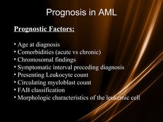
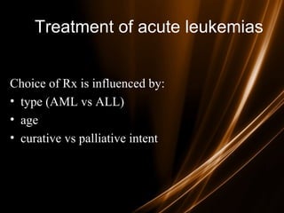
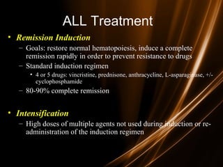
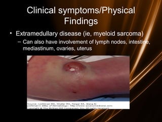
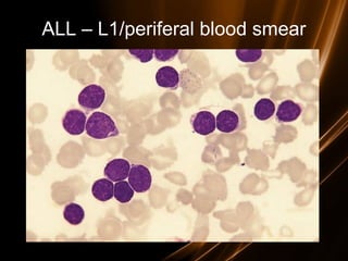
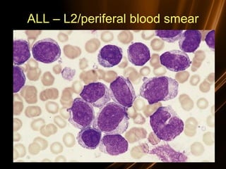
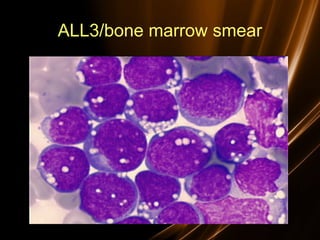
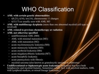
ACUTE
LEUKEMIA
Suraj Dhara
(Midnapore Medical College)
Leukemia
Group of malignant disorders of the
hematopoietic tissues characteristically
associated with increased numbers of white
cells in the bone marrow and / or peripheral
blood
Classification
• Classified based on cell type involved and
the clinical course
– Acute :
• ALL
• AML
– Chronic :
• CLL
• CML
Acute Leukemias
• Acute leukemias are clonal malignant hematopoietic
disorders resulting from genetic alterations in normal
hematopoietic stem cells.
• These alterations disrupt normal differentiation and/or
cause excessive proliferation of abnormal immature
“leukemic” cells or “blasts.”
• As the disease progresses, leukemic cells accumulate in
the bone marrow, blood, and organs, displacing normal
progenitor cells and suppressing normal hematopoiesis
Acute Leukemia - Essentials
• Short course of symptoms
• Fatigue, fever, easy bruising, bleeding
• Cytopenias - or pancytopenia
• More than 20% blasts in bone marrow
• Blasts in peripheral blood in 90% cases
AL - EPIDEMIOLOGY
• The frequency of AL varies between 1 and 6,5
cases to 100.000 population each year. The
frequency of AL varies with subtype and age.
• AL represents 10% of the neoplastic diseases and
is the principal cause of neoplastic for ages 0-35
years.
ALL - Epidemiology
• Approximately 3,000 new cases per year
• Mostly affects childrenchildren, accounts for 2/3 of childhood
leukemia (peak age 4 yearspeak age 4 years)
• Comprises less than 20% of leukemia in young adults
• May be B-cell, T-cell, or null-type (non-B, non-T cell)
•Only 20-40% of adults with ALL are cured with current
regimens.
AML - Epidemiology
• 2.3 per 100,000 people per year
• Higher among men than women (2.9 vs 1.9). The
difference is even more apparent in older patients.
• Most common leukemia in adults (80% of cases)
• Vast majority of patients 65 years or older65 years or older
• AML is more common in whites than in other
populations.
ALL - Etiology
• Uncertain, but several proposed linkages:
· Genetic - Philadelphia chromosome
· Viral infection (EBV, HIV)
· Exposure to high energy radiation (T-cell ALL)
· Toxic chemical exposure
· Smoking
AML - Etiology
• Primary AML
– Increased incidence
• Genetic fragility
– Bloom syndrome
– Faconi anemia
– Wiskott Aldrich
– Down, Klinefelter, Patau syndromes
• tobacco use?
• herbicides?, pesticides?
• benzene exposure
• Secondary AML
– XRT
– Topoisomerase II inhibitors (e.g etopisode), alkylating agents
– MDS
– other cell proliferation disorders
• CML, polycythemia vera, primary thrombocytosis, PNH
Clinical features
General :
Onset is abrupt & stormy
(usually present within 3 months)
– Bone marrow failure
(anemia, infection ,bleeding)
– Bone pain & tenderness
Clinical presentation:
• Will present with sign or symptoms related to :
– Pancytopenia:
∀↓WBC→infection  infectious syndrome
∀↓Hb →anemia  anemic syndrome
∀↓Platelets →bleeding – hemorragic syndrome
– Organ infiltration  tumoral syndrome
• Lymphadenopathy.
• Splenomegally.
• Hepatomegally.
• CNS:5-10% of patient with ALL
More common with ALL than AML.
Clinical features - specials
• L mediastinal tumoral mass
• L CNS infiltration
• M2 : Chloroma:-presents as a mass lesion ‘tumor
of leukemic cells’
• M3 : DIC
• M4/M5 : Infiltration of soft tissues,
gum infiltration, skin deposits
,Meningeal involvement-headache, vomiting, eye
symptoms
Gingival Infiltration in Monocytic
(AML M4 eos) Variant of AML
Gum hypertrophy
A
B
C
Chloromas
NEJM 1998
Clinical symptoms/Physical
Findings
• Extramedullary disease (ie, myeloid sarcoma)
– Can also have involvement of lymph nodes, intestine,
mediastinum, ovaries, uterus
Clinical features - specials
Skin Infiltration with AML (Leukemia Cutis)
Leukostasis
• Leukostasis – predominantly in those with WBC counts
> 100,000 (10% of patients); can also be seen in patients
with WBC > 50,000
– Most common in those with M4 or M5 leukemia
– Function of the blast cells being less deformable than mature
myeloid cells. As a result, intravascular plugs develop.
– High metabolic activity of blast cells and local production of
various cytokines contribute to underlying hypoxia
Leukostasis
Thornton, KA, Levis, M. FLT3 Mutation and Acute Myelogenous Leukemia with Leukostasis. N Engl J Med 2007; 357:1639. Copyright
©2007 Massachusetts Medical Society
Leukostasis
• Common symptoms
– Pulmonary: dyspnea, chest pain
– CNS: headaches, altered mentation, CN palsies,
ocular symptoms
– Priapism
– Myocardial Infarction
Acute leukemia - Diagnosis
–Lab evaluation
•The lab diagnosis is based on two things
–Finding a significant increase in the number of immature
cells in the bone marrow including blasts, promyelocytes,
promonocytes (>30% blasts is diagnostic)
–Identification of the cell lineage of the leukemic cells
Investigations
• CBC:
– 60% of pts have an elevated WBC.
– Most are anemic
– Most are thrombocytopenic
– 90%have blast in the periphral blood film.
• electrolytes:
– Hypo/hyper kalemia
– Hypomagnesimia
– hyperphosphatemia
• Hypermetabolism:
↑LDH.
↑uric acid.
• DIC:
– Most common with promyelocytic leukemia,small% monocytic leukemia&ALL
• Bone marrow biopsy and aspirate:
– 30%or more of all nucleated cells are blast.
• Radiology:
– CXR:mediastinal mass(T-cell ALL)
– Osteopenia or lytic lesion 50% of patients with ALL.(itractable pain).
Acute leukemia - Diagnosis
– Peripheral blood:
• Anemia (normochromic, normocytic)
• Decreased platlets
• Variable WBC count
– The degree of peripheral blood involvement determines classification:
»Leukemic – increased WBCs due to blasts
»Subleukemic – blasts without increased WBCs
»Aleukemic – decreased WBCs with no blasts
Acute Leukemias - CBC
• Hb – 5,6 g/dl
• Plt – 15.000/mmc
• WBC – 34.000/mmc
– PN – 10%
– L - 6%
– M – 3%
– Bl – 81%
• Hb – 3,5 g/dl
• Plt – 5.000/mmc
• WBC – 1.500/mmc
– PN - 15%
– L – 80%
– M – 5%
Acute leukemia - Diagnosis
• Bone marrow aspirate & trephine:
Hypercellular,
– blast cells ( > 20%),,
– presence of Auer rods - AML type
• Cytochemistry :
Special stains to differentiate AML from ALL ;
Positivity
with Sudan black & Myeloperoxidase (MPO) in
AML
Jemshidi trephine &
Salah aspiration needle
Acute leukemia - Diagnosis
• Diagnosis and classification of
the immature cells involved may
be done by :
–Morphology
–Cytochemistry
–Immunophenotyping
–Cytogenetic
Classification
• Criteria:
- Morphology :apperance of cell under microscope.
- Cytochemistry:chemical activity of the cell.
(myeloperoxidase , Sudan Black B)
- immunophenotyping: antigen pressent in the cell membrane
- Cytogenetics: chromosome of the cell
- Molecular biology:
• Classification:
3 groups of acute leukemias:
- acute myeloid leukemias AML(M1 –M6).
- acute lymphoblastic leukemias ALL (L1-L3).
- Biphenotypic leukemias or Acute undifferentiated
leukemia
Diagnosis
• Morphologic
– French American British Classification
• L1: small uniform blasts (pediatric ALL)
• L2: larger, more variable sized blasts (adult ALL)
• L3: uniform cells with basophilic and sometimes
vacuolated cytoplasm (mature B cell ALL)
L1 - 85% of childhood ALL
L2 - Majority of adult ALL
L3 - Includes Burkitt’s. < 5% of ALL
Immunologic Subtype FAB Type %of Cases Cytogenetic Abnl
Pre-B cell ALL L1, L2 75 t(9:22) t(4:11) t(1:19)
T-cell ALL L1, L2 20 14q11 or 7q34
B-cell ALL L3 5 t(8:14) t(8:22) t(2:8)
Classification of ALL
ALL – L1/periferal blood smear
ALL – L2/periferal blood smear
ALL3/bone marrow smear
FAB Classification of AML
• M0 undifferentiated acute myeloblastic leukemia (5%)
• M1 AML with minimal maturation (20%)
• M2 AML with maturation (30%)
– t(8;21)
• M3 Acute promyelocytic leukemia (5%)
– t(15;17)
• M4 Acute myelomonocytic leukemia (20%)
• M4 eos Acute myelomonocytic leukemia with eosinophilia (5%)
– inv (16)
• M5 Acute monocytic leukemia (10%)
– t(9;11)
• M6 Acute erythroid leukemia (3%)
• M7 Acute megakaryoblastic leukemia (3%)
WHO Classification
• AML with certain genetic abnormalities
– t(8;21), t(16), inv(16), chromosome 11 changes
– t(15;17) as usually seen with AML M3
• AML with multilineage dysplasia (more than one abnormal myeloid cell type is
involved)
• AML related to previous chemotherapy or radiation
• AML not otherwise specified
– undifferentiated AML (M0)
– AML with minimal maturation (M1)
– AML with maturation (M2)
– acute myelomonocytic leukemia (M4)
– acute monocytic leukemia (M5)
– acute erythroid leukemia (M6)
– acute megakaryoblastic leukemia (M7)
– acute basophilic leukemia
– acute panmyelosis with fibrosis
– myeloid sarcoma (also known as granulocytic sarcoma or chloroma)
• Undifferentiated or biphenotypic acute leukemias (leukemias that have both
lymphocytic and myeloid features. Sometimes called ALL with myeloid markers, AML
with lymphoid markers, or mixed lineage leukemias.)
AML2 – bone marrow smear
AML3 – bone marrow smear
Prognosis in ALL
parametersparameters GoodGood poorpoor
WBC lowlow High(>50x10High(>50x10 99
/l)/l)
Gender GirlsGirls BoysBoys
Immunophenotype C-ALLC-ALL B-ALLB-ALL
Age ChildChild Adult or infant.Adult or infant.
Cytogenetic Normal,hyperdiploid,Normal,hyperdiploid, Ph+,11q23rearrangemePh+,11q23rearrangeme
nts.nts.
Time to clear blast from
blood
< 1week< 1week >1week>1week
Time to remission <4weeks<4weeks >4weeks>4weeks
Cns disease at
presentation
AbsentAbsent PresentPresent
Minimal residual
disease.
Negative at 1-3 monthsNegative at 1-3 months Still positive at 3-6Still positive at 3-6
months.months.
Prognosis in ALL
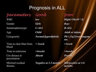
• Good risk includes
– (1) no adverse cytogenetics,
– (2) age younger than 30 years
– (3) WBC count of less than 30,000/mL, and
– (4) complete remission within 4 weeks.
• Intermediate risk
– does not meet the criteria for either good risk or poor risk.
• Poor risk includes
– (1) adverse cytogenetics [(t9;22), (4;11)],
– (2) age older than 60 years,
– (3) precursor B-cell WBCs with WBC count greater than
100,000/mL, or
– (4) failure to achieve complete remission within 4 weeks.
Prognosis in AML
Prognostic Factors:
• Age at diagnosis
• Comorbidities (acute vs chronic)
• Chromosomal findings
• Symptomatic interval preceding diagnosis
• Presenting Leukocyte count
• Circulating myeloblast count
• FAB classification
• Morphologic characteristics of the leukemic cell
Treatment of acute leukemias
Choice of Rx is influenced by:
• type (AML vs ALL)
• age
• curative vs palliative intent
Principles of treatment
• combination chemotherapy
– first goal is complete remission
– further Rx to prevent relapse
• supportive medical care
– transfusions, antibiotics, nutrition
• psychosocial support
– patient and family
Supportive measures:
-isolation in positive laminer flux room
-insertion of central line
-family and patient support by permanent social worker
-Alkaline diuresis to prevent tumor lysis syndrome
-oropharynx/GIT decontamination to prevent fungal infection
-IV antibiotics for infection
-Blood transfusion if anemia and thrombocytopenia.
Chemotherapy for acute
leukemias
• Phases of ALL treatment
– induction
– intensification
– CNS prophylaxis
– maintenance
• Phases of AML treatment
– induction
– consolidation (post-remission therapy)
post-remission therapy
ALL Treatment
• 1 – Remission Induction
• 2 – Intensification (Consolidation) Therapy
• 3 – Maintenance Therapy
• 4 – CNS Prophylaxis
• 5 – Allogeneic Stem Cell Transplant
ALL Treatment
• Remission Induction
– Goals: restore normal hematopoiesis, induce a complete
remission rapidly in order to prevent resistance to drugs
– Standard induction regimen
• 4 or 5 drugs: vincristine, prednisone, anthracycline, L-asparaginase, +/-
cyclophosphamide
– 80-90% complete remission
• Intensification
– High doses of multiple agents not used during induction or re-
administration of the induction regimen
ALL Treatment
• Maintenance Therapy
– Daily po 6MP, weekly MTX, monthly pulses of vincristine
and prednisone for 2-3 yrs
• CNS Prophylaxis
– Given during induction and intensification
– Intrathecal: MTX, Cytarabine, corticosteroids
– Systemic: high dose mtx, cytarabine, L-asparaginase
– +/- Cranial Irradiation
ALL Treatment
• Stem Cell Transplant
– Done during first CR
– Indications:
• Ph Chromosome
• t(4;11) mutation
• Poor initial response to induction therapy
• Other
– Adolescents benefit significantly from pediatric ALL
regimens vs. adult regimens
AML Treatment
• Remission induction therapy
– Commonly anthracycline (ie, daunorubicin, idarubicin) and cytarabine
→ (“3+7 regimen”)
• Cytarabine has ample CNS penetration so no need for prophylactic
intrathecal chemotx (also, ↓ risk in patients with AML compared to ALL)
• 60-80% achieve complete remission
• Postremission therapy
1. Consolidation
– longer survival than maintence alone
– typically high dose cytarabine
1. Maintenance – continue chemotx monthly for 4-12 months
– nonmyelosuppressive doses
COMPLICATIONS TREATMENT
Tumor Lysis Syndrome
• Characterized by metabolic derangements caused
by massive release of cellular components
following lysis of malignant cells
• Commonly seen in malignancies with high rates
of cell proliferation (esp. ALL, Burkitt’s
lymphoma); also can be seen with AML
Tumor Lysis Syndrome
• Tumor lysis syndrome - hyperphosphatemia,
hyperkalemia, hyperuricemia, hypocalcemia and uremia
– Retrospective study of 788 patients (433 adults) found
incidence of hyperuricemia and TLS to be 14.7%/3.4% in
patients with AML compared to 21.4%/5.2% in patients with
ALL and 19.6%/6.1% in patients with NHL
• Electrolyte abnormalities can occur without the entire
spectrum of TLS or even before tx is initiated
– Hyperuricemia
– Lactic acidosis
Tumor Lysis Syndrome
• Release of intracellular proteins →catobilized to hypoxanthine →
xanthine → uric acid → Crystalization of uric acid and in renal
tubules → impaired renal function
• Release of phosphate from malignant cells → calcium phosphate
precipitation and further renal impairment along with
hypocalcemia and resultant symptoms from ↓Ca
– Hyperphosphatemia: nausea, vomiting, diarrhea, seizures, lethargy
– Hypocalcemia: arrhythmia, hypotension, tetany, cramps
– Hyperkalemia: arrhythmia, cramps, paresthesia
Tumor Lysis Syndrome
• Prevention and management
– IV hydration : promotes excretion of uric acid and phosphate; improves renal blood
flow/GFR
– Allopurinol → competitive inhibitor for xanthine oxidase. Therefore, ↓ conversion of
purine metabolites to uric acid
• However, must consider buildup of xanthine crystals → acute obstructive uropathy
(HYDRATE!!!)
– Recominant urate oxidase (rasburicase)
• Promotes conversion of uric acid to allantoin (highly soluble; urinary excretion)
• Indicated in patients at high risk of TLS (Burkitt’s Lymphoma, B-ALL, ALL
(WBC >100,000), AML (WBC >50,000)
• Also indicated in patients that develop hyperuricemia despite allopurinol
– Dialysis can be used in severe cases
– Urine alkalization is NOT recommended – does not increase solubility of
xanthine/hypoxanthine with an increased propensity to develop xanthine-obstructive
uropathies (esp with allopurinol use)
DIC
• Common symptoms/findings
– in addition to weakness (anemia), infections/fever (malfunctioning WBCs)
– petechiae, ecchymoses, hematuria, bleeding from venipuncture sites
– migratory thrombophlebitis (Trousseau’s syndrome)
– nonbacterial thrombotic (marantic) endocarditis
– DVT/PE
• Lab findings
– Prolonged PT/INR, PTT
– microangiopathic anemia (schistocytes)
– thrombocytopenia
– elevated fibrin split products
– elevated D-dimer
– low fibrinogen
DIC
• Treatment
1. Supportive therapy
• Platelets
• Cryoprecipitate (fibrinogen)
• FFP
1. Treatment for obvious thrombosis (e.g
thrombophlebitis, mural thrombus)
• UFH or LMWH; often resistant to coumadin
• activated protein C
1. Treatment of underlying malignancy
• In the case of AML M3 → All-Trans Retinoic Acid (PML-RARα)
– Induces differentiation beyond promyelocyte phase
– Only with the more common t(15;17) translocation; t(11;17) and t(5;17) do not respond to
ATRA
• Remission rates of greater than 90% with AML M3 patient treated with ATRA and
chemotx (eg, anthracyclines (idarubicin)) with 60-70% disease free survival
• Arsenic trioxide in those that relapse – achieves complete remission in >90%
CNS Involvement
• Occurs in less than 5% of AML patients (highest
incidence in relapsed promyelocytic (M3) variant)
– Routine LP is not performed unless symptoms suggestive of
CNS pathology
• Common symptoms
– headache
– mental status changes
– CN palsies (commonly CN III or VI)
– CSF findings
• blast cells
• moderate increase in protein and moderate decrease in glucose
CNS Involvement
• Treatment
– Intrathecal chemotherapy (methotrexate or
cytarabine) +/- whole brain XRT
• addition of XRT depends on response to intrathecal
chemotx and whether there is cranial nerve involvement
• high relapse rate
– Commonly administer prophylactic intrathecal
chemotx in relapsed promyelocytic disease
THANK YOU

ACUTE LEUKEMIA

  • 1.
  • 2.
    Leukemia Group of malignantdisorders of the hematopoietic tissues characteristically associated with increased numbers of white cells in the bone marrow and / or peripheral blood
  • 3.
    Classification • Classified basedon cell type involved and the clinical course – Acute : • ALL • AML – Chronic : • CLL • CML
  • 4.
    Acute Leukemias • Acuteleukemias are clonal malignant hematopoietic disorders resulting from genetic alterations in normal hematopoietic stem cells. • These alterations disrupt normal differentiation and/or cause excessive proliferation of abnormal immature “leukemic” cells or “blasts.” • As the disease progresses, leukemic cells accumulate in the bone marrow, blood, and organs, displacing normal progenitor cells and suppressing normal hematopoiesis
  • 5.
    Acute Leukemia -Essentials • Short course of symptoms • Fatigue, fever, easy bruising, bleeding • Cytopenias - or pancytopenia • More than 20% blasts in bone marrow • Blasts in peripheral blood in 90% cases
  • 6.
    AL - EPIDEMIOLOGY •The frequency of AL varies between 1 and 6,5 cases to 100.000 population each year. The frequency of AL varies with subtype and age. • AL represents 10% of the neoplastic diseases and is the principal cause of neoplastic for ages 0-35 years.
  • 7.
    ALL - Epidemiology •Approximately 3,000 new cases per year • Mostly affects childrenchildren, accounts for 2/3 of childhood leukemia (peak age 4 yearspeak age 4 years) • Comprises less than 20% of leukemia in young adults • May be B-cell, T-cell, or null-type (non-B, non-T cell) •Only 20-40% of adults with ALL are cured with current regimens.
  • 8.
    AML - Epidemiology •2.3 per 100,000 people per year • Higher among men than women (2.9 vs 1.9). The difference is even more apparent in older patients. • Most common leukemia in adults (80% of cases) • Vast majority of patients 65 years or older65 years or older • AML is more common in whites than in other populations.
  • 9.
    ALL - Etiology •Uncertain, but several proposed linkages: · Genetic - Philadelphia chromosome · Viral infection (EBV, HIV) · Exposure to high energy radiation (T-cell ALL) · Toxic chemical exposure · Smoking
  • 10.
    AML - Etiology •Primary AML – Increased incidence • Genetic fragility – Bloom syndrome – Faconi anemia – Wiskott Aldrich – Down, Klinefelter, Patau syndromes • tobacco use? • herbicides?, pesticides? • benzene exposure • Secondary AML – XRT – Topoisomerase II inhibitors (e.g etopisode), alkylating agents – MDS – other cell proliferation disorders • CML, polycythemia vera, primary thrombocytosis, PNH
  • 11.
    Clinical features General : Onsetis abrupt & stormy (usually present within 3 months) – Bone marrow failure (anemia, infection ,bleeding) – Bone pain & tenderness
  • 12.
    Clinical presentation: • Willpresent with sign or symptoms related to : – Pancytopenia: ∀↓WBC→infection  infectious syndrome ∀↓Hb →anemia  anemic syndrome ∀↓Platelets →bleeding – hemorragic syndrome – Organ infiltration  tumoral syndrome • Lymphadenopathy. • Splenomegally. • Hepatomegally. • CNS:5-10% of patient with ALL More common with ALL than AML.
  • 13.
    Clinical features -specials • L mediastinal tumoral mass • L CNS infiltration • M2 : Chloroma:-presents as a mass lesion ‘tumor of leukemic cells’ • M3 : DIC • M4/M5 : Infiltration of soft tissues, gum infiltration, skin deposits ,Meningeal involvement-headache, vomiting, eye symptoms
  • 14.
    Gingival Infiltration inMonocytic (AML M4 eos) Variant of AML
  • 15.
  • 16.
  • 17.
    Clinical symptoms/Physical Findings • Extramedullarydisease (ie, myeloid sarcoma) – Can also have involvement of lymph nodes, intestine, mediastinum, ovaries, uterus
  • 18.
  • 19.
    Skin Infiltration withAML (Leukemia Cutis)
  • 20.
    Leukostasis • Leukostasis –predominantly in those with WBC counts > 100,000 (10% of patients); can also be seen in patients with WBC > 50,000 – Most common in those with M4 or M5 leukemia – Function of the blast cells being less deformable than mature myeloid cells. As a result, intravascular plugs develop. – High metabolic activity of blast cells and local production of various cytokines contribute to underlying hypoxia
  • 21.
    Leukostasis Thornton, KA, Levis,M. FLT3 Mutation and Acute Myelogenous Leukemia with Leukostasis. N Engl J Med 2007; 357:1639. Copyright ©2007 Massachusetts Medical Society
  • 22.
    Leukostasis • Common symptoms –Pulmonary: dyspnea, chest pain – CNS: headaches, altered mentation, CN palsies, ocular symptoms – Priapism – Myocardial Infarction
  • 23.
    Acute leukemia -Diagnosis –Lab evaluation •The lab diagnosis is based on two things –Finding a significant increase in the number of immature cells in the bone marrow including blasts, promyelocytes, promonocytes (>30% blasts is diagnostic) –Identification of the cell lineage of the leukemic cells
  • 24.
    Investigations • CBC: – 60%of pts have an elevated WBC. – Most are anemic – Most are thrombocytopenic – 90%have blast in the periphral blood film. • electrolytes: – Hypo/hyper kalemia – Hypomagnesimia – hyperphosphatemia • Hypermetabolism: ↑LDH. ↑uric acid. • DIC: – Most common with promyelocytic leukemia,small% monocytic leukemia&ALL • Bone marrow biopsy and aspirate: – 30%or more of all nucleated cells are blast. • Radiology: – CXR:mediastinal mass(T-cell ALL) – Osteopenia or lytic lesion 50% of patients with ALL.(itractable pain).
  • 25.
    Acute leukemia -Diagnosis – Peripheral blood: • Anemia (normochromic, normocytic) • Decreased platlets • Variable WBC count – The degree of peripheral blood involvement determines classification: »Leukemic – increased WBCs due to blasts »Subleukemic – blasts without increased WBCs »Aleukemic – decreased WBCs with no blasts
  • 26.
    Acute Leukemias -CBC • Hb – 5,6 g/dl • Plt – 15.000/mmc • WBC – 34.000/mmc – PN – 10% – L - 6% – M – 3% – Bl – 81% • Hb – 3,5 g/dl • Plt – 5.000/mmc • WBC – 1.500/mmc – PN - 15% – L – 80% – M – 5%
  • 27.
    Acute leukemia -Diagnosis • Bone marrow aspirate & trephine: Hypercellular, – blast cells ( > 20%),, – presence of Auer rods - AML type • Cytochemistry : Special stains to differentiate AML from ALL ; Positivity with Sudan black & Myeloperoxidase (MPO) in AML
  • 28.
    Jemshidi trephine & Salahaspiration needle
  • 29.
    Acute leukemia -Diagnosis • Diagnosis and classification of the immature cells involved may be done by : –Morphology –Cytochemistry –Immunophenotyping –Cytogenetic
  • 30.
    Classification • Criteria: - Morphology:apperance of cell under microscope. - Cytochemistry:chemical activity of the cell. (myeloperoxidase , Sudan Black B) - immunophenotyping: antigen pressent in the cell membrane - Cytogenetics: chromosome of the cell - Molecular biology: • Classification: 3 groups of acute leukemias: - acute myeloid leukemias AML(M1 –M6). - acute lymphoblastic leukemias ALL (L1-L3). - Biphenotypic leukemias or Acute undifferentiated leukemia
  • 31.
    Diagnosis • Morphologic – FrenchAmerican British Classification • L1: small uniform blasts (pediatric ALL) • L2: larger, more variable sized blasts (adult ALL) • L3: uniform cells with basophilic and sometimes vacuolated cytoplasm (mature B cell ALL)
  • 32.
    L1 - 85%of childhood ALL L2 - Majority of adult ALL L3 - Includes Burkitt’s. < 5% of ALL Immunologic Subtype FAB Type %of Cases Cytogenetic Abnl Pre-B cell ALL L1, L2 75 t(9:22) t(4:11) t(1:19) T-cell ALL L1, L2 20 14q11 or 7q34 B-cell ALL L3 5 t(8:14) t(8:22) t(2:8) Classification of ALL
  • 33.
  • 34.
  • 35.
  • 36.
    FAB Classification ofAML • M0 undifferentiated acute myeloblastic leukemia (5%) • M1 AML with minimal maturation (20%) • M2 AML with maturation (30%) – t(8;21) • M3 Acute promyelocytic leukemia (5%) – t(15;17) • M4 Acute myelomonocytic leukemia (20%) • M4 eos Acute myelomonocytic leukemia with eosinophilia (5%) – inv (16) • M5 Acute monocytic leukemia (10%) – t(9;11) • M6 Acute erythroid leukemia (3%) • M7 Acute megakaryoblastic leukemia (3%)
  • 37.
    WHO Classification • AMLwith certain genetic abnormalities – t(8;21), t(16), inv(16), chromosome 11 changes – t(15;17) as usually seen with AML M3 • AML with multilineage dysplasia (more than one abnormal myeloid cell type is involved) • AML related to previous chemotherapy or radiation • AML not otherwise specified – undifferentiated AML (M0) – AML with minimal maturation (M1) – AML with maturation (M2) – acute myelomonocytic leukemia (M4) – acute monocytic leukemia (M5) – acute erythroid leukemia (M6) – acute megakaryoblastic leukemia (M7) – acute basophilic leukemia – acute panmyelosis with fibrosis – myeloid sarcoma (also known as granulocytic sarcoma or chloroma) • Undifferentiated or biphenotypic acute leukemias (leukemias that have both lymphocytic and myeloid features. Sometimes called ALL with myeloid markers, AML with lymphoid markers, or mixed lineage leukemias.)
  • 38.
    AML2 – bonemarrow smear
  • 39.
    AML3 – bonemarrow smear
  • 40.
    Prognosis in ALL parametersparametersGoodGood poorpoor WBC lowlow High(>50x10High(>50x10 99 /l)/l) Gender GirlsGirls BoysBoys Immunophenotype C-ALLC-ALL B-ALLB-ALL Age ChildChild Adult or infant.Adult or infant. Cytogenetic Normal,hyperdiploid,Normal,hyperdiploid, Ph+,11q23rearrangemePh+,11q23rearrangeme nts.nts. Time to clear blast from blood < 1week< 1week >1week>1week Time to remission <4weeks<4weeks >4weeks>4weeks Cns disease at presentation AbsentAbsent PresentPresent Minimal residual disease. Negative at 1-3 monthsNegative at 1-3 months Still positive at 3-6Still positive at 3-6 months.months.
  • 41.
    Prognosis in ALL •Good risk includes – (1) no adverse cytogenetics, – (2) age younger than 30 years – (3) WBC count of less than 30,000/mL, and – (4) complete remission within 4 weeks. • Intermediate risk – does not meet the criteria for either good risk or poor risk. • Poor risk includes – (1) adverse cytogenetics [(t9;22), (4;11)], – (2) age older than 60 years, – (3) precursor B-cell WBCs with WBC count greater than 100,000/mL, or – (4) failure to achieve complete remission within 4 weeks.
  • 42.
    Prognosis in AML PrognosticFactors: • Age at diagnosis • Comorbidities (acute vs chronic) • Chromosomal findings • Symptomatic interval preceding diagnosis • Presenting Leukocyte count • Circulating myeloblast count • FAB classification • Morphologic characteristics of the leukemic cell
  • 43.
    Treatment of acuteleukemias Choice of Rx is influenced by: • type (AML vs ALL) • age • curative vs palliative intent
  • 44.
    Principles of treatment •combination chemotherapy – first goal is complete remission – further Rx to prevent relapse • supportive medical care – transfusions, antibiotics, nutrition • psychosocial support – patient and family
  • 45.
    Supportive measures: -isolation inpositive laminer flux room -insertion of central line -family and patient support by permanent social worker -Alkaline diuresis to prevent tumor lysis syndrome -oropharynx/GIT decontamination to prevent fungal infection -IV antibiotics for infection -Blood transfusion if anemia and thrombocytopenia.
  • 47.
    Chemotherapy for acute leukemias •Phases of ALL treatment – induction – intensification – CNS prophylaxis – maintenance • Phases of AML treatment – induction – consolidation (post-remission therapy) post-remission therapy
  • 48.
    ALL Treatment • 1– Remission Induction • 2 – Intensification (Consolidation) Therapy • 3 – Maintenance Therapy • 4 – CNS Prophylaxis • 5 – Allogeneic Stem Cell Transplant
  • 49.
    ALL Treatment • RemissionInduction – Goals: restore normal hematopoiesis, induce a complete remission rapidly in order to prevent resistance to drugs – Standard induction regimen • 4 or 5 drugs: vincristine, prednisone, anthracycline, L-asparaginase, +/- cyclophosphamide – 80-90% complete remission • Intensification – High doses of multiple agents not used during induction or re- administration of the induction regimen
  • 50.
    ALL Treatment • MaintenanceTherapy – Daily po 6MP, weekly MTX, monthly pulses of vincristine and prednisone for 2-3 yrs • CNS Prophylaxis – Given during induction and intensification – Intrathecal: MTX, Cytarabine, corticosteroids – Systemic: high dose mtx, cytarabine, L-asparaginase – +/- Cranial Irradiation
  • 51.
    ALL Treatment • StemCell Transplant – Done during first CR – Indications: • Ph Chromosome • t(4;11) mutation • Poor initial response to induction therapy • Other – Adolescents benefit significantly from pediatric ALL regimens vs. adult regimens
  • 52.
    AML Treatment • Remissioninduction therapy – Commonly anthracycline (ie, daunorubicin, idarubicin) and cytarabine → (“3+7 regimen”) • Cytarabine has ample CNS penetration so no need for prophylactic intrathecal chemotx (also, ↓ risk in patients with AML compared to ALL) • 60-80% achieve complete remission • Postremission therapy 1. Consolidation – longer survival than maintence alone – typically high dose cytarabine 1. Maintenance – continue chemotx monthly for 4-12 months – nonmyelosuppressive doses
  • 53.
  • 54.
    Tumor Lysis Syndrome •Characterized by metabolic derangements caused by massive release of cellular components following lysis of malignant cells • Commonly seen in malignancies with high rates of cell proliferation (esp. ALL, Burkitt’s lymphoma); also can be seen with AML
  • 55.
    Tumor Lysis Syndrome •Tumor lysis syndrome - hyperphosphatemia, hyperkalemia, hyperuricemia, hypocalcemia and uremia – Retrospective study of 788 patients (433 adults) found incidence of hyperuricemia and TLS to be 14.7%/3.4% in patients with AML compared to 21.4%/5.2% in patients with ALL and 19.6%/6.1% in patients with NHL • Electrolyte abnormalities can occur without the entire spectrum of TLS or even before tx is initiated – Hyperuricemia – Lactic acidosis
  • 56.
    Tumor Lysis Syndrome •Release of intracellular proteins →catobilized to hypoxanthine → xanthine → uric acid → Crystalization of uric acid and in renal tubules → impaired renal function • Release of phosphate from malignant cells → calcium phosphate precipitation and further renal impairment along with hypocalcemia and resultant symptoms from ↓Ca – Hyperphosphatemia: nausea, vomiting, diarrhea, seizures, lethargy – Hypocalcemia: arrhythmia, hypotension, tetany, cramps – Hyperkalemia: arrhythmia, cramps, paresthesia
  • 57.
    Tumor Lysis Syndrome •Prevention and management – IV hydration : promotes excretion of uric acid and phosphate; improves renal blood flow/GFR – Allopurinol → competitive inhibitor for xanthine oxidase. Therefore, ↓ conversion of purine metabolites to uric acid • However, must consider buildup of xanthine crystals → acute obstructive uropathy (HYDRATE!!!) – Recominant urate oxidase (rasburicase) • Promotes conversion of uric acid to allantoin (highly soluble; urinary excretion) • Indicated in patients at high risk of TLS (Burkitt’s Lymphoma, B-ALL, ALL (WBC >100,000), AML (WBC >50,000) • Also indicated in patients that develop hyperuricemia despite allopurinol – Dialysis can be used in severe cases – Urine alkalization is NOT recommended – does not increase solubility of xanthine/hypoxanthine with an increased propensity to develop xanthine-obstructive uropathies (esp with allopurinol use)
  • 58.
    DIC • Common symptoms/findings –in addition to weakness (anemia), infections/fever (malfunctioning WBCs) – petechiae, ecchymoses, hematuria, bleeding from venipuncture sites – migratory thrombophlebitis (Trousseau’s syndrome) – nonbacterial thrombotic (marantic) endocarditis – DVT/PE • Lab findings – Prolonged PT/INR, PTT – microangiopathic anemia (schistocytes) – thrombocytopenia – elevated fibrin split products – elevated D-dimer – low fibrinogen
  • 59.
    DIC • Treatment 1. Supportivetherapy • Platelets • Cryoprecipitate (fibrinogen) • FFP 1. Treatment for obvious thrombosis (e.g thrombophlebitis, mural thrombus) • UFH or LMWH; often resistant to coumadin • activated protein C 1. Treatment of underlying malignancy • In the case of AML M3 → All-Trans Retinoic Acid (PML-RARα) – Induces differentiation beyond promyelocyte phase – Only with the more common t(15;17) translocation; t(11;17) and t(5;17) do not respond to ATRA • Remission rates of greater than 90% with AML M3 patient treated with ATRA and chemotx (eg, anthracyclines (idarubicin)) with 60-70% disease free survival • Arsenic trioxide in those that relapse – achieves complete remission in >90%
  • 60.
    CNS Involvement • Occursin less than 5% of AML patients (highest incidence in relapsed promyelocytic (M3) variant) – Routine LP is not performed unless symptoms suggestive of CNS pathology • Common symptoms – headache – mental status changes – CN palsies (commonly CN III or VI) – CSF findings • blast cells • moderate increase in protein and moderate decrease in glucose
  • 61.
    CNS Involvement • Treatment –Intrathecal chemotherapy (methotrexate or cytarabine) +/- whole brain XRT • addition of XRT depends on response to intrathecal chemotx and whether there is cranial nerve involvement • high relapse rate – Commonly administer prophylactic intrathecal chemotx in relapsed promyelocytic disease
  • 62.

Editor's Notes

  • #32 Will not discuss morphologic much b/c not used as much and rely more on the later two
  • #50 Our patient received the 4 drug regimen with IT Ara-C and mtx -current intense combo therapy has led to CR in 80-90% of adult ALL cases -Intensification is given shortly after remission is achieved -Intensification  better outcomes, specifically event free survival in kids, adolescents, and adults
  • #51 CNS involvement dx by &amp;gt; or = to 5 wbc in csf with blasts present -cranial XRT has led to sz, dementia, intellectual dysfunction, grown retardation, and second cancers
  • #52 -adolescents probably do better b/c of the increased dose density of the chemo agents in peds -peds trials show CR in ~1 mo vs. in ~2 mo in adults -1st post remission chemo in peds is 2 d after and in adults is 7 d after