SlideShare a Scribd company logo
1 of 74
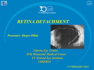
RETINA DETACHMENT
Presenter- Shayri Pillai
Liberia Eye Centre
JFK Memorial Medical Centre
LV Prasad Eye Institute
LIBERIA
17 FEBRUARY 2021
Retinal detachment (RD)
 RD refers to separation of the neurosensory retina from the RPE
 Results in the accumulation of SRF in the potential space between
the NSR and RPE
Innocuous peripheral retinal degenerations
 Tiny vesicles with indistinct boundaries on a greyish-white
background
 Retina appear thickened and less transparent adjacent to the
ora serrata and extends circumferentially and posteriorly with a
smooth undulating posterior border
 Present in essentially all adult eyes, increasing in extent with age
 May give rise to typical degenerative retinoschisis
 Discrete yellow–white patches of focal chorioretinal atrophy
that may have pigmented margins
 Found between the equator and the ora serrata
 More common in the inferior fundus
 Present to some extent in at least 25% of normal eyes
Microcystoid seen on scleral indentation Paving stone with adjacent
microcystoid
 An age-related change
 Fine network of perivascular pigmentation that sometimes extends
posterior to the equator
Honeycomb(reticular)
 Clustered or scattered small pale discrete lesions
 Hyperpigmented borders
 Similar to drusen at the posterior pole
 Usually occur in the eyes of older individuals
Peripheral drusen
 Clear-walled cysts usually small in size
 Derived from non-pigmented ciliary epithelium
 Present in 5–10% of eyes
 More common temporally
 They do not predispose to RD
Pars plana Cyst
Peripheral Lesions Predisposing to Retinal Detachment
Patients should be educated about the nature of symptoms and
the need to be reviewed urgently if these occur
Lattice degeneration
Prevalence
 Present in about 8% of the population
 Develops early in life, with a peak incidence during the second
and third decades
 Found more commonly in moderate myopes
 Most important degeneration directly related to RD
 Present in about 40% of eyes with RD
Pathology
 Discontinuity of the internal limiting membrane with variable
atrophy of the underlying NSR
 Vitreous overlying an area of lattice is synchytic but the
vitreous attachments around the margins are exaggerated
Vitreous changes associated with lattice
degeneration
Signs
 Bilateral, temporal and superior
 Spindle-shaped areas of retinal thinning, commonly located
between the equator and the posterior border of the vitreous
base
 Sclerosed vessels forming an arborizing network of white lines
is characteristic
 Some lesions may be associated with ‘snowflakes’, remnants of
degenerate Müller cells
 Associated hyperplasia of the RPE is common
 Small holes are common
 Associated hyperplasia of the RPE is common
 Small holes are common
Wide-field images of lattice degeneration
(A)Multiple lesions with small hole (B) sclerosed vessels forming a
characteristic white network; a vortex vein is seen
superonasally
Complications
 Do not occur in most eyes with lattice
 Tears may develop consequent to a posterior vitreous
detachment (PVD), when lattice is sometimes visible on the
flap of the tear
 Atrophic holes may rarely (2%) lead to RD
 Risk is higher in young myopes
Retinal detachment with lattice on the flap of the tear
Management
 Asymptomatic areas of lattice are generally not treated
prophylactically, unless particular risk factors are present
 RD in the fellow eye
 Treatment of the fellow eye when extensive lattice (more
than 6 clock hours) is present, or high myopia
 Routine annual review of eyes with lattice, with or without
asymptomatic round holes, particularly in young myopes
Snailtrack degeneration
 Sharply demarcated bands of tightly packed ‘snowflakes’
that give the peripheral retina a white frost-like appearance
 Marked vitreous traction is seldom present so that U-tears
rarely occurs
 Round holes are relatively common
 Prophylactic treatment is usually unnecessary, though
review every 1–2 years may be prudent as RD occurs in a
minority
A) Snailtrack degeneration; (B) and (C) wide-field images of lesions before
and after limited laser retinopexy
Cystic retinal tuft
 Known as a granular patch or retinal rosette, is a congenital
abnormality
 Consists of a small,round or oval, discrete elevated whitish
lesion
 Typically in the equatorial or peripheral retina, more commonly
temporally associated pigmentation at its base
 Comprised principally of glial tissue; strong vitreoretinal
adhesion is commonly present
 Small round holes and horseshoe tears can occur
 CRT are present in up to 5% of the population (bilateral
in 20%)
 Risk of RD in a given eye with CRT is probably well
under 1%
Cystic retinal tuft
(A) Isolated uncomplicated lesion (B) tuft with small round hole
Degenerative retinoschisis
Prevalence
 Present in about 5% of the population over the age of 20
years and is particularly prevalent in hypermetropia
Pathology
 Develop from microcystoid degeneration
 Gradual coalescence of degenerative cavities resulting in
separation or splitting of the NSR into inner and outer
layers with severing of neurones and complete loss of
visual function in the affected area
 In typical retinoschisis the split occurs in the outer
plexiform layer
 Reticular retinoschisis at the level of the nerve
fibre layer
Symptoms
 Photopsia and floaters are absent because there is no
vitreoretinal traction
 Rare for the patient to notice a visual field defect,
even with spread posterior to the equator
 Occasionally symptoms result from vitreous
haemorrhage or a progressive RD
Signs
 Bilateral in up to 80%
 Inner layer is thinner and tends to be more elevated
in the latter
 Early retinoschisis usually involves the extreme
inferotemporal periphery of both fundi, appearing as an
exaggeration of microcystoid degeneration with a smooth
immobile dome-shaped elevation of the retina
 Elevation is convex, smooth, thin and relatively immobile
unlike the opaque and corrugated appearance of a
rhegmatogenous RD
 Lesion progress circumferentially until it has involved the
entire periphery
 Typical form usually remains anterior to the equator
 Presence of a pigmented demarcation line is likely to
indicate the presence of associated RD
 Inner layer may show ‘snowflakes’(whitish remnants of
Müller cell footplates as well as sclerosis of blood
vessels, and the schisis cavity may be bridged by grey–
white tissue strands
A) Retinoschisis (B) composite image of the same lesion showing merging
microcystoid degeneration
 Inner layer breaks are small and round
 Outer layer breaks are usually larger, with rolled edges and
located behind the equator
 Microaneurysms and small telangiectases are common, in
the reticular type
Retinoschisis
(A) Inner and outer layer breaks (B) Large outer layer break;
retinal vessels in the inner layer can be
seen traversing the rolled edge
undiverted
Development of retinoschisis
(A) Histology showing intraretinal cavities bridged by Muller cells;(B) OCT
appearance showing separation principally in the outer plexiform layer; (C) OCT of
retinal detachment for comparison; (D) circumferential microcystoid degeneration
with progression to retinoschisis supero- and inferotemporally
Complications
 RD is rare; even in an eye with breaks in both layers the
incidence is only around 1%
 Detachment is almost always asymptomatic, infrequently
progressive and rarely requires surgery
 Posterior extension of RS to involve the fovea is very rare;
progression is generally very slow
 Vitreous hemorrhage is rare
Management
 Discussion of the symptoms is prudent in all patients,
especially those with double layer breaks
 A small peripheral RS discovered on incidental examination,
probably does not require routine review
 Large RS should be observed periodically
 Photography and visual field testing are useful, with
optical coherence tomography (OCT) imaging when
posterior extension is present
 OCT is also useful for distinguishing between RS and RD
 Retinopexy or surgical repair may be indicated for
relentless progression towards the fovea
 Prophylactic retinopexy of the posterior border of a large
bullous RS with substantial breaks to prevent progression to
symptomatic RD
 Recurrent vitreous hemorrhage may necessitate
Vitrectomy
 Progressive symptomatic RD should be addressed
promptly
 More than one procedure may be necessary
 Scleral buckling may be adequate for smaller RD with
small outer layer breaks
 Vitrectomy is generally indicated for more complex RD
Zonular traction tuft
 15% phenomenon caused by an aberrant zonular fibre
extending posteriorly to be attached to the retina near the
ora serrata
 Exerts traction on the retina at its base
 It is typically located nasally
 Risk of retinal tear formation is around 2%, and periodic
long-term review is generally recommended
White with pressure
 Refers to retinal areas in which a translucent white–grey
appearance can be induced by scleral indentation
 Each area has a fixed configuration
 It may also be observed along the posterior border of
islands of lattice degeneration, snailtrack degeneration
and the outer layer of acquired retinoschisis
 It is frequently seen in normal eyes and may be associated
with abnormally strong attachment of the vitreous gel
‘White without pressure’ (WWOP)
 Appears as WWP but is present without scleral
indentation
 WWOP corresponds to an area of fairly strong adhesion of
condensed vitreous
 Regular review should be considered for treated and
untreated eyes,
A) White with pressure (B) white without pressure (C) strong
attachment of condensed vitreous gel to an area of ‘white without pressure’
White without pressure wide-field images
(A) Pseudo-break (arrow) (B) retinal tear and adjacent pseudo-break
Myopic choroidal atrophy
 Diffuse or circumscribed choroidal depigmentation,
associated with thinning of the overlying retina
 Occurs typically in the posterior pole and equatorial area of
highly myopic eyes
 Retinal holes developing in the atrophic retina may occasionally
lead to RD
Myopic choroidal atrophy
Retinal break
 Defined as any full-thickness defect in the neurosensory retina
 Breaks are clinically significant in that they may allow liquid
from the vitreous cavity to enter the potential space between the
sensory retina and the RPE, thereby causing RRD
Some breaks are caused by atrophy of inner retinal layers
(holes); others result from vitreoretinal traction (tears)
Retinal breaks may be classified as:
 Flap, or horseshoe, tears
 Giant retinal tears
 Operculated holes
 Retinal dialyses
 Atrophic retinal holes
 Flap tear occurs when a strip of retina is pulled anteriorly by
vitreoretinal traction, often in the course of a posterior vitreous
detachment or trauma
 A tear is considered symptomatic when the patient reports
photopsias, floaters, or both
 A giant retinal tear extends 90° (3 clock-hours) or more
circumferentially and usually occurs along the posterior edge
of the vitreous base
Operculated hole occurs when traction is sufficient to tear a
piece of retina completely free from the adjacent retinal
surface
 Retinal dialysis is a circumferential, linear break that occurs
at the ora serrata, with vitreous base attached to the retina
posterior to the tear’s edge; it is commonly a consequence of
blunt trauma
 An atrophic hole is generally not associated with vitreoretinal
traction and has not been linked to an increased risk of
retinal detachment
Retinal tears. (A) Large U-tear in an area of lattice – laser retinopexy has been
performed; (B) operculated tear;(C) round holes; the blue arrows show probable atrophic
holes, the circle arrow shows a probable operculated hole with
localized subretinal fluid; (D) retinal dialysis;
(E) giant retinal tear (F) vitreous attached to the anterior edge of a giant tear
Schematic illustration of retinal tears and holes
.
Classification
RDs are classified as
 Rhegmatogenous
 Tractional or Exudative
 Most common are rhegmatogenous retinal detachments (RRDs)
 Term is derived from the Greek rhegma, meaning “break”
 RRDs are caused by fluid passing from the vitreous cavity
through a retinal break into the potential space between the
sensory retina and the RPE
 Tractional detachments are caused by proliferative membranes
that contract and elevate the retina
 TD are less common
 Combinations of tractional and rhegmatogenous pathophysiologic
components may also lead to retinal detachment
 Exudative, or secondary, detachments are caused by retinal
or choroidal diseases in which fluid leaks beneath the sensory
retina and accumulates there
Rhegmatogenous Retinal Detachment
 Incidence of 12.6 per 100,000 persons in a primarily white
population
Risk factors includes
 Myopia
 Family history
 Fellow-eye retinal tear or detachment
 Recent vitreous detachment
 Trauma
 Peripheral high-risk lesions
 Vitreoretinal degenerations
 Current or recent use of fluoroquinolones(controversial)
Symptoms
 Flashing lights and floaters associated with acute PVD
 Curtain-like relative peripheral visual field defect and can
progress to involve central vision
 Loss of central vision may be due to involvement of the
fovea by SRF or, infrequently, obstruction of the visual axis
by a large bullous RD
.
Identification of Retinal Breaks
Distribution of breaks in eyes with RD is approximately as
follows:
 60% superotemporal quadrant, 15% superonasal,
15% inferotemporal and 10% inferonasal
 The upper temporal region should therefore be examined in
detail if a break cannot be detected initially
 50% of eyes with RD have more than one break, often within
90°of each other
Configuration of SRF
 SRF spread is governed by gravity, by
anatomical limits (ora serrata and optic nerve) and by the
location of the primary retinal break
 If the primary break is located superiorly, the SRF first spreads
inferiorly on the same side of the fundus as the break and then s
uperiorly on the opposite side, so that the likely location of the
primary retinal break can be predicted
(modified from Lincoff’s rules):
Distribution of subretinal fluid in relation to the location of the primary retinal break
 A shallow inferior RD in which the SRF is slightly higher
on the temporal side points to a primary break located
inferiorly on that side
 A primary break located at 6 o’clock will cause an inferior
RD with equal fluid levels
 In a bullous inferior RD the primary break usually lies
above the horizontal meridian
 If the primary break is located in the upper nasal
quadrant the SRF will revolve around the optic disc and
then rise on the temporal side until it is level with the
primary break
 A subtotal RD with a superior wedge of attached retina
points to a primary break located in the periphery nearest
its highest border
 When the SRF crosses the vertical midline above, the
primary break is near to 12 o’clock, the lower edge of the
RD corresponding to the side of the break
Signs
 Relative afferent pupillary defect
 Intraocular pressure (IOP) is often lower by about 5 mmHg
compared with the normal eye
 Iritis
 ‘Tobacco dust’ consisting of pigment cells is commonly seen
in the anterior vitreous substantial vitreous blood or
inflammatory cells are also highly specific
A Shafer sign
present in
the anterior
vitreous
 Retinal breaks appear as discontinuities in the retinal surface
 They are usually red because of the color contrast between the
sensory retina and underlying choroid
 Retinal signs depend on the duration of RD and the
presence or absence of proliferative vitreoretinopathy (PVR)
Management of rhegmatogenous retinal detachment
The principles of surgery for retinal detachment are as follows:
Find all retinal breaks
Create a chorioretinal irritation around each break.
Close the retinal breaks
 In 90%–95% of RRDs, a definite retinal break can be found,
often with the help of Lincoff rules
 An occult break is presumed to be present
 Patients with RRD have photopsias, or floaters and flashes of
light
 Retina detaches progressively from the periphery to the optic
nerve head
 Convex borders and contours and a corrugated appearance,
especially in recent retinal detachments, and undulates with
eye movements
 Long-standing RRD,the retina may appear smooth and thin
 Fixed folds resulting from proliferative vitreoretinopathy (PVR) a
lmost always indicate an RRD
 Shifting fluid may occur, is uncommon and more typical of
serous retinal detachments
Fresh retinal detachment
 The RD has a convex configuration and a slightly opaque
and corrugated
 Appearance as a result of retinal oedema
 There is loss of the underlying choroidal pattern and retinal
blood vessels appear darker than in flat retina
Fresh retinal detachment.
(A) U-tear with superotemporal detachment, threatening the central macula;
note that substantially elevated retina appears dark on
the wide-field image (B) autofluorescence demonstrating
extent of fluid spread
(C) superior bullous detachment
(D) typical corrugated appearance of detached retina
(E) macular hole surrounded by shallow subretinal fluid confined to the posterior pole
Long-standing retinal detachment
Retinal thinning secondary to atrophy is a characteristic
finding, and should not lead to a misdiagnosis of retinoschisis
Intraretinal cysts may develop if the RD has
been present for about 1 year; these tend to disappear after
retinal reattachment
Subretinal demarcation lines (‘high water’ or ‘tide’ marks)
caused by proliferation of RPE cells at the junction of flat
Retinal cysts
(B) multiple cysts in chronic total detachment (red-free
wide-field image) (C) B-scan ultrasonogram demonstrating
cyst
(D) demarcation line (E) demarcation line surrounding
localized fluid associated with a small round asymptomatic
hole (wide-field image)
A.RRD with bullous configuration B.TRD with the cancave surface
.
.
REFERENCES
Albert’s Principles and practice of Ophthalmology-Retina
and Vitreous
Skuta,G.L. et. al. American Academy of Ophthalmology
Retina and Vitreous 2020 Edition. USA
Sihota, R. et al. Parsons’ Diseases of the Eye, 22nd Ed.,
2015
Bowling , B. Kanski’s Clinical Ophthalmology: A Systemic
Approach, 8th Ed. 2016. Australia.
Thank you!
Excellence Equity Efficiency
L V Prasad Eye Institute

More Related Content

What's hot

Choroidal neovascular membranes (CNVM)
Choroidal neovascular membranes (CNVM)Choroidal neovascular membranes (CNVM)
Choroidal neovascular membranes (CNVM)Md Riyaj Ali
 
Masquerade syndromes
Masquerade syndromesMasquerade syndromes
Masquerade syndromesBipin Bista
 
OPTICAL COHERENCE TOMOGRAPHY ANGIOGRAPHY
OPTICAL COHERENCE TOMOGRAPHY  ANGIOGRAPHY  OPTICAL COHERENCE TOMOGRAPHY  ANGIOGRAPHY
OPTICAL COHERENCE TOMOGRAPHY ANGIOGRAPHY Shruti Laddha
 
Orthoptic evaluation 1
Orthoptic evaluation 1Orthoptic evaluation 1
Orthoptic evaluation 1Pushkar Dhir
 
Proliferative vitreoretinopathy
Proliferative vitreoretinopathyProliferative vitreoretinopathy
Proliferative vitreoretinopathyPavanShroff
 
Pachychoroid spectrum diseases
Pachychoroid spectrum diseasesPachychoroid spectrum diseases
Pachychoroid spectrum diseasesYasuo Yanagi
 
Evaluation of preperimetric glaucoma
Evaluation of preperimetric glaucomaEvaluation of preperimetric glaucoma
Evaluation of preperimetric glaucomaMalvikaSuresh
 
Optical coherence tomography in glaucoma - Dr Shylesh Dabke
Optical coherence tomography in glaucoma - Dr Shylesh DabkeOptical coherence tomography in glaucoma - Dr Shylesh Dabke
Optical coherence tomography in glaucoma - Dr Shylesh DabkeShylesh Dabke
 
Lessons learned from DRCR protocols
Lessons learned from DRCR protocolsLessons learned from DRCR protocols
Lessons learned from DRCR protocolsSuhaib Ali
 
Idiopathic polypoidal choroidal vasculopathy
Idiopathic polypoidal choroidal vasculopathyIdiopathic polypoidal choroidal vasculopathy
Idiopathic polypoidal choroidal vasculopathyLaxmi Eye Institute
 
HRT and GDx VCC
HRT and GDx VCCHRT and GDx VCC
HRT and GDx VCCSSSIHMS-PG
 

What's hot (20)

Choroidal neovascular membranes (CNVM)
Choroidal neovascular membranes (CNVM)Choroidal neovascular membranes (CNVM)
Choroidal neovascular membranes (CNVM)
 
Masquerade syndromes
Masquerade syndromesMasquerade syndromes
Masquerade syndromes
 
OPTICAL COHERENCE TOMOGRAPHY ANGIOGRAPHY
OPTICAL COHERENCE TOMOGRAPHY  ANGIOGRAPHY  OPTICAL COHERENCE TOMOGRAPHY  ANGIOGRAPHY
OPTICAL COHERENCE TOMOGRAPHY ANGIOGRAPHY
 
Cone and Rod Dystrophy
Cone and Rod DystrophyCone and Rod Dystrophy
Cone and Rod Dystrophy
 
MACULAR DYSTROPHY
MACULAR DYSTROPHYMACULAR DYSTROPHY
MACULAR DYSTROPHY
 
Central Serous Retinopathy
Central Serous RetinopathyCentral Serous Retinopathy
Central Serous Retinopathy
 
Orthoptic evaluation 1
Orthoptic evaluation 1Orthoptic evaluation 1
Orthoptic evaluation 1
 
Proliferative vitreoretinopathy
Proliferative vitreoretinopathyProliferative vitreoretinopathy
Proliferative vitreoretinopathy
 
Pachychoroid spectrum diseases
Pachychoroid spectrum diseasesPachychoroid spectrum diseases
Pachychoroid spectrum diseases
 
Evaluation of preperimetric glaucoma
Evaluation of preperimetric glaucomaEvaluation of preperimetric glaucoma
Evaluation of preperimetric glaucoma
 
Disc edema
Disc edemaDisc edema
Disc edema
 
Optical coherence tomography in glaucoma - Dr Shylesh Dabke
Optical coherence tomography in glaucoma - Dr Shylesh DabkeOptical coherence tomography in glaucoma - Dr Shylesh Dabke
Optical coherence tomography in glaucoma - Dr Shylesh Dabke
 
Retinal artery occlusion
Retinal artery occlusionRetinal artery occlusion
Retinal artery occlusion
 
Aphakia
AphakiaAphakia
Aphakia
 
Retina drwaing
Retina drwaingRetina drwaing
Retina drwaing
 
Epiretinal membrane
Epiretinal membraneEpiretinal membrane
Epiretinal membrane
 
Lessons learned from DRCR protocols
Lessons learned from DRCR protocolsLessons learned from DRCR protocols
Lessons learned from DRCR protocols
 
Idiopathic polypoidal choroidal vasculopathy
Idiopathic polypoidal choroidal vasculopathyIdiopathic polypoidal choroidal vasculopathy
Idiopathic polypoidal choroidal vasculopathy
 
Macular hole
Macular holeMacular hole
Macular hole
 
HRT and GDx VCC
HRT and GDx VCCHRT and GDx VCC
HRT and GDx VCC
 

Similar to RETINA DETACHMENT CAUSES AND SIGNS

RETINAL DETACHMENT
RETINAL DETACHMENTRETINAL DETACHMENT
RETINAL DETACHMENTp K
 
3 mirror, retinal break.pptx
3 mirror, retinal break.pptx3 mirror, retinal break.pptx
3 mirror, retinal break.pptxTimothyLiew3
 
Retinal detachment
Retinal detachment Retinal detachment
Retinal detachment jyotigontia
 
Retinal detachment presentation
Retinal detachment presentationRetinal detachment presentation
Retinal detachment presentationNirav Modi
 
Tractional RD
Tractional RD Tractional RD
Tractional RD Nikhil Rp
 
Retinal Detachment_Pradeep Bastola.pptx
Retinal Detachment_Pradeep Bastola.pptxRetinal Detachment_Pradeep Bastola.pptx
Retinal Detachment_Pradeep Bastola.pptxDr. Pradeep Bastola
 
Rhegmatogenous retinal detachment (rrd)
Rhegmatogenous retinal detachment (rrd)Rhegmatogenous retinal detachment (rrd)
Rhegmatogenous retinal detachment (rrd)Pavan Mahajan
 
retinal_detachment 4.pptx
retinal_detachment 4.pptxretinal_detachment 4.pptx
retinal_detachment 4.pptxHarshika Malik
 
DISORDERS OF THE VITREOUS AND RETINAL DETACHMENT
DISORDERS OF THE VITREOUS AND RETINAL DETACHMENTDISORDERS OF THE VITREOUS AND RETINAL DETACHMENT
DISORDERS OF THE VITREOUS AND RETINAL DETACHMENTHossein Mirzaie
 
VASCULAR AND HEREDITARY RETINAL DISEASE
VASCULAR AND HEREDITARY RETINAL DISEASEVASCULAR AND HEREDITARY RETINAL DISEASE
VASCULAR AND HEREDITARY RETINAL DISEASEHossein Mirzaie
 
Rhegmatogenous retinal detachment
Rhegmatogenous retinal detachmentRhegmatogenous retinal detachment
Rhegmatogenous retinal detachmentSamuel Ponraj
 
Peripheral fundus & its disorders
Peripheral fundus & its disordersPeripheral fundus & its disorders
Peripheral fundus & its disordersRohit Rao
 
RETINOSCHISIS AND RETINAL DETACHMENT
RETINOSCHISIS AND  RETINAL DETACHMENTRETINOSCHISIS AND  RETINAL DETACHMENT
RETINOSCHISIS AND RETINAL DETACHMENTDrAbdelLatifsiam
 
Posterior segment manifestations of blunt trauma
Posterior segment manifestations of blunt traumaPosterior segment manifestations of blunt trauma
Posterior segment manifestations of blunt traumaSSSIHMS-PG
 
role of oct in diagnosis and ttt of AMD
role of oct in diagnosis and ttt of AMDrole of oct in diagnosis and ttt of AMD
role of oct in diagnosis and ttt of AMDDoaa Mahmoud
 

Similar to RETINA DETACHMENT CAUSES AND SIGNS (20)

RETINAL DETACHMENT
RETINAL DETACHMENTRETINAL DETACHMENT
RETINAL DETACHMENT
 
3 mirror, retinal break.pptx
3 mirror, retinal break.pptx3 mirror, retinal break.pptx
3 mirror, retinal break.pptx
 
Retinal detachment
Retinal detachment Retinal detachment
Retinal detachment
 
Retinal detachment presentation
Retinal detachment presentationRetinal detachment presentation
Retinal detachment presentation
 
Tractional RD
Tractional RD Tractional RD
Tractional RD
 
Retinal detachment
Retinal detachment Retinal detachment
Retinal detachment
 
Retinal Detachment_Pradeep Bastola.pptx
Retinal Detachment_Pradeep Bastola.pptxRetinal Detachment_Pradeep Bastola.pptx
Retinal Detachment_Pradeep Bastola.pptx
 
Retinal detachment
Retinal detachment  Retinal detachment
Retinal detachment
 
Rhegmatogenous retinal detachment (rrd)
Rhegmatogenous retinal detachment (rrd)Rhegmatogenous retinal detachment (rrd)
Rhegmatogenous retinal detachment (rrd)
 
retinal_detachment 4.pptx
retinal_detachment 4.pptxretinal_detachment 4.pptx
retinal_detachment 4.pptx
 
DISORDERS OF THE VITREOUS AND RETINAL DETACHMENT
DISORDERS OF THE VITREOUS AND RETINAL DETACHMENTDISORDERS OF THE VITREOUS AND RETINAL DETACHMENT
DISORDERS OF THE VITREOUS AND RETINAL DETACHMENT
 
VASCULAR AND HEREDITARY RETINAL DISEASE
VASCULAR AND HEREDITARY RETINAL DISEASEVASCULAR AND HEREDITARY RETINAL DISEASE
VASCULAR AND HEREDITARY RETINAL DISEASE
 
Rhegmatogenous retinal detachment
Rhegmatogenous retinal detachmentRhegmatogenous retinal detachment
Rhegmatogenous retinal detachment
 
RHEGMATOGENOUS Retinal detachment
 RHEGMATOGENOUS Retinal detachment RHEGMATOGENOUS Retinal detachment
RHEGMATOGENOUS Retinal detachment
 
Nw2013 RetinalDetachment
Nw2013 RetinalDetachmentNw2013 RetinalDetachment
Nw2013 RetinalDetachment
 
Retinoschisis
RetinoschisisRetinoschisis
Retinoschisis
 
Peripheral fundus & its disorders
Peripheral fundus & its disordersPeripheral fundus & its disorders
Peripheral fundus & its disorders
 
RETINOSCHISIS AND RETINAL DETACHMENT
RETINOSCHISIS AND  RETINAL DETACHMENTRETINOSCHISIS AND  RETINAL DETACHMENT
RETINOSCHISIS AND RETINAL DETACHMENT
 
Posterior segment manifestations of blunt trauma
Posterior segment manifestations of blunt traumaPosterior segment manifestations of blunt trauma
Posterior segment manifestations of blunt trauma
 
role of oct in diagnosis and ttt of AMD
role of oct in diagnosis and ttt of AMDrole of oct in diagnosis and ttt of AMD
role of oct in diagnosis and ttt of AMD
 

More from SHAYRI PILLAI

Eyelid laceration repair with defects.pptx
Eyelid laceration repair with defects.pptxEyelid laceration repair with defects.pptx
Eyelid laceration repair with defects.pptxSHAYRI PILLAI
 
Retinoscope theory.pptx
Retinoscope theory.pptxRetinoscope theory.pptx
Retinoscope theory.pptxSHAYRI PILLAI
 
Uveitis signs and symptoms.pptx
Uveitis signs and symptoms.pptxUveitis signs and symptoms.pptx
Uveitis signs and symptoms.pptxSHAYRI PILLAI
 
TASS AND ENDOPH.pptx
TASS AND ENDOPH.pptxTASS AND ENDOPH.pptx
TASS AND ENDOPH.pptxSHAYRI PILLAI
 
wound healing-Glaucoma filteration surgery.pptx
wound healing-Glaucoma filteration surgery.pptxwound healing-Glaucoma filteration surgery.pptx
wound healing-Glaucoma filteration surgery.pptxSHAYRI PILLAI
 
Indirect Ophthalmoscope.pptx
Indirect Ophthalmoscope.pptxIndirect Ophthalmoscope.pptx
Indirect Ophthalmoscope.pptxSHAYRI PILLAI
 
Anatomy -Lacrimal Apparatus.pptx
Anatomy -Lacrimal Apparatus.pptxAnatomy -Lacrimal Apparatus.pptx
Anatomy -Lacrimal Apparatus.pptxSHAYRI PILLAI
 
Colour Vision presentation.pptx
Colour Vision presentation.pptxColour Vision presentation.pptx
Colour Vision presentation.pptxSHAYRI PILLAI
 
Capsular opening.pptx
Capsular opening.pptxCapsular opening.pptx
Capsular opening.pptxSHAYRI PILLAI
 
AQUEOUS HUMOUR DYNAMICS.pptx
AQUEOUS HUMOUR DYNAMICS.pptxAQUEOUS HUMOUR DYNAMICS.pptx
AQUEOUS HUMOUR DYNAMICS.pptxSHAYRI PILLAI
 
Automated lensometer.pptx
Automated lensometer.pptxAutomated lensometer.pptx
Automated lensometer.pptxSHAYRI PILLAI
 

More from SHAYRI PILLAI (20)

Eyelid laceration repair with defects.pptx
Eyelid laceration repair with defects.pptxEyelid laceration repair with defects.pptx
Eyelid laceration repair with defects.pptx
 
VKC.pptx
VKC.pptxVKC.pptx
VKC.pptx
 
pxf.pptx
pxf.pptxpxf.pptx
pxf.pptx
 
OSSN.ppt
OSSN.pptOSSN.ppt
OSSN.ppt
 
Retinoscope theory.pptx
Retinoscope theory.pptxRetinoscope theory.pptx
Retinoscope theory.pptx
 
Uveitis signs and symptoms.pptx
Uveitis signs and symptoms.pptxUveitis signs and symptoms.pptx
Uveitis signs and symptoms.pptx
 
Retinoblastoma.pptx
Retinoblastoma.pptxRetinoblastoma.pptx
Retinoblastoma.pptx
 
PCR.pptx
PCR.pptxPCR.pptx
PCR.pptx
 
TASS AND ENDOPH.pptx
TASS AND ENDOPH.pptxTASS AND ENDOPH.pptx
TASS AND ENDOPH.pptx
 
Prisms.pptx
Prisms.pptxPrisms.pptx
Prisms.pptx
 
wound healing-Glaucoma filteration surgery.pptx
wound healing-Glaucoma filteration surgery.pptxwound healing-Glaucoma filteration surgery.pptx
wound healing-Glaucoma filteration surgery.pptx
 
Indirect Ophthalmoscope.pptx
Indirect Ophthalmoscope.pptxIndirect Ophthalmoscope.pptx
Indirect Ophthalmoscope.pptx
 
dacrocystitis.pptx
dacrocystitis.pptxdacrocystitis.pptx
dacrocystitis.pptx
 
Anatomy -Lacrimal Apparatus.pptx
Anatomy -Lacrimal Apparatus.pptxAnatomy -Lacrimal Apparatus.pptx
Anatomy -Lacrimal Apparatus.pptx
 
Colour Vision presentation.pptx
Colour Vision presentation.pptxColour Vision presentation.pptx
Colour Vision presentation.pptx
 
Capsular opening.pptx
Capsular opening.pptxCapsular opening.pptx
Capsular opening.pptx
 
Color Vision.pptx
Color Vision.pptxColor Vision.pptx
Color Vision.pptx
 
AGM.pptx
AGM.pptxAGM.pptx
AGM.pptx
 
AQUEOUS HUMOUR DYNAMICS.pptx
AQUEOUS HUMOUR DYNAMICS.pptxAQUEOUS HUMOUR DYNAMICS.pptx
AQUEOUS HUMOUR DYNAMICS.pptx
 
Automated lensometer.pptx
Automated lensometer.pptxAutomated lensometer.pptx
Automated lensometer.pptx
 

Recently uploaded

Call Girl Chennai Indira 9907093804 Independent Call Girls Service Chennai
Call Girl Chennai Indira 9907093804 Independent Call Girls Service ChennaiCall Girl Chennai Indira 9907093804 Independent Call Girls Service Chennai
Call Girl Chennai Indira 9907093804 Independent Call Girls Service ChennaiNehru place Escorts
 
Call Girls Service Surat Samaira ❤️🍑 8250192130 👄 Independent Escort Service ...
Call Girls Service Surat Samaira ❤️🍑 8250192130 👄 Independent Escort Service ...Call Girls Service Surat Samaira ❤️🍑 8250192130 👄 Independent Escort Service ...
Call Girls Service Surat Samaira ❤️🍑 8250192130 👄 Independent Escort Service ...CALL GIRLS
 
VIP Call Girls Pune Vani 9907093804 Short 1500 Night 6000 Best call girls Ser...
VIP Call Girls Pune Vani 9907093804 Short 1500 Night 6000 Best call girls Ser...VIP Call Girls Pune Vani 9907093804 Short 1500 Night 6000 Best call girls Ser...
VIP Call Girls Pune Vani 9907093804 Short 1500 Night 6000 Best call girls Ser...Miss joya
 
Call Girls Service Bellary Road Just Call 7001305949 Enjoy College Girls Service
Call Girls Service Bellary Road Just Call 7001305949 Enjoy College Girls ServiceCall Girls Service Bellary Road Just Call 7001305949 Enjoy College Girls Service
Call Girls Service Bellary Road Just Call 7001305949 Enjoy College Girls Servicenarwatsonia7
 
Aspirin presentation slides by Dr. Rewas Ali
Aspirin presentation slides by Dr. Rewas AliAspirin presentation slides by Dr. Rewas Ali
Aspirin presentation slides by Dr. Rewas AliRewAs ALI
 
Russian Call Girls in Chennai Pallavi 9907093804 Independent Call Girls Servi...
Russian Call Girls in Chennai Pallavi 9907093804 Independent Call Girls Servi...Russian Call Girls in Chennai Pallavi 9907093804 Independent Call Girls Servi...
Russian Call Girls in Chennai Pallavi 9907093804 Independent Call Girls Servi...Nehru place Escorts
 
Russian Call Girls in Bangalore Manisha 7001305949 Independent Escort Service...
Russian Call Girls in Bangalore Manisha 7001305949 Independent Escort Service...Russian Call Girls in Bangalore Manisha 7001305949 Independent Escort Service...
Russian Call Girls in Bangalore Manisha 7001305949 Independent Escort Service...narwatsonia7
 
Call Girls Chennai Megha 9907093804 Independent Call Girls Service Chennai
Call Girls Chennai Megha 9907093804 Independent Call Girls Service ChennaiCall Girls Chennai Megha 9907093804 Independent Call Girls Service Chennai
Call Girls Chennai Megha 9907093804 Independent Call Girls Service ChennaiNehru place Escorts
 
Vip Call Girls Anna Salai Chennai 👉 8250192130 ❣️💯 Top Class Girls Available
Vip Call Girls Anna Salai Chennai 👉 8250192130 ❣️💯 Top Class Girls AvailableVip Call Girls Anna Salai Chennai 👉 8250192130 ❣️💯 Top Class Girls Available
Vip Call Girls Anna Salai Chennai 👉 8250192130 ❣️💯 Top Class Girls AvailableNehru place Escorts
 
Call Girls In Andheri East Call 9920874524 Book Hot And Sexy Girls
Call Girls In Andheri East Call 9920874524 Book Hot And Sexy GirlsCall Girls In Andheri East Call 9920874524 Book Hot And Sexy Girls
Call Girls In Andheri East Call 9920874524 Book Hot And Sexy Girlsnehamumbai
 
VIP Call Girls Indore Kirti 💚😋 9256729539 🚀 Indore Escorts
VIP Call Girls Indore Kirti 💚😋  9256729539 🚀 Indore EscortsVIP Call Girls Indore Kirti 💚😋  9256729539 🚀 Indore Escorts
VIP Call Girls Indore Kirti 💚😋 9256729539 🚀 Indore Escortsaditipandeya
 
Artifacts in Nuclear Medicine with Identifying and resolving artifacts.
Artifacts in Nuclear Medicine with Identifying and resolving artifacts.Artifacts in Nuclear Medicine with Identifying and resolving artifacts.
Artifacts in Nuclear Medicine with Identifying and resolving artifacts.MiadAlsulami
 
Call Girl Bangalore Nandini 7001305949 Independent Escort Service Bangalore
Call Girl Bangalore Nandini 7001305949 Independent Escort Service BangaloreCall Girl Bangalore Nandini 7001305949 Independent Escort Service Bangalore
Call Girl Bangalore Nandini 7001305949 Independent Escort Service Bangalorenarwatsonia7
 
Call Girls Doddaballapur Road Just Call 7001305949 Top Class Call Girl Servic...
Call Girls Doddaballapur Road Just Call 7001305949 Top Class Call Girl Servic...Call Girls Doddaballapur Road Just Call 7001305949 Top Class Call Girl Servic...
Call Girls Doddaballapur Road Just Call 7001305949 Top Class Call Girl Servic...narwatsonia7
 
Ahmedabad Call Girls CG Road 🔝9907093804 Short 1500 💋 Night 6000
Ahmedabad Call Girls CG Road 🔝9907093804  Short 1500  💋 Night 6000Ahmedabad Call Girls CG Road 🔝9907093804  Short 1500  💋 Night 6000
Ahmedabad Call Girls CG Road 🔝9907093804 Short 1500 💋 Night 6000aliya bhat
 
Low Rate Call Girls Pune Esha 9907093804 Short 1500 Night 6000 Best call girl...
Low Rate Call Girls Pune Esha 9907093804 Short 1500 Night 6000 Best call girl...Low Rate Call Girls Pune Esha 9907093804 Short 1500 Night 6000 Best call girl...
Low Rate Call Girls Pune Esha 9907093804 Short 1500 Night 6000 Best call girl...Miss joya
 
Call Girls Horamavu WhatsApp Number 7001035870 Meeting With Bangalore Escorts
Call Girls Horamavu WhatsApp Number 7001035870 Meeting With Bangalore EscortsCall Girls Horamavu WhatsApp Number 7001035870 Meeting With Bangalore Escorts
Call Girls Horamavu WhatsApp Number 7001035870 Meeting With Bangalore Escortsvidya singh
 
Hi,Fi Call Girl In Mysore Road - 7001305949 | 24x7 Service Available Near Me
Hi,Fi Call Girl In Mysore Road - 7001305949 | 24x7 Service Available Near MeHi,Fi Call Girl In Mysore Road - 7001305949 | 24x7 Service Available Near Me
Hi,Fi Call Girl In Mysore Road - 7001305949 | 24x7 Service Available Near Menarwatsonia7
 

Recently uploaded (20)

Call Girl Chennai Indira 9907093804 Independent Call Girls Service Chennai
Call Girl Chennai Indira 9907093804 Independent Call Girls Service ChennaiCall Girl Chennai Indira 9907093804 Independent Call Girls Service Chennai
Call Girl Chennai Indira 9907093804 Independent Call Girls Service Chennai
 
Call Girls Service Surat Samaira ❤️🍑 8250192130 👄 Independent Escort Service ...
Call Girls Service Surat Samaira ❤️🍑 8250192130 👄 Independent Escort Service ...Call Girls Service Surat Samaira ❤️🍑 8250192130 👄 Independent Escort Service ...
Call Girls Service Surat Samaira ❤️🍑 8250192130 👄 Independent Escort Service ...
 
VIP Call Girls Pune Vani 9907093804 Short 1500 Night 6000 Best call girls Ser...
VIP Call Girls Pune Vani 9907093804 Short 1500 Night 6000 Best call girls Ser...VIP Call Girls Pune Vani 9907093804 Short 1500 Night 6000 Best call girls Ser...
VIP Call Girls Pune Vani 9907093804 Short 1500 Night 6000 Best call girls Ser...
 
Call Girls Service Bellary Road Just Call 7001305949 Enjoy College Girls Service
Call Girls Service Bellary Road Just Call 7001305949 Enjoy College Girls ServiceCall Girls Service Bellary Road Just Call 7001305949 Enjoy College Girls Service
Call Girls Service Bellary Road Just Call 7001305949 Enjoy College Girls Service
 
Aspirin presentation slides by Dr. Rewas Ali
Aspirin presentation slides by Dr. Rewas AliAspirin presentation slides by Dr. Rewas Ali
Aspirin presentation slides by Dr. Rewas Ali
 
Russian Call Girls in Chennai Pallavi 9907093804 Independent Call Girls Servi...
Russian Call Girls in Chennai Pallavi 9907093804 Independent Call Girls Servi...Russian Call Girls in Chennai Pallavi 9907093804 Independent Call Girls Servi...
Russian Call Girls in Chennai Pallavi 9907093804 Independent Call Girls Servi...
 
Russian Call Girls in Bangalore Manisha 7001305949 Independent Escort Service...
Russian Call Girls in Bangalore Manisha 7001305949 Independent Escort Service...Russian Call Girls in Bangalore Manisha 7001305949 Independent Escort Service...
Russian Call Girls in Bangalore Manisha 7001305949 Independent Escort Service...
 
Escort Service Call Girls In Sarita Vihar,, 99530°56974 Delhi NCR
Escort Service Call Girls In Sarita Vihar,, 99530°56974 Delhi NCREscort Service Call Girls In Sarita Vihar,, 99530°56974 Delhi NCR
Escort Service Call Girls In Sarita Vihar,, 99530°56974 Delhi NCR
 
Call Girls Chennai Megha 9907093804 Independent Call Girls Service Chennai
Call Girls Chennai Megha 9907093804 Independent Call Girls Service ChennaiCall Girls Chennai Megha 9907093804 Independent Call Girls Service Chennai
Call Girls Chennai Megha 9907093804 Independent Call Girls Service Chennai
 
Vip Call Girls Anna Salai Chennai 👉 8250192130 ❣️💯 Top Class Girls Available
Vip Call Girls Anna Salai Chennai 👉 8250192130 ❣️💯 Top Class Girls AvailableVip Call Girls Anna Salai Chennai 👉 8250192130 ❣️💯 Top Class Girls Available
Vip Call Girls Anna Salai Chennai 👉 8250192130 ❣️💯 Top Class Girls Available
 
sauth delhi call girls in Bhajanpura 🔝 9953056974 🔝 escort Service
sauth delhi call girls in Bhajanpura 🔝 9953056974 🔝 escort Servicesauth delhi call girls in Bhajanpura 🔝 9953056974 🔝 escort Service
sauth delhi call girls in Bhajanpura 🔝 9953056974 🔝 escort Service
 
Call Girls In Andheri East Call 9920874524 Book Hot And Sexy Girls
Call Girls In Andheri East Call 9920874524 Book Hot And Sexy GirlsCall Girls In Andheri East Call 9920874524 Book Hot And Sexy Girls
Call Girls In Andheri East Call 9920874524 Book Hot And Sexy Girls
 
VIP Call Girls Indore Kirti 💚😋 9256729539 🚀 Indore Escorts
VIP Call Girls Indore Kirti 💚😋  9256729539 🚀 Indore EscortsVIP Call Girls Indore Kirti 💚😋  9256729539 🚀 Indore Escorts
VIP Call Girls Indore Kirti 💚😋 9256729539 🚀 Indore Escorts
 
Artifacts in Nuclear Medicine with Identifying and resolving artifacts.
Artifacts in Nuclear Medicine with Identifying and resolving artifacts.Artifacts in Nuclear Medicine with Identifying and resolving artifacts.
Artifacts in Nuclear Medicine with Identifying and resolving artifacts.
 
Call Girl Bangalore Nandini 7001305949 Independent Escort Service Bangalore
Call Girl Bangalore Nandini 7001305949 Independent Escort Service BangaloreCall Girl Bangalore Nandini 7001305949 Independent Escort Service Bangalore
Call Girl Bangalore Nandini 7001305949 Independent Escort Service Bangalore
 
Call Girls Doddaballapur Road Just Call 7001305949 Top Class Call Girl Servic...
Call Girls Doddaballapur Road Just Call 7001305949 Top Class Call Girl Servic...Call Girls Doddaballapur Road Just Call 7001305949 Top Class Call Girl Servic...
Call Girls Doddaballapur Road Just Call 7001305949 Top Class Call Girl Servic...
 
Ahmedabad Call Girls CG Road 🔝9907093804 Short 1500 💋 Night 6000
Ahmedabad Call Girls CG Road 🔝9907093804  Short 1500  💋 Night 6000Ahmedabad Call Girls CG Road 🔝9907093804  Short 1500  💋 Night 6000
Ahmedabad Call Girls CG Road 🔝9907093804 Short 1500 💋 Night 6000
 
Low Rate Call Girls Pune Esha 9907093804 Short 1500 Night 6000 Best call girl...
Low Rate Call Girls Pune Esha 9907093804 Short 1500 Night 6000 Best call girl...Low Rate Call Girls Pune Esha 9907093804 Short 1500 Night 6000 Best call girl...
Low Rate Call Girls Pune Esha 9907093804 Short 1500 Night 6000 Best call girl...
 
Call Girls Horamavu WhatsApp Number 7001035870 Meeting With Bangalore Escorts
Call Girls Horamavu WhatsApp Number 7001035870 Meeting With Bangalore EscortsCall Girls Horamavu WhatsApp Number 7001035870 Meeting With Bangalore Escorts
Call Girls Horamavu WhatsApp Number 7001035870 Meeting With Bangalore Escorts
 
Hi,Fi Call Girl In Mysore Road - 7001305949 | 24x7 Service Available Near Me
Hi,Fi Call Girl In Mysore Road - 7001305949 | 24x7 Service Available Near MeHi,Fi Call Girl In Mysore Road - 7001305949 | 24x7 Service Available Near Me
Hi,Fi Call Girl In Mysore Road - 7001305949 | 24x7 Service Available Near Me
 

RETINA DETACHMENT CAUSES AND SIGNS

  • 1. RETINA DETACHMENT Presenter- Shayri Pillai Liberia Eye Centre JFK Memorial Medical Centre LV Prasad Eye Institute LIBERIA 17 FEBRUARY 2021
  • 2. Retinal detachment (RD)  RD refers to separation of the neurosensory retina from the RPE  Results in the accumulation of SRF in the potential space between the NSR and RPE
  • 3. Innocuous peripheral retinal degenerations  Tiny vesicles with indistinct boundaries on a greyish-white background  Retina appear thickened and less transparent adjacent to the ora serrata and extends circumferentially and posteriorly with a smooth undulating posterior border  Present in essentially all adult eyes, increasing in extent with age  May give rise to typical degenerative retinoschisis
  • 4.  Discrete yellow–white patches of focal chorioretinal atrophy that may have pigmented margins  Found between the equator and the ora serrata  More common in the inferior fundus  Present to some extent in at least 25% of normal eyes
  • 5. Microcystoid seen on scleral indentation Paving stone with adjacent microcystoid
  • 6.  An age-related change  Fine network of perivascular pigmentation that sometimes extends posterior to the equator Honeycomb(reticular)
  • 7.  Clustered or scattered small pale discrete lesions  Hyperpigmented borders  Similar to drusen at the posterior pole  Usually occur in the eyes of older individuals Peripheral drusen
  • 8.  Clear-walled cysts usually small in size  Derived from non-pigmented ciliary epithelium  Present in 5–10% of eyes  More common temporally  They do not predispose to RD Pars plana Cyst
  • 9. Peripheral Lesions Predisposing to Retinal Detachment Patients should be educated about the nature of symptoms and the need to be reviewed urgently if these occur
  • 10. Lattice degeneration Prevalence  Present in about 8% of the population  Develops early in life, with a peak incidence during the second and third decades  Found more commonly in moderate myopes  Most important degeneration directly related to RD  Present in about 40% of eyes with RD
  • 11. Pathology  Discontinuity of the internal limiting membrane with variable atrophy of the underlying NSR  Vitreous overlying an area of lattice is synchytic but the vitreous attachments around the margins are exaggerated Vitreous changes associated with lattice degeneration
  • 12. Signs  Bilateral, temporal and superior  Spindle-shaped areas of retinal thinning, commonly located between the equator and the posterior border of the vitreous base  Sclerosed vessels forming an arborizing network of white lines is characteristic  Some lesions may be associated with ‘snowflakes’, remnants of degenerate Müller cells  Associated hyperplasia of the RPE is common  Small holes are common
  • 13.  Associated hyperplasia of the RPE is common  Small holes are common Wide-field images of lattice degeneration (A)Multiple lesions with small hole (B) sclerosed vessels forming a characteristic white network; a vortex vein is seen superonasally
  • 14. Complications  Do not occur in most eyes with lattice  Tears may develop consequent to a posterior vitreous detachment (PVD), when lattice is sometimes visible on the flap of the tear  Atrophic holes may rarely (2%) lead to RD  Risk is higher in young myopes Retinal detachment with lattice on the flap of the tear
  • 15. Management  Asymptomatic areas of lattice are generally not treated prophylactically, unless particular risk factors are present  RD in the fellow eye  Treatment of the fellow eye when extensive lattice (more than 6 clock hours) is present, or high myopia  Routine annual review of eyes with lattice, with or without asymptomatic round holes, particularly in young myopes
  • 16. Snailtrack degeneration  Sharply demarcated bands of tightly packed ‘snowflakes’ that give the peripheral retina a white frost-like appearance  Marked vitreous traction is seldom present so that U-tears rarely occurs  Round holes are relatively common  Prophylactic treatment is usually unnecessary, though review every 1–2 years may be prudent as RD occurs in a minority
  • 17. A) Snailtrack degeneration; (B) and (C) wide-field images of lesions before and after limited laser retinopexy
  • 18. Cystic retinal tuft  Known as a granular patch or retinal rosette, is a congenital abnormality  Consists of a small,round or oval, discrete elevated whitish lesion  Typically in the equatorial or peripheral retina, more commonly temporally associated pigmentation at its base  Comprised principally of glial tissue; strong vitreoretinal adhesion is commonly present  Small round holes and horseshoe tears can occur
  • 19.  CRT are present in up to 5% of the population (bilateral in 20%)  Risk of RD in a given eye with CRT is probably well under 1% Cystic retinal tuft (A) Isolated uncomplicated lesion (B) tuft with small round hole
  • 20. Degenerative retinoschisis Prevalence  Present in about 5% of the population over the age of 20 years and is particularly prevalent in hypermetropia
  • 21. Pathology  Develop from microcystoid degeneration  Gradual coalescence of degenerative cavities resulting in separation or splitting of the NSR into inner and outer layers with severing of neurones and complete loss of visual function in the affected area  In typical retinoschisis the split occurs in the outer plexiform layer  Reticular retinoschisis at the level of the nerve fibre layer
  • 22. Symptoms  Photopsia and floaters are absent because there is no vitreoretinal traction  Rare for the patient to notice a visual field defect, even with spread posterior to the equator  Occasionally symptoms result from vitreous haemorrhage or a progressive RD
  • 23. Signs  Bilateral in up to 80%  Inner layer is thinner and tends to be more elevated in the latter  Early retinoschisis usually involves the extreme inferotemporal periphery of both fundi, appearing as an exaggeration of microcystoid degeneration with a smooth immobile dome-shaped elevation of the retina  Elevation is convex, smooth, thin and relatively immobile unlike the opaque and corrugated appearance of a rhegmatogenous RD
  • 24.  Lesion progress circumferentially until it has involved the entire periphery  Typical form usually remains anterior to the equator  Presence of a pigmented demarcation line is likely to indicate the presence of associated RD  Inner layer may show ‘snowflakes’(whitish remnants of Müller cell footplates as well as sclerosis of blood vessels, and the schisis cavity may be bridged by grey– white tissue strands
  • 25. A) Retinoschisis (B) composite image of the same lesion showing merging microcystoid degeneration
  • 26.  Inner layer breaks are small and round  Outer layer breaks are usually larger, with rolled edges and located behind the equator  Microaneurysms and small telangiectases are common, in the reticular type
  • 27. Retinoschisis (A) Inner and outer layer breaks (B) Large outer layer break; retinal vessels in the inner layer can be seen traversing the rolled edge undiverted
  • 28. Development of retinoschisis (A) Histology showing intraretinal cavities bridged by Muller cells;(B) OCT appearance showing separation principally in the outer plexiform layer; (C) OCT of retinal detachment for comparison; (D) circumferential microcystoid degeneration with progression to retinoschisis supero- and inferotemporally
  • 29. Complications  RD is rare; even in an eye with breaks in both layers the incidence is only around 1%  Detachment is almost always asymptomatic, infrequently progressive and rarely requires surgery  Posterior extension of RS to involve the fovea is very rare; progression is generally very slow  Vitreous hemorrhage is rare
  • 30. Management  Discussion of the symptoms is prudent in all patients, especially those with double layer breaks  A small peripheral RS discovered on incidental examination, probably does not require routine review  Large RS should be observed periodically  Photography and visual field testing are useful, with optical coherence tomography (OCT) imaging when posterior extension is present  OCT is also useful for distinguishing between RS and RD
  • 31.  Retinopexy or surgical repair may be indicated for relentless progression towards the fovea  Prophylactic retinopexy of the posterior border of a large bullous RS with substantial breaks to prevent progression to symptomatic RD  Recurrent vitreous hemorrhage may necessitate Vitrectomy  Progressive symptomatic RD should be addressed promptly
  • 32.  More than one procedure may be necessary  Scleral buckling may be adequate for smaller RD with small outer layer breaks  Vitrectomy is generally indicated for more complex RD
  • 33. Zonular traction tuft  15% phenomenon caused by an aberrant zonular fibre extending posteriorly to be attached to the retina near the ora serrata  Exerts traction on the retina at its base  It is typically located nasally  Risk of retinal tear formation is around 2%, and periodic long-term review is generally recommended
  • 34. White with pressure  Refers to retinal areas in which a translucent white–grey appearance can be induced by scleral indentation  Each area has a fixed configuration  It may also be observed along the posterior border of islands of lattice degeneration, snailtrack degeneration and the outer layer of acquired retinoschisis  It is frequently seen in normal eyes and may be associated with abnormally strong attachment of the vitreous gel
  • 35. ‘White without pressure’ (WWOP)  Appears as WWP but is present without scleral indentation  WWOP corresponds to an area of fairly strong adhesion of condensed vitreous  Regular review should be considered for treated and untreated eyes,
  • 36. A) White with pressure (B) white without pressure (C) strong attachment of condensed vitreous gel to an area of ‘white without pressure’
  • 37. White without pressure wide-field images (A) Pseudo-break (arrow) (B) retinal tear and adjacent pseudo-break
  • 38. Myopic choroidal atrophy  Diffuse or circumscribed choroidal depigmentation, associated with thinning of the overlying retina  Occurs typically in the posterior pole and equatorial area of highly myopic eyes  Retinal holes developing in the atrophic retina may occasionally lead to RD
  • 40. Retinal break  Defined as any full-thickness defect in the neurosensory retina  Breaks are clinically significant in that they may allow liquid from the vitreous cavity to enter the potential space between the sensory retina and the RPE, thereby causing RRD Some breaks are caused by atrophy of inner retinal layers (holes); others result from vitreoretinal traction (tears)
  • 41. Retinal breaks may be classified as:  Flap, or horseshoe, tears  Giant retinal tears  Operculated holes  Retinal dialyses  Atrophic retinal holes
  • 42.  Flap tear occurs when a strip of retina is pulled anteriorly by vitreoretinal traction, often in the course of a posterior vitreous detachment or trauma  A tear is considered symptomatic when the patient reports photopsias, floaters, or both  A giant retinal tear extends 90° (3 clock-hours) or more circumferentially and usually occurs along the posterior edge of the vitreous base Operculated hole occurs when traction is sufficient to tear a piece of retina completely free from the adjacent retinal surface
  • 43.  Retinal dialysis is a circumferential, linear break that occurs at the ora serrata, with vitreous base attached to the retina posterior to the tear’s edge; it is commonly a consequence of blunt trauma  An atrophic hole is generally not associated with vitreoretinal traction and has not been linked to an increased risk of retinal detachment
  • 44. Retinal tears. (A) Large U-tear in an area of lattice – laser retinopexy has been performed; (B) operculated tear;(C) round holes; the blue arrows show probable atrophic holes, the circle arrow shows a probable operculated hole with localized subretinal fluid; (D) retinal dialysis;
  • 45. (E) giant retinal tear (F) vitreous attached to the anterior edge of a giant tear
  • 46. Schematic illustration of retinal tears and holes
  • 47. .
  • 48. Classification RDs are classified as  Rhegmatogenous  Tractional or Exudative  Most common are rhegmatogenous retinal detachments (RRDs)  Term is derived from the Greek rhegma, meaning “break”  RRDs are caused by fluid passing from the vitreous cavity through a retinal break into the potential space between the sensory retina and the RPE
  • 49.  Tractional detachments are caused by proliferative membranes that contract and elevate the retina  TD are less common  Combinations of tractional and rhegmatogenous pathophysiologic components may also lead to retinal detachment  Exudative, or secondary, detachments are caused by retinal or choroidal diseases in which fluid leaks beneath the sensory retina and accumulates there
  • 50. Rhegmatogenous Retinal Detachment  Incidence of 12.6 per 100,000 persons in a primarily white population Risk factors includes  Myopia  Family history  Fellow-eye retinal tear or detachment  Recent vitreous detachment  Trauma  Peripheral high-risk lesions  Vitreoretinal degenerations  Current or recent use of fluoroquinolones(controversial)
  • 51. Symptoms  Flashing lights and floaters associated with acute PVD  Curtain-like relative peripheral visual field defect and can progress to involve central vision  Loss of central vision may be due to involvement of the fovea by SRF or, infrequently, obstruction of the visual axis by a large bullous RD
  • 52. .
  • 53. Identification of Retinal Breaks Distribution of breaks in eyes with RD is approximately as follows:  60% superotemporal quadrant, 15% superonasal, 15% inferotemporal and 10% inferonasal  The upper temporal region should therefore be examined in detail if a break cannot be detected initially  50% of eyes with RD have more than one break, often within 90°of each other
  • 54. Configuration of SRF  SRF spread is governed by gravity, by anatomical limits (ora serrata and optic nerve) and by the location of the primary retinal break  If the primary break is located superiorly, the SRF first spreads inferiorly on the same side of the fundus as the break and then s uperiorly on the opposite side, so that the likely location of the primary retinal break can be predicted (modified from Lincoff’s rules):
  • 55. Distribution of subretinal fluid in relation to the location of the primary retinal break
  • 56.  A shallow inferior RD in which the SRF is slightly higher on the temporal side points to a primary break located inferiorly on that side  A primary break located at 6 o’clock will cause an inferior RD with equal fluid levels  In a bullous inferior RD the primary break usually lies above the horizontal meridian
  • 57.  If the primary break is located in the upper nasal quadrant the SRF will revolve around the optic disc and then rise on the temporal side until it is level with the primary break  A subtotal RD with a superior wedge of attached retina points to a primary break located in the periphery nearest its highest border  When the SRF crosses the vertical midline above, the primary break is near to 12 o’clock, the lower edge of the RD corresponding to the side of the break
  • 58. Signs  Relative afferent pupillary defect  Intraocular pressure (IOP) is often lower by about 5 mmHg compared with the normal eye  Iritis  ‘Tobacco dust’ consisting of pigment cells is commonly seen in the anterior vitreous substantial vitreous blood or inflammatory cells are also highly specific
  • 59. A Shafer sign present in the anterior vitreous
  • 60.  Retinal breaks appear as discontinuities in the retinal surface  They are usually red because of the color contrast between the sensory retina and underlying choroid  Retinal signs depend on the duration of RD and the presence or absence of proliferative vitreoretinopathy (PVR)
  • 61. Management of rhegmatogenous retinal detachment The principles of surgery for retinal detachment are as follows: Find all retinal breaks Create a chorioretinal irritation around each break. Close the retinal breaks
  • 62.  In 90%–95% of RRDs, a definite retinal break can be found, often with the help of Lincoff rules  An occult break is presumed to be present  Patients with RRD have photopsias, or floaters and flashes of light
  • 63.  Retina detaches progressively from the periphery to the optic nerve head  Convex borders and contours and a corrugated appearance, especially in recent retinal detachments, and undulates with eye movements  Long-standing RRD,the retina may appear smooth and thin  Fixed folds resulting from proliferative vitreoretinopathy (PVR) a lmost always indicate an RRD  Shifting fluid may occur, is uncommon and more typical of serous retinal detachments
  • 64. Fresh retinal detachment  The RD has a convex configuration and a slightly opaque and corrugated  Appearance as a result of retinal oedema  There is loss of the underlying choroidal pattern and retinal blood vessels appear darker than in flat retina
  • 65. Fresh retinal detachment. (A) U-tear with superotemporal detachment, threatening the central macula; note that substantially elevated retina appears dark on the wide-field image (B) autofluorescence demonstrating extent of fluid spread
  • 66. (C) superior bullous detachment (D) typical corrugated appearance of detached retina (E) macular hole surrounded by shallow subretinal fluid confined to the posterior pole
  • 67. Long-standing retinal detachment Retinal thinning secondary to atrophy is a characteristic finding, and should not lead to a misdiagnosis of retinoschisis Intraretinal cysts may develop if the RD has been present for about 1 year; these tend to disappear after retinal reattachment Subretinal demarcation lines (‘high water’ or ‘tide’ marks) caused by proliferation of RPE cells at the junction of flat Retinal cysts
  • 68. (B) multiple cysts in chronic total detachment (red-free wide-field image) (C) B-scan ultrasonogram demonstrating cyst
  • 69. (D) demarcation line (E) demarcation line surrounding localized fluid associated with a small round asymptomatic hole (wide-field image)
  • 70. A.RRD with bullous configuration B.TRD with the cancave surface
  • 71. .
  • 72. .
  • 73. REFERENCES Albert’s Principles and practice of Ophthalmology-Retina and Vitreous Skuta,G.L. et. al. American Academy of Ophthalmology Retina and Vitreous 2020 Edition. USA Sihota, R. et al. Parsons’ Diseases of the Eye, 22nd Ed., 2015 Bowling , B. Kanski’s Clinical Ophthalmology: A Systemic Approach, 8th Ed. 2016. Australia.
  • 74. Thank you! Excellence Equity Efficiency L V Prasad Eye Institute