Downloaded 260 times


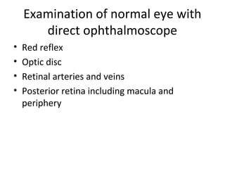


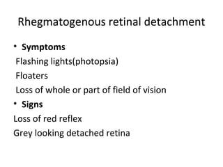


This document provides an overview of vitreoretinal diseases and the anatomy of the vitreous and retina. It discusses examination of the normal eye, symptoms of vitreoretinal disorders, and abnormal fundus features seen on examination. Specific conditions covered include retinal detachment, age-related macular degeneration, diabetic retinopathy, and effects of systemic diseases like hypertension and AIDS. Management approaches for various vitreoretinal diseases are also summarized.