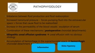
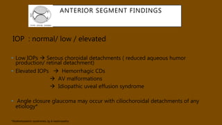
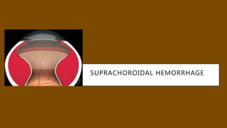
CHOROIDAL EFFUSIONS AND
DETACHMENT..
Dr.Sameera
Vitreo-Retina
MMJEI ,Hubli
CONTENTS
• Definition
• Anatomy
• Types
• Causes
• Diagnosis
• Management
• Prevention
SYNONYMS…
• First described by von Graefe in 1858
• Ciliochoroidal detachment
• Suprachoroidal effusion
• Supraciliary effusion
• the accumulation of fluid in the suprachoroidal space
CHOROID -FUNCTIONS
• Vasculature : major supply for the outer retina
• Choroidal blood flow : cool and warm the retina
• Contains secretory cells : modulation of vascularization and in
growth of the sclera
• Dramatic changes in choroidal thickness : move the retina
forward and back, bringing the photoreceptors into the plane of
focus
• Important role in the drainage of the aqueous humor from the
anterior chamber, via the uveoscleral pathway
ANATOMY
• Four layers
• Haller's layer - outermost layer consisting of
larger diameter blood vessels
• Sattler's layer - layer of medium diameter
blood vessels
• Choriocapillaris - layer of capillaries
• Bruch's membrane (synonyms: Lamina basalis,
Complexus basalis, Lamina vitra) - innermost
layer
NORMAL EYE
• Choroid lies in close approximation to the sclera
• Suprachoroidal space  the space between the choroid and the
sclera : a potential space
• Approximately :10 microlitre fluid seen
ABNORMAL….
• Fluid originates in the choriocapillaries and seeps across the
spongy choroidal tissue into the potential suprachoroidal
space
• Severe cases : fluid escape into the subretinal space 
secondary serous retinal detachment
TYPES..
• Clinically : depending on the characteristics of the fluid
contained between the tissue planes
•  SEROUS
•  HEMORRHAGIC
PATHOPHYSIOLOGY
• Choroidal effusions represent tissue edema
• Starling’s hypothesis  elucidates the balance of
hydrostatic and osmotic gradients between the choroidal
capillaries and interstitial space of the eye
 intraocular pressure (IOP) :hydrostatic pressure in the
interstitial space
 process that shifts flow from the choroidal capillaries
into the interstitium may lead to an effusion
Under normal circumstances : IOP > Pressure in
suprachoroidal space > atmospheric pressure
PATHOPHYSIOLOGY
• Imbalance between fluid production and fluid reabsorption
• Increased transmural pressure - forces pumping fluid into the extravascular
space > forces that cause fluid to be reabsorbed
• Increase in the permeability of the blood vessels : exudation of serum
• Combination of these mechanisms ( postoperative choroidal detachments)
• Idiopathic uveal effusion syndrome  uveal effusion with no obvious
etiology
• Accumulation of hemorrhage in the suprachoroidal space  hemorrhagic
choroidal detachment, e.g., after trauma or surgery
Globe Hypotony
Inflammation
PATHOPHYSIOLOGY
• INFLAMMATION – increases colloid leakage into suprachoroidal space
• Another theory suggests that a tear in the ciliary body allows aqueous
humor to flow into the suprachoroidal space*
* Fuchs E. Ablosung der Aderhaut nach staaroperation. Albrecht von Graefes Arch Ophthalmol. 1900;51:199-
224
EPIDEMIOLOGY
• Incidence following surgery varies between 0.05-6%
• No racial / sexual predilection
• Hemorrhagic detachments are seen more often in elderly
patients
RISK FACTORS-GLAUCOMA
• Antimetabolites—particularly mitomycin C (MMC)—during
trabeculectomy
(5- 33% )
• Latanoprost use in eyes with prior cataract extraction
• Increased risk of subclinical choroidal effusions after laser iridotomy
• Increased association of choroidal effusion with nanophthalmos and with
Sturge-Weber syndrome
• Sturge-Weber syndrome : higher risk in the presence of choroidal
hemangiomas
HISTORY
• Serous choroidal detachments : painless decrease in vision
History  intraocular surgery
 trauma
 anti-glaucoma medications
 panretinal photocoagulation (PRP)/ cryotherapy
• Hemorrhagic detachments: sudden onset of severe pain / loss of vision
 Valsalva maneuver (straining at stools, coughing, sneezing)
 Anticoagulants and aspirin may facilitate bleeding
OTHER SYMPTOMS
• Small, peripheral effusions : asymptomatic
• Large effusions : refractive changes from anterior displacement of
the lens-iris diaphragm and resultant myopia
• Absolute scotoma at the site of effusion
ANTERIOR SEGMENT FINDINGS
• Inflammatory conditions: Congestion /AC cells/ KPs
• Associated with increased resistance to venous drainage : signs of
vascular congestion  dilated episcleral veins /blood in Schlemm’s
canal
• Shallow anterior chamber: Nanophthalmic eyes
Massive choroidal effusions
Ciliochoroidal effusions
IOP : normal/ low / elevated
• Low IOPs  Serous choroidal detachments ( reduced aqueous humor
production/ retinal detachment)
• Elevated IOPs  Hemorrhagic CDs
 AV malformations
 Idiopathic uveal effusion syndrome
• Angle closure glaucoma may occur with ciliochoroidal detachments of any
etiology*
*Myelodysplastic syndromes, Ig A nephropathy
ANTERIOR SEGMENT FINDINGS
POSTERIOR SEGMENT FINDINGS
Detachment typically begins in the periphery  an
ultrabiomicroscopy (UBM) exam
• More fluid  the choroid detaches in a distinct, mound-like
fashion
• Mounds demarcated by the fibrous attachments that correspond
to the sites of the vortex veins
• Very large detachments  the lobes can contact in the visual axis :
appositional or ‘kissing choroidals’
• Annular : involving the circumference of the globe 360°
POSTERIOR SEGMENT FINDINGS
• Inflammatory choroidal effusions : vitreous cells
• Visualization of the ora serrata without scleral depression :pre
equatorial choroidal detachment
• Ciliochoroidal edema/detachment without evidence of intraocular
surgery or trauma
??? Neoplastic/vascular/inflammatory cause
POSTERIOR SEGMENT FINDINGS
• Chronic effusions  blood-ocular barrier
break down at the RPE  Exudative RD /
Shifting SRF
• Depigmentation and multifocal hyperplasia of
the RPE  leopard spots in the fundus
• Linear areas of RPE hypertrophy and
hyperplasia  Verhoeffs lines
INVESTIGATIONS- B SCAN
• Most important imaging modality
• Distincts hemorrhagic versus serous effusions
• Hemorrhagic choroidal effusions (acoustically empty) vs tumors
• Delineate the extent of the choroidal detachment
• Assess the thickness of the choroid in early uveal effusion
syndrome
• Evaluate for posterior scleral thickening / retrobulbar edema
around the optic nerve  T-sign in posterior scleritis
INVESTIGATIONS- B SCAN
1) TOPOGRAPHIC
• • Smooth dome or flat elevation
• • No disc insertion
• • Inserts at Ora or ciliary body
2) QUANTITATIVE
• • Steeply rising ,thick , double peaked spike
• • 100% amplitude
3)KINETIC
• • Mild to none after movement
INVESTIGATIONS-UBM
• Assess anterior choroid/ciliary body
• Any fluid / anterior rotation of the ciliary body associated with
angle closure glaucoma
• Small effusions at ciliary body
 Detachment at scleral spur
CLASSIFICATION
MANAGEMENT
• Conservative
• Postoperative serous choroidal detachments :resolve on
their own within days
• Cycloplegics and corticosteroids
• Excessive leakage from a wound or after glaucoma
filtering surgery  pressure patching / glue /bandage
contact lens
MANAGEMENT
• Oral fluids can be given to increase aqueous humor flow
• Acetazolamide : absorption of suprachoroidal fluid
• ?? paradoxical
PREVENTION
• During surgery
 Good scleral flap ,sutures
 judicious use of antimetabolites
 meticulous closure of the conjunctiva
• High-risk patients
 Prophylactic sclerotomies at the time of glaucoma filtration
surgery
DRUGS
• Topiramate
• Warfarin
• Sulfonamides
• Tetracycline
• Diuretics
• Aspirin
UVEAL EFFUSION SYNDROME
• Described for the first time in 1963 by RJ Brockhurst ,Schepens
• Primary underlying cause :congenital anomaly of the sclera / the vortex veins
• Three types
• Type 1 : Nanophthalmic eyes  small eyeball (average axial length 16 mm)
,high hypermetropic (average +16 diopters)
• Type 2 : Non-nanophthalmic eyes with clinically abnormal sclera  normal
eyeball size (average axial length 21 mm) , small refractive error
• Type 3 : Non-nanophthalmic eyes with clinically normal sclera
PATHOGENESIS OF UVEAL EFFUSION
SYNDROME
• Suprachoroidal space : no lymphatics
• Protein escape from the choriocapillaris : osmotic fluid retention
• Healthy eyes : proteins can diffuse across the sclera
• Form of mucopolysaccharidosis -deposition of dermatan sulfates in sclera
• Egress impaired in Uveal effusion eyes
UVEAL EFFUSION SYNDROME
• U/L or B/L loss of vision  ciliochoroidal and subsequent retinal
detachments
• Metamorphopsia and scotomas caused by shallow macular
detachments
• Bilateral involvement in over 60% of cases
UVEAL EFFUSION SYNDROME
• A/S : episcleral vascular dilation/ blood in Schlemm’s canal
• No inflammatory signs
• IOP : usually normal
• Few vitreous cells +/-
• CD, Exudative RD
• Angiography : leopard-skin appearance of hyperfluorescence and
hypofluorescence
• OCT : focal thickening of the RPE corresponding to the areas of
leopard spots
NANOPHTHALMOS
• Small eye (less than 21 mm in axial length) but with an otherwise
normal structure
• Bilateral; both males and females
• Syndromic /Non -syndromic
• Thickened abnormal sclera  disruption of the normal
arrangement of scleral collagen bundles and a ‘fraying’ of the
collagen fibrils
• Abnormal formation and packing of the collagen bundles
• Hamper the escape of aqueous by the uveo-scleral route
• Outflow obstruction of the vortex veins
NANOPHTHALMOS
• High hypermetropes
• Shallow AC
• Angle closure glaucoma
• Fundus: Foveal hypoplasia/Rudimentary foveal avascular zone/Foveal
schisis
Choroidal effusions/Exudative RD
DIFFERENTIAL DIAGNOSIS
• Choroidal melanoma
• Multifocal, atypical central serous retinopathy
• Vogt-Koyanagi-Harada disease
• Posterior scleritis
• Hypertension
• Metastasis
MANAGEMENT
• Small peripheral uveal effusions :Observe
• Fluid involves or threatens the fovea :
 vortex vein decompression
 full-thickness scleral decompression (Gass et al)
Sclerectomy/Sclerotomy
• Uveal effusion with out Nanophthalmos: ? Vitrectomy – Quick
retinal reattachment
MANAGEMENT
• Pre-operative B-scan: identify the area of maximal effusion
• 360° peritomy / dissection of quadrants
• Isolation of extraocular muscles: care not to avulse vortex veins
• Creation of a lamellar sclerectomy: approximately ~2 × 2 mm
• 50% scleral thickness, with or without vortex vein decompression
• May or may not perform full thickness sclerotomies in sclerectomy
bed to drain choroidal fluid
• Perform in all four quadrants
• Drain subretinal fluid in severe cases at a separate sclerotomy site
• Restore IOP with saline or gas/air injection
• Consider sub-Tenon’s steroids at the end of surgery
VIDEO
OTHER EFFUSIONS
• HUNTER’S SYNDROME
systemic mucopolysaccharidosis : short stature, joint abnormalities
and mental retardation
Circular peripheral choroidal detachments /exudative RDs
• ARTERIOVENOUS FISTULAS
proptosis (often pulsatile), episcleral venous dilation
palsy of the cranial nerves in the cavernous sinus and the orbit
INFLAMMATIONS
• Scleritis:
generalized inflammation of the choroid  leakage from the choriocapillaris
localized or an annular choroidal detachment
Females
Severe pain, Ant scleritis+/-, Raised IOP
Disc oedema,Macular oedema , T-sign
Mx: Steroids
SUPRACHOROIDAL HEMORRHAGE
SUPRACHOROIDAL HEMORRHAGE (SCH)
• One of the most dreaded complications
• Could result in total visual loss and phthisis
SUPRACHOROIDAL HEMORRHAGE (SCH)
• Limited hemorrhage
• Massive hemorrhage
• Appositional (“kissing”).
• Expulsive
MECHANISM
• Impeding vortex vein outflow
- RBH,Retrobulbar anaesthesia,Scleral buckle,Pressure during
surgery
• Fluctuation in intra ocular fluid dynamics and pressure
Hypotony – serous effusion – tension on ciliary vessels* – rupture
-sudden compression and decompression events
• Rarely –spontaneous
RISK FACTORS
• Systemic : Advanced age, Arteriosclerosis,DM,HT,
Anticoagulation,IHD
• Ocular: Glaucoma, Increased axial length, Previous surgery
(Vitrectomy),Previous laser photocoagulation, Aphakia,
Uveitis,Recent trauma
• Intra operative: High IOP,High Myopia, Open sky procedures, Intra
operative tachycardia, Sudden decrease of IOP, Vitreous loss,
INTRA OPERATIVE SYMPTOMS AND SIGNS
• Sudden onset of severe intra operative pain
• Shallowing of anterior chamber/excessive iris movement or
prolapse
• Forward movement of lens and vitreous
• Darkening/loss of red reflex
• Excessive bleeding of conjunctiva/episclera
• Vitreous hemorrhage/RD
• Expulsion of intra ocular contents
• Tachycardia
DELAYED NON EXPULSIVE SUPRACHOROIDAL
HEMORRHAGE
• Decreased vision/Pain
• Shallow anterior chamber with mild cells and flare
• Smooth orange- brown elevation of the retina and
choroid
• B scan: Smooth dome shaped highly reflective solid
appearing mass
FUNDUS FINDINGS
Anterior to the equator
Extends in an annular fashion
Postequatorial
unilobulated or multilobulated.
FUNDUS FINDINGS
• Localised choroidal hemorrhage  choroidal melanoma
(DDs) pigmented choroidal nevus
subretinal pigment epithelial
hamartoma
Acoustic hollowness
SEROUS CD VS HEMORRHAGIC CD
• Low IOP
• Transiiluminates
• No pain
• Pre equatorial
• Resolves with in 3 weeks
• Good visual acuity
• High IOP
• No transillumination
• Painful
• More voluminous
posteriorly
• Resolves 6-25 days; resorbs
~ 4 weeks to months
• Poor visual acuity
SEROUS CD VS HEMORRHAGIC CD
MANAGEMENT: DELAYED HEMORRHAGE
• Limited hemorrhage
 Conservative: Cycloplegics and topical steroids
Usually resolves with in 1-2 months
• Delayed massive hemorrhage
 Systemic corticosteroids with serial B scans ( liquefaction of
hemorrhage
- low reflective mobile echoes )
 Surgery :7-14 days post hemorrhage
WHEN TO OPERATE ON A CHOROIDAL
HEMORRHAGE….
• Massive hemorrhage with severe pain
• Kissing choroidals
• Corneo lenticular touch
• Persistently high IOP
• Persistently flat AC
• Massive hemorrhage under macula/ subretinal/vitreous cavity
• Retinal/Vitreous incarceration
• Preferably after liquefaction
INTRA OPERATIVE MANAGEMENT
• Direct digital pressure on open wounds
• Rapid wound closure
• Post PK: Immediate cardinal sutures to be put
• Reform AC with viscoelastic
• Posterior sclerotomy done only if wound apposition not
possible
• Post operatively : Control IOP/Inflammation/Pain
SECONDARY MANAGEMENT
• Associted RD/VH
• VR traction
• Dislocated lens/fragments :treated accordingly
• PFCL injected to flatten the retina and drain the
hemorrhage
• Silicon oil/Long acting gas for a long term tamponade
CHOROIDAL HEMORRHAGE IN TRAUMA
• Intraocular structural damage +
• High likelihood of RD and associated PVR
• B-scan : hemorrhage tend to be more diffuse and less
elevated
SUPRA CHOROIDAL DRAINAGE
• Optimal drainage site : highest point of choroidal elevation, as
determined by echography or ophthalmoscopy
• Sclerotomies : radial and created with a cut –down incision
• Isolation of the rectus muscle using silk ties/ traction sutures :
good exposure of the sclera
DRAINAGE
• Conjunctival peritomy  2 to 3 mm radial incision made in the sclera : 3 to 4
mm posterior to the limbus
• Incision deepened until the suprachoroidal space is entered  fluid is
released
• Fluid : clear and yellowish in serous CDs
• dark red with blood clots in hemorrhagic CDs
• Incision’s edges may be pulled apart by forceps or cautery to facilitate fluid
egress
• Sclerotomy site left open with closure of overlying conjunctiva
• Eye should be kept pressurized with injection of BSS / viscoelastic in AC /
ACmaintainer
VIDEO
SURGICAL TECHNIQUE
• Trocar in tangential angle inserted into the
suprachoroidal space
• Place the anterior chamber infusion, the
choroidal fluid/hemorrhage drains
spontaneously through the cannulas
• Pars plana vitrectomy +/-
WORD TO THE WISE….
• Non-valved cannulas ; preferable to drain the infero-temporal quadrant first
• Serous CD : one drainage site
• Hemorrhagic CD : more than one drainage site may be needed
“Dry tap”
• Hemorrhagic CD  clot blocking the cannula
Turn the cannula side to side /gentle scleral depression with a cotton swab
• Serous CD  intra-scleral cannula position / cannula is too anterior and is
blocked by choroid, retina or vitreous (if so, sclerotomy placement at 7 mm from
limbus)
• Avoid 3 and 9 o’clock meridians : Traumatise ciliary nerve
OUTCOME
• Good prognosis
Delayed/limited hemorrhage
seen after a week
Hemorrhage after cataract
surgery
Good visual acuity
• Poor prognosis
RD
Hemorrhage all quadrants
Retinal incarceration
Extention to posterior post
pole
THE SUPRACHOROIDAL SPACE: FROM POTENTIAL SPACE TO A
SPACE WITH POTENTIAL
• Suprachoroidal buckling
 illuminated catheter inserted into the SCS and
navigated to any desired location
 long-lasting hyaluronic acid filler can be injected to
create internal choroidal indentation
 Vitrectomy +/-
 myopic tractional maculopathies, RD
SCS AS A ROUTE FOR DRUG DELIVERY
• bypasses the internal limiting membrane barrier and
outer blood-retina-barrier
• preferred route for drug delivery targeting RPE
• Triamcinolone (Uveitis)
• “SAPPHIRE” : SCS steroid vs Aflibercept
INTERESTING FACTS….
• Choroid supply the inner retina (Guinea pigs) as well..!!
• In humans, the choroid is approximately 200 μm thick at
birth and decreases to about 80 μm by age 90
• Choroids are thick at night….. Thin at day.. !
• Teleosts : choroidal gland  supplemental oxygen
carrier
REFERENCES
THANK YOU…
DRAINAGE OF CHOROIDAL
HEMORRHAGE
Choroidal detachment  -Nov 2017

Choroidal detachment -Nov 2017

  • 1.
  • 2.
    CONTENTS • Definition • Anatomy •Types • Causes • Diagnosis • Management • Prevention
  • 3.
    SYNONYMS… • First describedby von Graefe in 1858 • Ciliochoroidal detachment • Suprachoroidal effusion • Supraciliary effusion • the accumulation of fluid in the suprachoroidal space
  • 4.
    CHOROID -FUNCTIONS • Vasculature: major supply for the outer retina • Choroidal blood flow : cool and warm the retina • Contains secretory cells : modulation of vascularization and in growth of the sclera • Dramatic changes in choroidal thickness : move the retina forward and back, bringing the photoreceptors into the plane of focus • Important role in the drainage of the aqueous humor from the anterior chamber, via the uveoscleral pathway
  • 5.
    ANATOMY • Four layers •Haller's layer - outermost layer consisting of larger diameter blood vessels • Sattler's layer - layer of medium diameter blood vessels • Choriocapillaris - layer of capillaries • Bruch's membrane (synonyms: Lamina basalis, Complexus basalis, Lamina vitra) - innermost layer
  • 6.
    NORMAL EYE • Choroidlies in close approximation to the sclera • Suprachoroidal space  the space between the choroid and the sclera : a potential space • Approximately :10 microlitre fluid seen
  • 7.
    ABNORMAL…. • Fluid originatesin the choriocapillaries and seeps across the spongy choroidal tissue into the potential suprachoroidal space • Severe cases : fluid escape into the subretinal space  secondary serous retinal detachment
  • 8.
    TYPES.. • Clinically :depending on the characteristics of the fluid contained between the tissue planes •  SEROUS •  HEMORRHAGIC
  • 9.
    PATHOPHYSIOLOGY • Choroidal effusionsrepresent tissue edema • Starling’s hypothesis  elucidates the balance of hydrostatic and osmotic gradients between the choroidal capillaries and interstitial space of the eye  intraocular pressure (IOP) :hydrostatic pressure in the interstitial space  process that shifts flow from the choroidal capillaries into the interstitium may lead to an effusion Under normal circumstances : IOP > Pressure in suprachoroidal space > atmospheric pressure
  • 10.
    PATHOPHYSIOLOGY • Imbalance betweenfluid production and fluid reabsorption • Increased transmural pressure - forces pumping fluid into the extravascular space > forces that cause fluid to be reabsorbed • Increase in the permeability of the blood vessels : exudation of serum • Combination of these mechanisms ( postoperative choroidal detachments) • Idiopathic uveal effusion syndrome  uveal effusion with no obvious etiology • Accumulation of hemorrhage in the suprachoroidal space  hemorrhagic choroidal detachment, e.g., after trauma or surgery Globe Hypotony Inflammation
  • 11.
    PATHOPHYSIOLOGY • INFLAMMATION –increases colloid leakage into suprachoroidal space • Another theory suggests that a tear in the ciliary body allows aqueous humor to flow into the suprachoroidal space* * Fuchs E. Ablosung der Aderhaut nach staaroperation. Albrecht von Graefes Arch Ophthalmol. 1900;51:199- 224
  • 12.
    EPIDEMIOLOGY • Incidence followingsurgery varies between 0.05-6% • No racial / sexual predilection • Hemorrhagic detachments are seen more often in elderly patients
  • 13.
    RISK FACTORS-GLAUCOMA • Antimetabolites—particularlymitomycin C (MMC)—during trabeculectomy (5- 33% ) • Latanoprost use in eyes with prior cataract extraction • Increased risk of subclinical choroidal effusions after laser iridotomy • Increased association of choroidal effusion with nanophthalmos and with Sturge-Weber syndrome • Sturge-Weber syndrome : higher risk in the presence of choroidal hemangiomas
  • 14.
    HISTORY • Serous choroidaldetachments : painless decrease in vision History  intraocular surgery  trauma  anti-glaucoma medications  panretinal photocoagulation (PRP)/ cryotherapy • Hemorrhagic detachments: sudden onset of severe pain / loss of vision  Valsalva maneuver (straining at stools, coughing, sneezing)  Anticoagulants and aspirin may facilitate bleeding
  • 15.
    OTHER SYMPTOMS • Small,peripheral effusions : asymptomatic • Large effusions : refractive changes from anterior displacement of the lens-iris diaphragm and resultant myopia • Absolute scotoma at the site of effusion
  • 16.
    ANTERIOR SEGMENT FINDINGS •Inflammatory conditions: Congestion /AC cells/ KPs • Associated with increased resistance to venous drainage : signs of vascular congestion  dilated episcleral veins /blood in Schlemm’s canal • Shallow anterior chamber: Nanophthalmic eyes Massive choroidal effusions Ciliochoroidal effusions
  • 17.
    IOP : normal/low / elevated • Low IOPs  Serous choroidal detachments ( reduced aqueous humor production/ retinal detachment) • Elevated IOPs  Hemorrhagic CDs  AV malformations  Idiopathic uveal effusion syndrome • Angle closure glaucoma may occur with ciliochoroidal detachments of any etiology* *Myelodysplastic syndromes, Ig A nephropathy ANTERIOR SEGMENT FINDINGS
  • 18.
    POSTERIOR SEGMENT FINDINGS Detachmenttypically begins in the periphery  an ultrabiomicroscopy (UBM) exam • More fluid  the choroid detaches in a distinct, mound-like fashion • Mounds demarcated by the fibrous attachments that correspond to the sites of the vortex veins • Very large detachments  the lobes can contact in the visual axis : appositional or ‘kissing choroidals’ • Annular : involving the circumference of the globe 360°
  • 19.
    POSTERIOR SEGMENT FINDINGS •Inflammatory choroidal effusions : vitreous cells • Visualization of the ora serrata without scleral depression :pre equatorial choroidal detachment • Ciliochoroidal edema/detachment without evidence of intraocular surgery or trauma ??? Neoplastic/vascular/inflammatory cause
  • 20.
    POSTERIOR SEGMENT FINDINGS •Chronic effusions  blood-ocular barrier break down at the RPE  Exudative RD / Shifting SRF • Depigmentation and multifocal hyperplasia of the RPE  leopard spots in the fundus • Linear areas of RPE hypertrophy and hyperplasia  Verhoeffs lines
  • 21.
    INVESTIGATIONS- B SCAN •Most important imaging modality • Distincts hemorrhagic versus serous effusions • Hemorrhagic choroidal effusions (acoustically empty) vs tumors • Delineate the extent of the choroidal detachment • Assess the thickness of the choroid in early uveal effusion syndrome • Evaluate for posterior scleral thickening / retrobulbar edema around the optic nerve  T-sign in posterior scleritis
  • 22.
    INVESTIGATIONS- B SCAN 1)TOPOGRAPHIC • • Smooth dome or flat elevation • • No disc insertion • • Inserts at Ora or ciliary body 2) QUANTITATIVE • • Steeply rising ,thick , double peaked spike • • 100% amplitude 3)KINETIC • • Mild to none after movement
  • 23.
    INVESTIGATIONS-UBM • Assess anteriorchoroid/ciliary body • Any fluid / anterior rotation of the ciliary body associated with angle closure glaucoma • Small effusions at ciliary body  Detachment at scleral spur
  • 24.
  • 25.
    MANAGEMENT • Conservative • Postoperativeserous choroidal detachments :resolve on their own within days • Cycloplegics and corticosteroids • Excessive leakage from a wound or after glaucoma filtering surgery  pressure patching / glue /bandage contact lens
  • 26.
    MANAGEMENT • Oral fluidscan be given to increase aqueous humor flow • Acetazolamide : absorption of suprachoroidal fluid • ?? paradoxical
  • 27.
    PREVENTION • During surgery Good scleral flap ,sutures  judicious use of antimetabolites  meticulous closure of the conjunctiva • High-risk patients  Prophylactic sclerotomies at the time of glaucoma filtration surgery
  • 28.
    DRUGS • Topiramate • Warfarin •Sulfonamides • Tetracycline • Diuretics • Aspirin
  • 29.
    UVEAL EFFUSION SYNDROME •Described for the first time in 1963 by RJ Brockhurst ,Schepens • Primary underlying cause :congenital anomaly of the sclera / the vortex veins • Three types • Type 1 : Nanophthalmic eyes  small eyeball (average axial length 16 mm) ,high hypermetropic (average +16 diopters) • Type 2 : Non-nanophthalmic eyes with clinically abnormal sclera  normal eyeball size (average axial length 21 mm) , small refractive error • Type 3 : Non-nanophthalmic eyes with clinically normal sclera
  • 30.
    PATHOGENESIS OF UVEALEFFUSION SYNDROME • Suprachoroidal space : no lymphatics • Protein escape from the choriocapillaris : osmotic fluid retention • Healthy eyes : proteins can diffuse across the sclera • Form of mucopolysaccharidosis -deposition of dermatan sulfates in sclera • Egress impaired in Uveal effusion eyes
  • 31.
    UVEAL EFFUSION SYNDROME •U/L or B/L loss of vision  ciliochoroidal and subsequent retinal detachments • Metamorphopsia and scotomas caused by shallow macular detachments • Bilateral involvement in over 60% of cases
  • 32.
    UVEAL EFFUSION SYNDROME •A/S : episcleral vascular dilation/ blood in Schlemm’s canal • No inflammatory signs • IOP : usually normal • Few vitreous cells +/- • CD, Exudative RD • Angiography : leopard-skin appearance of hyperfluorescence and hypofluorescence • OCT : focal thickening of the RPE corresponding to the areas of leopard spots
  • 34.
    NANOPHTHALMOS • Small eye(less than 21 mm in axial length) but with an otherwise normal structure • Bilateral; both males and females • Syndromic /Non -syndromic • Thickened abnormal sclera  disruption of the normal arrangement of scleral collagen bundles and a ‘fraying’ of the collagen fibrils • Abnormal formation and packing of the collagen bundles • Hamper the escape of aqueous by the uveo-scleral route • Outflow obstruction of the vortex veins
  • 35.
    NANOPHTHALMOS • High hypermetropes •Shallow AC • Angle closure glaucoma • Fundus: Foveal hypoplasia/Rudimentary foveal avascular zone/Foveal schisis Choroidal effusions/Exudative RD
  • 36.
    DIFFERENTIAL DIAGNOSIS • Choroidalmelanoma • Multifocal, atypical central serous retinopathy • Vogt-Koyanagi-Harada disease • Posterior scleritis • Hypertension • Metastasis
  • 37.
    MANAGEMENT • Small peripheraluveal effusions :Observe • Fluid involves or threatens the fovea :  vortex vein decompression  full-thickness scleral decompression (Gass et al) Sclerectomy/Sclerotomy • Uveal effusion with out Nanophthalmos: ? Vitrectomy – Quick retinal reattachment
  • 38.
    MANAGEMENT • Pre-operative B-scan:identify the area of maximal effusion • 360° peritomy / dissection of quadrants • Isolation of extraocular muscles: care not to avulse vortex veins • Creation of a lamellar sclerectomy: approximately ~2 × 2 mm • 50% scleral thickness, with or without vortex vein decompression • May or may not perform full thickness sclerotomies in sclerectomy bed to drain choroidal fluid • Perform in all four quadrants • Drain subretinal fluid in severe cases at a separate sclerotomy site • Restore IOP with saline or gas/air injection • Consider sub-Tenon’s steroids at the end of surgery
  • 39.
  • 40.
    OTHER EFFUSIONS • HUNTER’SSYNDROME systemic mucopolysaccharidosis : short stature, joint abnormalities and mental retardation Circular peripheral choroidal detachments /exudative RDs • ARTERIOVENOUS FISTULAS proptosis (often pulsatile), episcleral venous dilation palsy of the cranial nerves in the cavernous sinus and the orbit
  • 41.
    INFLAMMATIONS • Scleritis: generalized inflammationof the choroid  leakage from the choriocapillaris localized or an annular choroidal detachment Females Severe pain, Ant scleritis+/-, Raised IOP Disc oedema,Macular oedema , T-sign Mx: Steroids
  • 42.
  • 43.
    SUPRACHOROIDAL HEMORRHAGE (SCH) •One of the most dreaded complications • Could result in total visual loss and phthisis
  • 44.
    SUPRACHOROIDAL HEMORRHAGE (SCH) •Limited hemorrhage • Massive hemorrhage • Appositional (“kissing”). • Expulsive
  • 45.
    MECHANISM • Impeding vortexvein outflow - RBH,Retrobulbar anaesthesia,Scleral buckle,Pressure during surgery • Fluctuation in intra ocular fluid dynamics and pressure Hypotony – serous effusion – tension on ciliary vessels* – rupture -sudden compression and decompression events • Rarely –spontaneous
  • 46.
    RISK FACTORS • Systemic: Advanced age, Arteriosclerosis,DM,HT, Anticoagulation,IHD • Ocular: Glaucoma, Increased axial length, Previous surgery (Vitrectomy),Previous laser photocoagulation, Aphakia, Uveitis,Recent trauma • Intra operative: High IOP,High Myopia, Open sky procedures, Intra operative tachycardia, Sudden decrease of IOP, Vitreous loss,
  • 47.
    INTRA OPERATIVE SYMPTOMSAND SIGNS • Sudden onset of severe intra operative pain • Shallowing of anterior chamber/excessive iris movement or prolapse • Forward movement of lens and vitreous • Darkening/loss of red reflex • Excessive bleeding of conjunctiva/episclera • Vitreous hemorrhage/RD • Expulsion of intra ocular contents • Tachycardia
  • 48.
    DELAYED NON EXPULSIVESUPRACHOROIDAL HEMORRHAGE • Decreased vision/Pain • Shallow anterior chamber with mild cells and flare • Smooth orange- brown elevation of the retina and choroid • B scan: Smooth dome shaped highly reflective solid appearing mass
  • 49.
    FUNDUS FINDINGS Anterior tothe equator Extends in an annular fashion Postequatorial unilobulated or multilobulated.
  • 50.
    FUNDUS FINDINGS • Localisedchoroidal hemorrhage  choroidal melanoma (DDs) pigmented choroidal nevus subretinal pigment epithelial hamartoma Acoustic hollowness
  • 51.
    SEROUS CD VSHEMORRHAGIC CD • Low IOP • Transiiluminates • No pain • Pre equatorial • Resolves with in 3 weeks • Good visual acuity • High IOP • No transillumination • Painful • More voluminous posteriorly • Resolves 6-25 days; resorbs ~ 4 weeks to months • Poor visual acuity
  • 52.
    SEROUS CD VSHEMORRHAGIC CD
  • 53.
    MANAGEMENT: DELAYED HEMORRHAGE •Limited hemorrhage  Conservative: Cycloplegics and topical steroids Usually resolves with in 1-2 months • Delayed massive hemorrhage  Systemic corticosteroids with serial B scans ( liquefaction of hemorrhage - low reflective mobile echoes )  Surgery :7-14 days post hemorrhage
  • 54.
    WHEN TO OPERATEON A CHOROIDAL HEMORRHAGE…. • Massive hemorrhage with severe pain • Kissing choroidals • Corneo lenticular touch • Persistently high IOP • Persistently flat AC • Massive hemorrhage under macula/ subretinal/vitreous cavity • Retinal/Vitreous incarceration • Preferably after liquefaction
  • 55.
    INTRA OPERATIVE MANAGEMENT •Direct digital pressure on open wounds • Rapid wound closure • Post PK: Immediate cardinal sutures to be put • Reform AC with viscoelastic • Posterior sclerotomy done only if wound apposition not possible • Post operatively : Control IOP/Inflammation/Pain
  • 56.
    SECONDARY MANAGEMENT • AssocitedRD/VH • VR traction • Dislocated lens/fragments :treated accordingly • PFCL injected to flatten the retina and drain the hemorrhage • Silicon oil/Long acting gas for a long term tamponade
  • 57.
    CHOROIDAL HEMORRHAGE INTRAUMA • Intraocular structural damage + • High likelihood of RD and associated PVR • B-scan : hemorrhage tend to be more diffuse and less elevated
  • 58.
    SUPRA CHOROIDAL DRAINAGE •Optimal drainage site : highest point of choroidal elevation, as determined by echography or ophthalmoscopy • Sclerotomies : radial and created with a cut –down incision • Isolation of the rectus muscle using silk ties/ traction sutures : good exposure of the sclera
  • 59.
    DRAINAGE • Conjunctival peritomy 2 to 3 mm radial incision made in the sclera : 3 to 4 mm posterior to the limbus • Incision deepened until the suprachoroidal space is entered  fluid is released • Fluid : clear and yellowish in serous CDs • dark red with blood clots in hemorrhagic CDs • Incision’s edges may be pulled apart by forceps or cautery to facilitate fluid egress • Sclerotomy site left open with closure of overlying conjunctiva • Eye should be kept pressurized with injection of BSS / viscoelastic in AC / ACmaintainer
  • 60.
  • 61.
    SURGICAL TECHNIQUE • Trocarin tangential angle inserted into the suprachoroidal space • Place the anterior chamber infusion, the choroidal fluid/hemorrhage drains spontaneously through the cannulas • Pars plana vitrectomy +/-
  • 62.
    WORD TO THEWISE…. • Non-valved cannulas ; preferable to drain the infero-temporal quadrant first • Serous CD : one drainage site • Hemorrhagic CD : more than one drainage site may be needed “Dry tap” • Hemorrhagic CD  clot blocking the cannula Turn the cannula side to side /gentle scleral depression with a cotton swab • Serous CD  intra-scleral cannula position / cannula is too anterior and is blocked by choroid, retina or vitreous (if so, sclerotomy placement at 7 mm from limbus) • Avoid 3 and 9 o’clock meridians : Traumatise ciliary nerve
  • 63.
    OUTCOME • Good prognosis Delayed/limitedhemorrhage seen after a week Hemorrhage after cataract surgery Good visual acuity • Poor prognosis RD Hemorrhage all quadrants Retinal incarceration Extention to posterior post pole
  • 64.
    THE SUPRACHOROIDAL SPACE:FROM POTENTIAL SPACE TO A SPACE WITH POTENTIAL • Suprachoroidal buckling  illuminated catheter inserted into the SCS and navigated to any desired location  long-lasting hyaluronic acid filler can be injected to create internal choroidal indentation  Vitrectomy +/-  myopic tractional maculopathies, RD
  • 65.
    SCS AS AROUTE FOR DRUG DELIVERY • bypasses the internal limiting membrane barrier and outer blood-retina-barrier • preferred route for drug delivery targeting RPE • Triamcinolone (Uveitis) • “SAPPHIRE” : SCS steroid vs Aflibercept
  • 66.
    INTERESTING FACTS…. • Choroidsupply the inner retina (Guinea pigs) as well..!! • In humans, the choroid is approximately 200 μm thick at birth and decreases to about 80 μm by age 90 • Choroids are thick at night….. Thin at day.. ! • Teleosts : choroidal gland  supplemental oxygen carrier
  • 67.
  • 68.
  • 69.

Editor's Notes

  • #10 A decrease in IOP allows fluid to accumulate in the interstitial spaces, while inflammation increases the permeability of the choroidal capillaries 
  • #15 some cases of postsurgical choroidal effusions, forward rotation of the lens–iris diaphragm can precipitate angle closure with relative pupillary block. This is another indication for surgical relief of the choroidal effusion. proposed mechanism is inflammation, that increases the permeability of the choriocapillaris
  • #20 In some instances, the choroidal detachments and associated retinal detachments may be so massive as to cause contact between the posterior lens surface and the retina. The exudative retinal detachments in these instances are characterized by shifting subretinal fluid, i.e., the subretinal fluid has a tendency to shift and settle into the dependent area on shifting head position and this phenomenon can be mapped out by visual field testing.11,12 This is, in part, because of the high protein (more than 26.5 g/dL
  • #24 The first peak may represent the surface of the overlying detached retina or the anterior surface of the choroid. Alternatively, the double peak may represent both the anterior and posterior surfaces of the choroid
  • #28 ?? paradoxical - aqueous humor suppressant Vasoconstrictive properties : lessen fluid extravasation from choroidal vessels
  • #38 posterior scleral thickness of greater than 2 mm is abnormal
  • #40 local episcleral instillation of corticosteroids prior to conjunctival closure, to prevent closure of the sclerectomies.9 Some surgeons also report re-formation of the anterior chamber using BSS through a paracentesis site as the effusion is drained, or the injection of SF6 gas or air into the vitreous cavity for maintaining IOP. Some authors have also suggested that, in cases of large exudative retinal detachments, subretinal fluid be drained through a separate sclerotomy site. This site is made in full-thickness sclera, diathermizing the scleral edges to fishmouth the sclera and better expose the choroidal knuckle. A 4–0 polyester mattress suture is then preplaced to close the sclerotomy at the end, and the choroidal knuckle is perforated using a sharp perforating needle (e.g., a 30 gauge 0.5 in needle). After drainage, the drainage sclerotomy is closed using the 4–0 mattress suture.