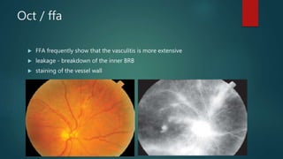
vasculitis
DR ABHISHEK GHELANI
presentation
 Asymptomatic
 decrease in vision / floaters
 scotomata
signs
 Sheathing or cuffing of blood vessels
 vitreous cells
 macular oedema
 cotton-wool spots
 retinal oedema
 Haemorrhages
 telangiectasis, microaneurysms, and neovascularization - late
Oct / ffa
 FFA frequently show that the vasculitis is more extensive
 leakage - breakdown of the inner BRB
 staining of the vessel wall
oct
 Cme
 Reproducible
 In micrometres
 Treatment response
OCULAR VASCULITIS
VERSUS RETINAL VASCULITIS
 more global concept of ocular vasculitis including retinal vasculitis is
useful
 encompass episcleritis, scleritis, (PUK), retinal vasculitis, choroidal
vasculitis, optic nerve vasculitis
Systemic vasculitis
 Vasculitis – histologically proven inflammation of vessel wall
 Primary
 Large vessel
 Medium
 small
 secondary vessel changes without histologic evidence
 Secondary vasculitis / vasculopathy
Ocular vasculitis
 Primary vasculitis limited to the eye
 Secondary vasculitis limited to eye
 Systemic primary vasculitis involving the eye
 Systemic secondary vasculitis involving the eye
PRIMARY VASCULITIS LIMITED TO
THE EYE
 Episcleritis without any systemic involvement
 Scleritis and peripheral ulcerative keratitis (PUK) without systemic involvement
 Retinal vasculitis
 Idiopathic retinal vasculitis – Eale’s
 pars planitis
 Frosted branch angiitis
 IRVAN
 Acute multifocal haemorrhagic retinal vasculitis
 Choroidal vasculitis
 MEWDS
 APMPPE
 MFC
 Serpiginous choroiditis
Eale’s
 Obliterative periphlebitis
 venous sheathing are the commonest clinical presentation
 Starts at or around equator and
progresses posteriorly
 compensatory phenomena like collaterals, microaneurysms, capillary
telangiectasia, corkscrew vessels, and venous beading
 can lead to neovascularization - peri[heral
 recurrent vitreous haemorrhage
 Traction retinal detachment
 affect healthy young adults in the third and fourth decades
 Men> women
 Etiopathogenesis – type III hypersensitivity reaction ?hypersensitivity to
tuberculoprotein
 (HLA) B5 (B51), DR1, and DR4
 systemic steroids
 panretinal photocoagulation
 vitrectomy – endolaser in non-resolving VH with FVP
Secondary inflammatory vasculopathy
limited to eye
 Episcleritis, Scleritis and PUK secondary to local infection
 Retinal inflammatory vasculopathy
 Immune mediated
–Birdshot chorioretinopathy retinal vasculitis
– Ocular sarcoidosis
 Infectious or para-infectious
– Necrotic herpetic retinopathies (herpes simplex virus, varicella-zoster virus)
– Toxoplasma
– – DUSN (Diffuse unilateral subacute neuroretinitis, due to parasites, Toxocara canis)
 Neoplasms
– Primary ocular lymphoma
 Choroidal inflammatory vasculopathy or vasculitis
secondary choriocapillaropathy
adjacent to retinitis or choroiditis)
Secondary stromal vasculitis
-Immune mediated
– Birdshot choroiditis (choroidal disease of birdshot chorioretinopathy)
– Sympathetic ophthalmia
– Toxoplasmic retinochoroiditis
– Vogt-Koyanagi-Harada disease
– ocular Sarcoidosis
-Infectious
Systemic primary vasculitis involving
the eye
 Giant cell arteritis
 Takayasu arteritis
 Polyarteritis nodosa
 Kawasaki disease
 Wegener’s granulomatosis
 Churg-Strauss syndrome
 Henoch-Schönlein purpura
 Cutaneous leucocytoclastic angiitis
 Essential cryoglobulinaemic vasculitis
Giant cell arteritis
 affects large and medium-sized arteries
 50 years or older
 headache and tenderness of the temporal artery or scalp
 Jaw or tongue claudication
 malaise, anorexia and weight loss, fever, neck pain, joint and muscle pain,
and ear pain
 Visual symptoms may include transient or permanent visual loss, diplopia,
and eye pain
 (AAION) is the most common cause of vision loss but
 central retinal artery occlusion, cilioretinal artery occlusion
 posterior ischemic optic neuropathy
 ocular ischemic syndrome also occur
 posterior ciliary arteries, choroidal ischaemia
 anterior segment ischaemia (uveitis and episcleritis
 extraocular muscle palsies
 Westergren ESR (mean 70 mm/hr; often >100 mm/hr) - may be normal in
up to 16% of cases
 CRP level
 Temporal artery biopsy – CONFIRMATORY (false –ve 3 – 9 %)
 IV MP (1 g/day for the first 3–5 days)
 suspected GCA without loss of vision, oral prednisone
 continue therapy for at least 1–2 years
Polyarteritis nodosa
 medium-sized and small muscular arteries
 40 and 60 years and affects men 3 times more
 hepatitis B ?
 Ocular involvement is present in up to 20%
 fatigue, fever, weight loss, and arthralgia
 mononeuritis multiplex is the most common
 Renal involvement – HT - may manifest as hypertensive retinopathy
 small-bowel ischemia and infarction
 Scleritis, PUK, episcleritis, conjunctivitis and conjunctival vasculitis
 choroidal vasculitis most common ocular involvement (posterior ciliary
arteries, large and small choroidal vessels → choroidal ischaemia
 Cranial nerve palsies, amaurosis fugax, homonymous hemianopia, Horner
syndrome
 The 5- year mortality rate of untreated PAN is 90%
 Combinaton steroid + IMT – 80%
Wegener’s granulomatosis
 Granulomatosis with polyangiitis
 classic triad
 Involvement of the paranasal sinuses is the most characteristic
 followed by pulmonary and renal disease
 Dermatologic involvement - one-half of patients, purpura (lower
extremities) ulcers and subcutaneous nodules
 Nervous system - one-third of patients - peripheral neuropathies;
mononeuritis multiplex and less frequently cranial neuropathies, seizures,
stroke syndromes, and cerebral vasculitis
 Ocular involvement in 50 %
 Orbit most common - contiguous extension
 Dacryocystitis
 Scleritis of any type – 40%
 Ant, int., post. Uveitis
 Retinal involvement 10% - cotton-wool spots, intraretinal hemorrhages,
branch or central retinal artery or vein occlusion
 Retinitis - 20% of patients; may accompany retinal vasculitis - retinal
neovascularization, vitreous hemorrhage, neovascular glaucoma
 Tissue biopsy
 chest x-ray - nodular, diffuse, or cavitary lesions
 proteinuria or hematuria
 elevated (ESR) and CRP level
 ANCA - GPA, MPA, eosinophilic granulomatosis with polyangiitis (Churg-
Strauss syndrome), renal limited vasculitis, and pauci-
immunoglomerulonephritis
Immunofluorescence pattern of ANCA
 c-ANCA (PR3 - ANCA)
 present in up to 95% of patients of GPA
 p-ANCA (Myeloperoxidase) - MPA, renal limited vasculitis, and pauci-
immunoglomerulonephritis
 Without therapy, the 1-year mortality rate is 80%.
 cyclophosphamide and corticosteroids 93% successfully achieve remission
with resolution of ocular manifestations
Systemic secondary vasculitis
involving the eye
 Immune mediated
HLA B27-associated uveitis
Rheumatoid arthritis , JIA
Multiple sclerosis
Sarcoidosis
Systemic lupus erythematosus
Behçet’s disease
Susac’s syndrome
 Infectious
Mycobacteria
Spirochaetes
Herpes zoster
HTLV vasculitis
toxocara
Lyme disease
Bartonella henselae
Whipple’ disease
Rickettsial diseases
HLA-B27–related diseases
 Ankylosing spondylitis
 Reactive arthritis syndrome
 Inflammatory bowel disease
 Psoriatic arthritis
Ankylosing spondylitis
 Asymptomatic
 lower back pain and morning stiffness, (Often, persons with anterior uveitis
lack symptoms of back disease)
 90% - HLA B27 +ve
 Sacroiliac imaging
 Pulmonary apical fibrosis and cardiovascular disease (aortic valvular
insufficiency) may also develop
 NSAIDs
 Sulfasalazine
 anti-TNF drugs
 exercise, physical therapy, and smoking cessation
Reactive arthritis syndrome
 Reiter syndrome
 triad of nonspecific urethritis, polyarthritis, and conjunctival inflammation,
often accompanied by nongranulomatous anterior uveitis
 keratoderma blennorrhagicum:
 circinate balanitis
 HLA-B27 – 95%
 triggered by episodes of diarrhea or dysentery
 Ureaplasma urealyticum, Chlamydia, Shigella, Salmonella, and Yersinia
 Arthritis begins within 30 days of infection in 80% of patients
 knees, ankles, feet, and wrists
 Asymmetrical Oligoarticular
 Eye involvement - 20%. Conjunctivitis is the most common
 Punctate and subepithelial keratitis
 Acute nongranulomatous anterior uveitis occurs in up to 10% of patients
and may become bilateral and chronic
Juvenile idiopathic arthritis
 Pedia ant uveitis – most common syst. Assoc. is JIA
 arthritis begins before age 16 and lasts for at least 6 weeks
 Risk factors for chronic uveitis in JIA patients - female sex, oligoarticular
onset, and the presence of ANA
 Most patients test negative for rheumatoid factor
 Ocular involvement in JIA JIA can be classified into 3 types
 Systemic onset (Still disease) - 10 – 15%
under age 5 years,
fever, rash, lymphadenopathy, and hepatosplenomegaly
Joint involvement may be minimal
fewer than 6% of patients have uveitis
 Polyarticular onset - 40%
involvement of > 4 joints in the first 6 months of the disease
 Oligoarticular onset - 40%–50%
(80%–90%) of patients with JIA-associated uveitis
Multiple sclerosis
 Uveitis is 10 times more common in MS
 Bilateral intermediate uveitis is the most common manifestation
 cross-reactivity between myelin-associated glycoprotein and Müller cells
SLE
 connective tissue disorder
 women of childbearing age
 polyclonal B-lymphocyte activation, hypergammaglobulinemia, immune
complex deposition - end-organ damage
 ANA - anti-ssDNA and anti-dsDNA
 antibodies to cytoplasmic components (anti- Sm, anti-Ro, and anti-La)
 antiphospholipid antibodies
 Ocular manifestations - in 50%
 cutaneous lesions on the eyelids (discoid lupus erythematosus),
 Scleritis, episcleritis
 secondary Sjögren syndrome in 20% of patients
 neuro-ophthalmic lesions (cranial nerve palsies, optic neuropathy, and
retrochiasmal and cerebral visual disorders)
 retinal vasculopathy
 in rare cases, uveitis
Lupus retinopathy
 Cotton-wool spots with or without intraretinal hemorrhages occur
independently of hypertension and are due to the microangiopathy
 arterial and venous thrombosis - related to anti phospholipild
autoantibodies induced hypercoagulable state
 Lupus choroidopathy - choroidal infarction and choroidal
neovascularization
 NSAIDs, corticosteroids, IMT, plasmapheresis, and systemic
antihypertensive
 antiplatelet therapy or systemic anticoagulation

Vasculitis

  • 1.
  • 2.
    presentation  Asymptomatic  decreasein vision / floaters  scotomata
  • 3.
    signs  Sheathing orcuffing of blood vessels  vitreous cells  macular oedema  cotton-wool spots  retinal oedema  Haemorrhages  telangiectasis, microaneurysms, and neovascularization - late
  • 4.
    Oct / ffa FFA frequently show that the vasculitis is more extensive  leakage - breakdown of the inner BRB  staining of the vessel wall
  • 5.
    oct  Cme  Reproducible In micrometres  Treatment response
  • 6.
    OCULAR VASCULITIS VERSUS RETINALVASCULITIS  more global concept of ocular vasculitis including retinal vasculitis is useful  encompass episcleritis, scleritis, (PUK), retinal vasculitis, choroidal vasculitis, optic nerve vasculitis
  • 7.
    Systemic vasculitis  Vasculitis– histologically proven inflammation of vessel wall  Primary  Large vessel  Medium  small  secondary vessel changes without histologic evidence  Secondary vasculitis / vasculopathy
  • 8.
    Ocular vasculitis  Primaryvasculitis limited to the eye  Secondary vasculitis limited to eye  Systemic primary vasculitis involving the eye  Systemic secondary vasculitis involving the eye
  • 9.
    PRIMARY VASCULITIS LIMITEDTO THE EYE  Episcleritis without any systemic involvement  Scleritis and peripheral ulcerative keratitis (PUK) without systemic involvement  Retinal vasculitis  Idiopathic retinal vasculitis – Eale’s  pars planitis  Frosted branch angiitis  IRVAN  Acute multifocal haemorrhagic retinal vasculitis  Choroidal vasculitis  MEWDS  APMPPE  MFC  Serpiginous choroiditis
  • 10.
    Eale’s  Obliterative periphlebitis venous sheathing are the commonest clinical presentation  Starts at or around equator and progresses posteriorly  compensatory phenomena like collaterals, microaneurysms, capillary telangiectasia, corkscrew vessels, and venous beading
  • 12.
     can leadto neovascularization - peri[heral  recurrent vitreous haemorrhage  Traction retinal detachment  affect healthy young adults in the third and fourth decades  Men> women  Etiopathogenesis – type III hypersensitivity reaction ?hypersensitivity to tuberculoprotein  (HLA) B5 (B51), DR1, and DR4
  • 13.
     systemic steroids panretinal photocoagulation  vitrectomy – endolaser in non-resolving VH with FVP
  • 14.
    Secondary inflammatory vasculopathy limitedto eye  Episcleritis, Scleritis and PUK secondary to local infection  Retinal inflammatory vasculopathy  Immune mediated –Birdshot chorioretinopathy retinal vasculitis – Ocular sarcoidosis  Infectious or para-infectious – Necrotic herpetic retinopathies (herpes simplex virus, varicella-zoster virus) – Toxoplasma – – DUSN (Diffuse unilateral subacute neuroretinitis, due to parasites, Toxocara canis)  Neoplasms – Primary ocular lymphoma
  • 15.
     Choroidal inflammatoryvasculopathy or vasculitis secondary choriocapillaropathy adjacent to retinitis or choroiditis) Secondary stromal vasculitis -Immune mediated – Birdshot choroiditis (choroidal disease of birdshot chorioretinopathy) – Sympathetic ophthalmia – Toxoplasmic retinochoroiditis – Vogt-Koyanagi-Harada disease – ocular Sarcoidosis -Infectious
  • 16.
    Systemic primary vasculitisinvolving the eye  Giant cell arteritis  Takayasu arteritis  Polyarteritis nodosa  Kawasaki disease  Wegener’s granulomatosis  Churg-Strauss syndrome  Henoch-Schönlein purpura  Cutaneous leucocytoclastic angiitis  Essential cryoglobulinaemic vasculitis
  • 17.
    Giant cell arteritis affects large and medium-sized arteries  50 years or older  headache and tenderness of the temporal artery or scalp  Jaw or tongue claudication  malaise, anorexia and weight loss, fever, neck pain, joint and muscle pain, and ear pain  Visual symptoms may include transient or permanent visual loss, diplopia, and eye pain
  • 18.
     (AAION) isthe most common cause of vision loss but  central retinal artery occlusion, cilioretinal artery occlusion  posterior ischemic optic neuropathy  ocular ischemic syndrome also occur  posterior ciliary arteries, choroidal ischaemia  anterior segment ischaemia (uveitis and episcleritis  extraocular muscle palsies
  • 19.
     Westergren ESR(mean 70 mm/hr; often >100 mm/hr) - may be normal in up to 16% of cases  CRP level  Temporal artery biopsy – CONFIRMATORY (false –ve 3 – 9 %)  IV MP (1 g/day for the first 3–5 days)  suspected GCA without loss of vision, oral prednisone  continue therapy for at least 1–2 years
  • 20.
    Polyarteritis nodosa  medium-sizedand small muscular arteries  40 and 60 years and affects men 3 times more  hepatitis B ?  Ocular involvement is present in up to 20%  fatigue, fever, weight loss, and arthralgia  mononeuritis multiplex is the most common  Renal involvement – HT - may manifest as hypertensive retinopathy  small-bowel ischemia and infarction
  • 21.
     Scleritis, PUK,episcleritis, conjunctivitis and conjunctival vasculitis  choroidal vasculitis most common ocular involvement (posterior ciliary arteries, large and small choroidal vessels → choroidal ischaemia  Cranial nerve palsies, amaurosis fugax, homonymous hemianopia, Horner syndrome  The 5- year mortality rate of untreated PAN is 90%  Combinaton steroid + IMT – 80%
  • 22.
    Wegener’s granulomatosis  Granulomatosiswith polyangiitis  classic triad  Involvement of the paranasal sinuses is the most characteristic  followed by pulmonary and renal disease  Dermatologic involvement - one-half of patients, purpura (lower extremities) ulcers and subcutaneous nodules  Nervous system - one-third of patients - peripheral neuropathies; mononeuritis multiplex and less frequently cranial neuropathies, seizures, stroke syndromes, and cerebral vasculitis
  • 23.
     Ocular involvementin 50 %  Orbit most common - contiguous extension  Dacryocystitis  Scleritis of any type – 40%  Ant, int., post. Uveitis  Retinal involvement 10% - cotton-wool spots, intraretinal hemorrhages, branch or central retinal artery or vein occlusion  Retinitis - 20% of patients; may accompany retinal vasculitis - retinal neovascularization, vitreous hemorrhage, neovascular glaucoma
  • 24.
     Tissue biopsy chest x-ray - nodular, diffuse, or cavitary lesions  proteinuria or hematuria  elevated (ESR) and CRP level  ANCA - GPA, MPA, eosinophilic granulomatosis with polyangiitis (Churg- Strauss syndrome), renal limited vasculitis, and pauci- immunoglomerulonephritis
  • 25.
    Immunofluorescence pattern ofANCA  c-ANCA (PR3 - ANCA)  present in up to 95% of patients of GPA  p-ANCA (Myeloperoxidase) - MPA, renal limited vasculitis, and pauci- immunoglomerulonephritis  Without therapy, the 1-year mortality rate is 80%.  cyclophosphamide and corticosteroids 93% successfully achieve remission with resolution of ocular manifestations
  • 26.
    Systemic secondary vasculitis involvingthe eye  Immune mediated HLA B27-associated uveitis Rheumatoid arthritis , JIA Multiple sclerosis Sarcoidosis Systemic lupus erythematosus Behçet’s disease Susac’s syndrome
  • 27.
     Infectious Mycobacteria Spirochaetes Herpes zoster HTLVvasculitis toxocara Lyme disease Bartonella henselae Whipple’ disease Rickettsial diseases
  • 28.
    HLA-B27–related diseases  Ankylosingspondylitis  Reactive arthritis syndrome  Inflammatory bowel disease  Psoriatic arthritis
  • 29.
    Ankylosing spondylitis  Asymptomatic lower back pain and morning stiffness, (Often, persons with anterior uveitis lack symptoms of back disease)  90% - HLA B27 +ve  Sacroiliac imaging  Pulmonary apical fibrosis and cardiovascular disease (aortic valvular insufficiency) may also develop  NSAIDs  Sulfasalazine  anti-TNF drugs  exercise, physical therapy, and smoking cessation
  • 30.
    Reactive arthritis syndrome Reiter syndrome  triad of nonspecific urethritis, polyarthritis, and conjunctival inflammation, often accompanied by nongranulomatous anterior uveitis  keratoderma blennorrhagicum:  circinate balanitis  HLA-B27 – 95%  triggered by episodes of diarrhea or dysentery  Ureaplasma urealyticum, Chlamydia, Shigella, Salmonella, and Yersinia  Arthritis begins within 30 days of infection in 80% of patients
  • 31.
     knees, ankles,feet, and wrists  Asymmetrical Oligoarticular  Eye involvement - 20%. Conjunctivitis is the most common  Punctate and subepithelial keratitis  Acute nongranulomatous anterior uveitis occurs in up to 10% of patients and may become bilateral and chronic
  • 32.
    Juvenile idiopathic arthritis Pedia ant uveitis – most common syst. Assoc. is JIA  arthritis begins before age 16 and lasts for at least 6 weeks  Risk factors for chronic uveitis in JIA patients - female sex, oligoarticular onset, and the presence of ANA  Most patients test negative for rheumatoid factor  Ocular involvement in JIA JIA can be classified into 3 types  Systemic onset (Still disease) - 10 – 15% under age 5 years, fever, rash, lymphadenopathy, and hepatosplenomegaly Joint involvement may be minimal fewer than 6% of patients have uveitis
  • 33.
     Polyarticular onset- 40% involvement of > 4 joints in the first 6 months of the disease  Oligoarticular onset - 40%–50% (80%–90%) of patients with JIA-associated uveitis
  • 34.
    Multiple sclerosis  Uveitisis 10 times more common in MS  Bilateral intermediate uveitis is the most common manifestation  cross-reactivity between myelin-associated glycoprotein and Müller cells
  • 35.
    SLE  connective tissuedisorder  women of childbearing age  polyclonal B-lymphocyte activation, hypergammaglobulinemia, immune complex deposition - end-organ damage  ANA - anti-ssDNA and anti-dsDNA  antibodies to cytoplasmic components (anti- Sm, anti-Ro, and anti-La)  antiphospholipid antibodies
  • 36.
     Ocular manifestations- in 50%  cutaneous lesions on the eyelids (discoid lupus erythematosus),  Scleritis, episcleritis  secondary Sjögren syndrome in 20% of patients  neuro-ophthalmic lesions (cranial nerve palsies, optic neuropathy, and retrochiasmal and cerebral visual disorders)  retinal vasculopathy  in rare cases, uveitis
  • 37.
    Lupus retinopathy  Cotton-woolspots with or without intraretinal hemorrhages occur independently of hypertension and are due to the microangiopathy  arterial and venous thrombosis - related to anti phospholipild autoantibodies induced hypercoagulable state  Lupus choroidopathy - choroidal infarction and choroidal neovascularization  NSAIDs, corticosteroids, IMT, plasmapheresis, and systemic antihypertensive  antiplatelet therapy or systemic anticoagulation