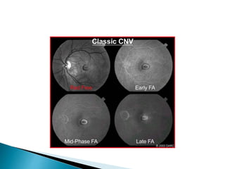
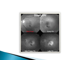
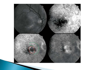
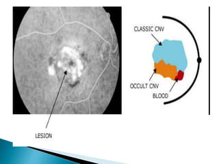
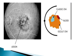
Dr. K. Vasantha M.S., F.R.C.S. Edin
Director RIO Chennai (Rtd)
 Defective vision
 Metamorphopsia
 Central or paracentral scotoma
 Sometimes CNV may be found just on routine
examination
 Soft drusen as CNV is most commonly seen in age
related macular degeneration
 Proliferation of pigmentary epithelium
 Elevation of retinal pigmentary epithelium (RPE) by the
CNV, pigment proliferation or serous fluid
 Cystic changes in the sensory retina
 New vessels themselves also may be seen as yellow
green discoloration
 Pigment ring may be seen around this lesion
 Sub RPE or sub retinal hemorrhage which may be
present just near the periphery of the CNV extensive
enough to obscure the CNV
 A ring of lipid precipitates either partial or total may be
seen. As this occurs around CNV one should suspect
CNV if this is seen in a case of soft drusen
 Classic CNV
 Occult CNV
 Retinal angiomatosis proliferans
 These types are recognized by doing a FFA
 An area of well demarcated hyper fluorescence in the
early phase
 These are discrete and bright
 Pooling of the dye due to leakage in the sub sensory
retinal layer
 Here the new vessels extend between the basement
membrane and RPE
 Signs of pigment epithelial detachment
 Late leakage from undetermined source
 The hyper fluorescence seen here is not as discrete and
bright as in classic CNV, but it is stippled
 Late leakage from undetermined source appear as
pooling of the dye in which the point source of leakage
which can be seen as discrete hyper fluorescent spot is
not seen here
 Here the capillary proliferation has a telangiectatic
pattern
 It is often bilateral and more common in females
 Associated morbidities like hypertension, cardio
vascular diseases and hyper cholestrolemia are more
common
 Reticular drusen is also more common
 The neovascularization is seen both in the deep retina
and the choroid
 Intra retinal hemorrhage
 Sub retinal fibrosis occurs earlier in this type as both
retina and choroid are involved
 There will be a connection between the retinal vessels
and the choroid
 Stage 1: lesion is intra retinal only
 Stage 2: sub retinal lesion with or without PED. In this
stage the perfusing vessel may be seen. If seen it can be
occluded by laser treatment
 Stage 3: with choroidal neovascularization
 This type occurs in around 35% of cases
 FFA: intra retinal and sub retinal leakage with indistinct
margins
 Vascularized PED with or without vascular anastomosis
 RPE tears
 ICG: Hot spot at the center of PED and in the intra and
sub retinal layers
 OCT: focal hyper reflective lesions due to exudation in
intra retinal and sub retinal layers
 Tuft of vessels will be seen in the outer retinal layer with
feeder vessels from inner retinal layers
Ref- AJO 2017 September
 Since most of these cases have age related macular
degeneration we have to look for those changes
 Eyes with intermediate AMD show thinning of ganglion
cell complex and decreased normalized ellipsoid zone
reflectivity
 Will show the vascular network pattern even in the
presence of hemorrhage
 Hyper flow lesion in the outer retina with glomerulous or
Medusa shape, along with a dark halo
 This halo is due to occlusion by blood or due to capillary
alteration causing flow impairment, steal or atrophy
 Fan, octopus or spider like feeder vessels also seen
 If vessels are seen in the sub retinal space with OCTA it
means it is abnormal
The boundaries may not be clearly delineated if
 Blood is thick enough to obscure
 Blocked fluorescence due to hyper plastic pigment or
fibrous proliferations
 In serous detachment of the RPE there will be an hyper
fluorescence which will obscure the leak from the CNV
 Tears or rips in the RPE can occur either spontaneously
or following LASER treatment
 If it occurs vision will be grossly affected
 FFA will show hyper fluorescence due to exposed
choroid in the torn area with blocked fluorescence in the
area where the pigment cells are heaped up.
 With these tears RD does not occur probably because
the choroid removes the fluid from the sub retinal space
This often occurs in age related macular degenerations
which is the commonest cause for CNV
 Fibro vascular PED which shows slow filling with
stippled fluorescence in FFA
 Serous PED will make it difficult to differentiate occult
and classic CNV
 Hemorrhagic PED may be mistaken for melanoma – B
scan is to be done. PED with hemorrhage will not show
the low internal reflectivity shown by melanoma
 Drusenoid PED with large areas of confluent soft drusen.
This will be small and shallow with irregular outline.
Pigment clumping also is seen overlying the druse.
 The FFA shows a faint pattern with no signs of bright
fluorescence seen in serous detachments
 Appears as yellow white fibrous lesion with variable
amount of pigmentation
 It is often seen in the central portion of the CNV with
active lesion around
Will be poor in
 Type 2 CNV
 Poor baseline vision
 Vascular sub retinal hyper reflective material
 Disruption of external limiting membrane
 Intra retinal fluid
 Photodynamic therapy is not done routinely after the
advent of VEGF. It generates free oxygen radicals which
causes cytotoxic damage to the new vessels
 Ranibizumab: Anchor and Marina study said monthly
injections of Ranibizumab was effective
 PrONTO study said that with OCT based retreatment (pro
re nata) a mean improvement of 11.1 letters at 2 years
was possible with a mean of 9.9 injections
 SAILOR study – 3 initial monthly injections followed by
PRN dosing at 3 month follow up
 SUSTAIN study also advises PRN dosing of ranizumab
Bevacizumab:
 CATT (Comparison of AMD Treatment Trial) compared
monthly and prn Bevacizumab and Ranibizumab. It
showed non inferiority of Bevacizumab
 LUCAS study: Lucentis compared to Avastin that
Bevacizumab is almost as effective as Ranibizumab.
 As both 1 and 2 receptors of VEGF are bound it is more
effective but costly
 VIEW-1 and VIEW-2 trials compared monthly and every 2
months injections of Ranibizumab and Aflibercept. It
showed that Aflibercept is better than Ranibizumab
 Conbercept, designated ankyrin repeat proteins,
Sphingosin-1 phosphate antibody, Platelet derived
growth factor antibody are other drugs under
investigation
 Gene therapy: Viral vectors are used to introduce genes
and treat CNV
 Improved technology to deliver the drugs and reduce the
number of injections like
 Encapsulated cell technology, reservoir devices,
Suprachoroidal injections, drugs incorporated colloids,
transscleral delivery and ultra sound delivery
If hemorrhage is present
 Macro aneurysm
 Pathologic myopia with lacquer cracks
 Choroidal rupture
 Choroidal tumors when hemorrhage occurs in a
peripheral scar anterior to the posterior pole
 Ocular histoplasmosis
 Geographic atrophy with rupture of chorio capillaries
If there is retinal angiomatosis proliferans rule out
 Para foveal telangiectasis
 Diabetic macular edema
 Macular branch retinal vein occlusion

Choroidal neovascularization

  • 1.
    Dr. K. VasanthaM.S., F.R.C.S. Edin Director RIO Chennai (Rtd)
  • 2.
     Defective vision Metamorphopsia  Central or paracentral scotoma  Sometimes CNV may be found just on routine examination
  • 3.
     Soft drusenas CNV is most commonly seen in age related macular degeneration  Proliferation of pigmentary epithelium  Elevation of retinal pigmentary epithelium (RPE) by the CNV, pigment proliferation or serous fluid  Cystic changes in the sensory retina
  • 4.
     New vesselsthemselves also may be seen as yellow green discoloration  Pigment ring may be seen around this lesion  Sub RPE or sub retinal hemorrhage which may be present just near the periphery of the CNV extensive enough to obscure the CNV  A ring of lipid precipitates either partial or total may be seen. As this occurs around CNV one should suspect CNV if this is seen in a case of soft drusen
  • 5.
     Classic CNV Occult CNV  Retinal angiomatosis proliferans  These types are recognized by doing a FFA
  • 6.
     An areaof well demarcated hyper fluorescence in the early phase  These are discrete and bright  Pooling of the dye due to leakage in the sub sensory retinal layer  Here the new vessels extend between the basement membrane and RPE
  • 8.
     Signs ofpigment epithelial detachment  Late leakage from undetermined source  The hyper fluorescence seen here is not as discrete and bright as in classic CNV, but it is stippled  Late leakage from undetermined source appear as pooling of the dye in which the point source of leakage which can be seen as discrete hyper fluorescent spot is not seen here
  • 13.
     Here thecapillary proliferation has a telangiectatic pattern  It is often bilateral and more common in females  Associated morbidities like hypertension, cardio vascular diseases and hyper cholestrolemia are more common  Reticular drusen is also more common  The neovascularization is seen both in the deep retina and the choroid
  • 14.
     Intra retinalhemorrhage  Sub retinal fibrosis occurs earlier in this type as both retina and choroid are involved  There will be a connection between the retinal vessels and the choroid
  • 15.
     Stage 1:lesion is intra retinal only  Stage 2: sub retinal lesion with or without PED. In this stage the perfusing vessel may be seen. If seen it can be occluded by laser treatment  Stage 3: with choroidal neovascularization  This type occurs in around 35% of cases
  • 16.
     FFA: intraretinal and sub retinal leakage with indistinct margins  Vascularized PED with or without vascular anastomosis  RPE tears  ICG: Hot spot at the center of PED and in the intra and sub retinal layers  OCT: focal hyper reflective lesions due to exudation in intra retinal and sub retinal layers
  • 17.
     Tuft ofvessels will be seen in the outer retinal layer with feeder vessels from inner retinal layers
  • 18.
    Ref- AJO 2017September  Since most of these cases have age related macular degeneration we have to look for those changes  Eyes with intermediate AMD show thinning of ganglion cell complex and decreased normalized ellipsoid zone reflectivity
  • 19.
     Will showthe vascular network pattern even in the presence of hemorrhage  Hyper flow lesion in the outer retina with glomerulous or Medusa shape, along with a dark halo  This halo is due to occlusion by blood or due to capillary alteration causing flow impairment, steal or atrophy  Fan, octopus or spider like feeder vessels also seen  If vessels are seen in the sub retinal space with OCTA it means it is abnormal
  • 20.
    The boundaries maynot be clearly delineated if  Blood is thick enough to obscure  Blocked fluorescence due to hyper plastic pigment or fibrous proliferations  In serous detachment of the RPE there will be an hyper fluorescence which will obscure the leak from the CNV
  • 21.
     Tears orrips in the RPE can occur either spontaneously or following LASER treatment  If it occurs vision will be grossly affected  FFA will show hyper fluorescence due to exposed choroid in the torn area with blocked fluorescence in the area where the pigment cells are heaped up.  With these tears RD does not occur probably because the choroid removes the fluid from the sub retinal space
  • 23.
    This often occursin age related macular degenerations which is the commonest cause for CNV  Fibro vascular PED which shows slow filling with stippled fluorescence in FFA  Serous PED will make it difficult to differentiate occult and classic CNV  Hemorrhagic PED may be mistaken for melanoma – B scan is to be done. PED with hemorrhage will not show the low internal reflectivity shown by melanoma
  • 24.
     Drusenoid PEDwith large areas of confluent soft drusen. This will be small and shallow with irregular outline. Pigment clumping also is seen overlying the druse.  The FFA shows a faint pattern with no signs of bright fluorescence seen in serous detachments
  • 25.
     Appears asyellow white fibrous lesion with variable amount of pigmentation  It is often seen in the central portion of the CNV with active lesion around
  • 26.
    Will be poorin  Type 2 CNV  Poor baseline vision  Vascular sub retinal hyper reflective material  Disruption of external limiting membrane  Intra retinal fluid
  • 27.
     Photodynamic therapyis not done routinely after the advent of VEGF. It generates free oxygen radicals which causes cytotoxic damage to the new vessels  Ranibizumab: Anchor and Marina study said monthly injections of Ranibizumab was effective  PrONTO study said that with OCT based retreatment (pro re nata) a mean improvement of 11.1 letters at 2 years was possible with a mean of 9.9 injections  SAILOR study – 3 initial monthly injections followed by PRN dosing at 3 month follow up
  • 28.
     SUSTAIN studyalso advises PRN dosing of ranizumab Bevacizumab:  CATT (Comparison of AMD Treatment Trial) compared monthly and prn Bevacizumab and Ranibizumab. It showed non inferiority of Bevacizumab  LUCAS study: Lucentis compared to Avastin that Bevacizumab is almost as effective as Ranibizumab.
  • 29.
     As both1 and 2 receptors of VEGF are bound it is more effective but costly  VIEW-1 and VIEW-2 trials compared monthly and every 2 months injections of Ranibizumab and Aflibercept. It showed that Aflibercept is better than Ranibizumab  Conbercept, designated ankyrin repeat proteins, Sphingosin-1 phosphate antibody, Platelet derived growth factor antibody are other drugs under investigation
  • 30.
     Gene therapy:Viral vectors are used to introduce genes and treat CNV  Improved technology to deliver the drugs and reduce the number of injections like  Encapsulated cell technology, reservoir devices, Suprachoroidal injections, drugs incorporated colloids, transscleral delivery and ultra sound delivery
  • 31.
    If hemorrhage ispresent  Macro aneurysm  Pathologic myopia with lacquer cracks  Choroidal rupture  Choroidal tumors when hemorrhage occurs in a peripheral scar anterior to the posterior pole  Ocular histoplasmosis  Geographic atrophy with rupture of chorio capillaries
  • 32.
    If there isretinal angiomatosis proliferans rule out  Para foveal telangiectasis  Diabetic macular edema  Macular branch retinal vein occlusion