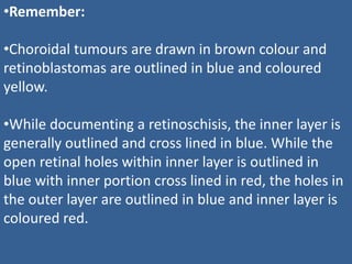
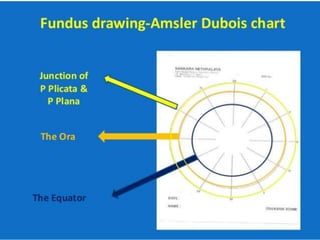
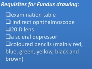
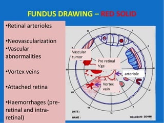
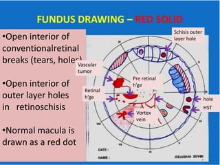
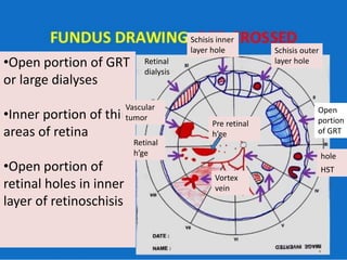
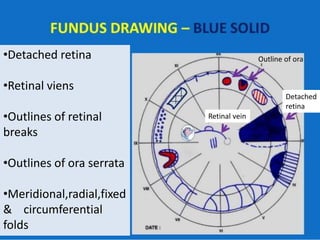
This document provides guidance on how to draw fundus diagrams. It lists the requisites needed which include an examination table, indirect ophthalmoscope, 20D lens, scleral depressor, and colored pencils. It then provides detailed instructions on how to represent various retinal structures, abnormalities, tumors, detachments and other findings using different colors and markings. Remember that choroidal tumors are drawn in brown, retinoblastomas are outlined in blue and colored yellow, and retinoschisis involves outlining the inner layer in blue and cross hatching or coloring the open portions.
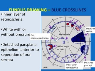
![•VR traction tufts
•Retinal granular tags & tufts
•Outline of flat neo-
vascularization
•Outline of lattice degeneration
[inner ‘x’]
•Outline of thin areas of retina
•Intra-retinal cysts [with
overlying curvilinear stripes to
show configuration]
lattice Thin area
of retina
Flat
neovascularization
Detached
retina
Outline of ora
Retinal vein
Shifting fluid
of RD](https://image.slidesharecdn.com/retinadrwaing-190814033131/85/Retina-drwaing-7-320.jpg)