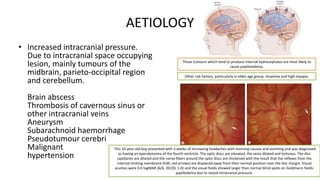
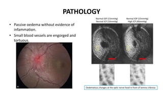
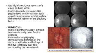
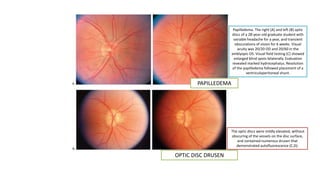
PAPILLOEDEMA
Nikitha Crasta, MBBS 2015
PAPILLOEDEMA
• Oedema of the optic disc or nerve
head due to raised intracranial
pressure.
• Optic nerve is enclosed up to the
lamina cribrosa within the meningeal
sheaths common to the brain.
– Rise in ICT is equally evident around the
nerve.
– Development of oedema at the optic disc.
– Hydrostatic, non inflammatory
phenomenon.
AETIOLOGY
• Increased intracranial pressure.
Due to intracranial space occupying
lesion, mainly tumours of the
midbrain, parieto-occipital region
and cerebellum.
Brain abscess
Thrombosis of cavernous sinus or
other intracranial veins
Aneurysm
Subarachnoid haemorrhage
Pseudotumour cerebri
Malignant
hypertension
Those tumours which tend to produce internal hydrocephalus are most likely to
cause papilloedema.
Other risk factors, particularly in older age group- Anaemia and high myopia.
This 10-year-old boy presented with 3 weeks of increasing headaches with morning nausea and vomiting and was diagnosed
as having an ependymoma of the fourth ventricle. The optic discs are elevated, the veins dilated and tortuous. The disc
capillaries are dilated and the nerve fibers around the optic discs are thickened with the result that the reflexes from the
internal limiting membrane (ILM, red arrows) are displaced away from their normal position near the disc margin. Visual
acuities were 0.0 logMAR (6/6, 20/20, 1.0) and the visual fields showed larger than normal blind spots on Goldmann fields:
papilledema due to raised intracranial pressure.
(a) Left optic nerve sheath meningioma with disc edema and shunt vessels. (b) Axial computed
tomography of the orbits with contrast showing enhancement along the left optic nerve (arrows).
PSEUDOTUMOUR CEREBRI
• P/K/A- Benign intracranial
hypertension.
• Raised ICT w/o presence of ICSOL
• Obese females (20s-30s)
• Transient blurring of vision and
photopsia.
• Loss of visual function (High grade or
atrophic papilledema or peripapillary
subretinal hemorrhage.
This 10-year-old girl with idiopathic intracranial hypertension (pseudotumor cerebri) had a
recurrence of symptoms of headache following cessation of treatment. Both optic discs
show slight elevation and indistinct margins due to thickening of the nerve fiber layer
around the disc. The lumbar puncture opening pressure was 35 cm of CSF. Visual acuity and
color vision were normal. The visual fields showed marginally enlarged blind spots.
Elevated cerebrospinal fluid
pressure.
Axoplasmic stasis in the optic
nerve head.
Swelling of the optic disc and
secondary vascular changes at
the disc surface.
PRINCIPAL PATHOPHYSIOLOGY- Blockage of axoplasmic transport
PATHOLOGY
• Passive oedema without evidence of
infammation.
• Small blood vessels are engorged and
tortuous.
Oedematous changes at the optic nerve head in front of lamina cribrosa.
• Nerve fibres in ONH are swollen and
axoplasmic stasis is noted.
• Physiological cup gets filled in and
internal limiting membrane is raised.
• Nerve fibres become swollen and
varicose and ultimately degenerate.
• Numerous cystoid bodies in front of
lamina cribrosa.
• Engorgement of axons.
Swollen axons are filled with
mitochondria.
Mitochondria are structurally swollen
and disrupted. Cotton-wool spots. A, Clinical appearance of cotton-wool spots. B, Microinfarct of nerve
fiber layer produces aggregates of ruptured and enlarged axons (cytoid bodies). Cytoid
bodies, observed clinically as cotton-wool spots, lie just under internal limiting
membrane. C, Nucleoid of cytoid body consists of dense mass of filamentous material, the
edge of which is more detailed in inset. D, Marked swelling of axon (a) in nerve fiber layer
two hours after experimental central retinal artery occlusion (m, axonal mitochondrion).
PATHOLOGICAL CHANGES IN SURROUNDING RETINA AND MACULAR REGION
• Oedema in the nerve fibre layer
raises the internal limiting
membrane in folds.
• Retinal exudates distributed
radially along the folds, present
in the macular region- Macular
fan or Macular star.
STAGES OF PAPILLOEDEMA
• EARLY STAGE
Fullness and tortuosity of veins
Venous pulsation on optic disc is
absent
• LATE STAGE
Hard exudates on retina between
disc and macula
Macular star and macular fan
SYMPTOMS
• Early stages- Asymptomatic
• On time- Constant compression
leading to transient ischemia
without obscuring visual
functions (4-6 weeks)
• >6 weeks- Nerve fibres
degenerate
Transient obscuration of vision
Enlargement of blind spot
Contraction of visual field
Relative scotoma
• Transient attacks of blurred
vision- Bilateral or monocular
black outs lasting for a few
seconds often precipitated by
changes in posture.
• Chronic papilloedema- Severe
loss of central visual acuity.
• As atrophy sets in-
– Complete blindness
– Pupils are large and
immobile
– Headache which is worse in
a recumbent position
– Nausea and vomiting
– Diplopia (Non specific
paresis of the sixth nerve)
This 7-year-old boy with a brain stem glioma presented with diplopia associated with bilateral
sixth nerve palsies that had started gradually about 2 months before and more recent symptoms
of raised intracranial pressure. The optic discs are grossly elevated with markedly dilated
capillaries and veins, numerous cotton wool spots, and the right eye has a macular star, as a result
of protein/lipid accumulation from transudation from disc capillaries. Although the blind spots
were very large on Goldmann fields, the visual acuity was 0.1 (6/7.5, 20/25, 0.8) right and 0.0 left.
SIGNS
• Blurring of optic disc margins-
– Starts at upper and lower margins
and extends around the nasal side,
temporal side usually visible and
sharp
– Hyperaemic
– Progressive edema reduces size of
the physiological cup
• Veins become congested and
turgescent (absent pulsations)
• Small arterioles become
prominent, appearing as red
streaks.
• Eventually the disc becomes
elevated into a mound higher
than the surrounding retina and
mushrooms out so that the
vessles bends sharply over its
margins
Definite parallax elicited with IDO
• Flame shaped and punctate
hemorrhages
• Surface of the disc loses its
reddish hue, now opaque.
Note circumferential retinal folds a.k.a Paton’s lines.
Veins that are enormously dilated may be buried for large
tracts of their course in the swolen and oedematous retina.
• Macular star- Radiating,
oedematous folds around the
macula (incomplete)
• Macular fan- fan shaped folds on
the side towards the disc
• Cotton wool spots due to retinal
microinfarcts- scattered
throughout posterior half of
fundus
• Longstanding cases- Atrophic
changes when the nerve fibres
can no longer withstand the
pressure and degenerate.
Optic atrophy secondary to long standing papilloedema
• Usually bilateral, not necesssarily
equal on both sides.
• Foster-Kennedy syndrome- U/L
papilledema with or without optic
atrophy on groove or orbital surface
if the frontal lobe or of the pituitary
body.
• DIAGNOSIS:
Indirect ophthalmoscopy- difficult
to assess in early cases for disc
changes
Fluorescien angiography
demonstrates dilatation of the
surface capillaries and leakage of
the dye (vertically oval pool
surrounding the nerve head)
(a) True disc edema with (b) leakage on fluorescein angiography (late phase).
DIFFERENTIAL DIAGNOSIS
• ISCHAEMIC OPTIC NEUROPATHY-
– Arteritic a/w GCA or Non Arteritic a/w
vasculopathy
– Profound, sudden visual loss
– Swollen disc, characteristic pallid
appearance
– Self limiting disease, >55 years, women
– Non arteritic ischaemic optic atrophy,
involvement of short posterior ciliary
branches leading to swelling of optic
nerve head and later to atrophy and
sometimes cupping.
(a) Right anterior ischemic optic neuropathy with mild disc edema and a few
peripapillary hemorrhages. (b) Corresponding inferior altitudinal visual field
defect on a 30–2 Humphrey visual field test.
• OPTIC DISC DRUSEN
– Bilateral, presence of drusen bodies
that evolve over several years
– Small optic nerve head, composede
of calcified concentrically laminated
globular aggregates
– A/w angiod streaks, subretinal
neovascular membranes, vitreous
hemorrhage and retinitis
pigmentosa
– Angiographically, the vessels of
ONH are normal.
The optic discs were mildly elevated, without obscuring of the vessels on the disc surface, and contained numerous
drusen that demonstrated autofluorescence (C,D).
PAPILLEDEMA
OPTIC DISC DRUSEN
Papilledema. The right (A) and left (B) optic
discs of a 28-year-old graduate student with
variable headache for a year, and transient
obscurations of vision for 6 weeks. Visual
acuity was 20/20 OD and 20/60 in the
amblyopic OS. Visual field testing (C) showed
enlarged blind spots bilaterally. Evaluation
revealed marked hydrocephalus. Resolution
of the papilledema followed placement of a
ventriculoperitoneal shunt.
The optic discs were mildly elevated, without
obscuring of the vessels on the disc surface,
and contained numerous drusen that
demonstrated autofluorescence (C,D).
• OPTIC NEURITIS
– Moderate swelling shelving off
gradually into the surrounding
retina.
– Visual symptoms are marked
– Differentiating feature: Acute
depression of central vision,
presence of definite afferent
pupillary defect and the absence of
signs of an ICSOL. Papillitis. The right (A) and left (B) optic discs of a 43-year-old woman who had experienced pain
with movement of the left eye for 2 weeks, and blurred vision of that eye for 1 week. Visual acuity
was 20/20 OD and 20/50 OS. There was a moderate relative afferent pupil defect OS. Visual field
testing (C) showed diffusely reduced sensitivity in the central field of the left eye, especially
nasally. The right optic disc was normal, and the left was mildly swollen. Without treatment, the
patient subsequently had improvement of the visual field abnormality and pupillary sluggishness,
though the visual acuity remained impaired a year later.
(a) Right anterior optic neuritis with moderate disc edema. (b) Axial T1-weighted magnetic resonance imaging of
the orbits with contrast and fat suppression, showing enhancement of the right optic nerve (arrow).
• PSEUDONEURITIS/
PSEUOPAPILLEDEMA/ PSEUDO DISC
OEDEMA
– Occurs in hypermetropic eyes, lamina
cribrosa is small, crowded nerve fibres
are heaped up
– Swelling and blurred margins due to
ophthalmoscopic reflexes
– FFA reveals no leakage
ORBITAL LESIONS AND DISC OEDEMA
Conditions causing stasis in the orbit
Tumours of the optic nerve,
meningioma, cellulitis
(a) Pseudoedema with (b) no leakage on fluorescein
angiography (there is late staining only).
TREATMENT
• Relief of causal pressure
• Timely decompression or
removal of tumour- Relief of
general symptoms of raised ICT,
vision improves rapidly if the
nerves have not been
irretrievably damaged and
papilledema subsides
• Surgical option for
pseudotumour cerebri-
Lumbar peritoneal shunt, or local
decompressionby making
multiple slits or cutting a window
in the optic nerve sheaths (dura
and arachnoid) in the orbit.
THANK YOU

PAPILLEDEMA

  • 1.
  • 2.
    PAPILLOEDEMA • Oedema ofthe optic disc or nerve head due to raised intracranial pressure. • Optic nerve is enclosed up to the lamina cribrosa within the meningeal sheaths common to the brain. – Rise in ICT is equally evident around the nerve. – Development of oedema at the optic disc. – Hydrostatic, non inflammatory phenomenon.
  • 3.
    AETIOLOGY • Increased intracranialpressure. Due to intracranial space occupying lesion, mainly tumours of the midbrain, parieto-occipital region and cerebellum. Brain abscess Thrombosis of cavernous sinus or other intracranial veins Aneurysm Subarachnoid haemorrhage Pseudotumour cerebri Malignant hypertension Those tumours which tend to produce internal hydrocephalus are most likely to cause papilloedema. Other risk factors, particularly in older age group- Anaemia and high myopia. This 10-year-old boy presented with 3 weeks of increasing headaches with morning nausea and vomiting and was diagnosed as having an ependymoma of the fourth ventricle. The optic discs are elevated, the veins dilated and tortuous. The disc capillaries are dilated and the nerve fibers around the optic discs are thickened with the result that the reflexes from the internal limiting membrane (ILM, red arrows) are displaced away from their normal position near the disc margin. Visual acuities were 0.0 logMAR (6/6, 20/20, 1.0) and the visual fields showed larger than normal blind spots on Goldmann fields: papilledema due to raised intracranial pressure.
  • 4.
    (a) Left opticnerve sheath meningioma with disc edema and shunt vessels. (b) Axial computed tomography of the orbits with contrast showing enhancement along the left optic nerve (arrows).
  • 5.
    PSEUDOTUMOUR CEREBRI • P/K/A-Benign intracranial hypertension. • Raised ICT w/o presence of ICSOL • Obese females (20s-30s) • Transient blurring of vision and photopsia. • Loss of visual function (High grade or atrophic papilledema or peripapillary subretinal hemorrhage. This 10-year-old girl with idiopathic intracranial hypertension (pseudotumor cerebri) had a recurrence of symptoms of headache following cessation of treatment. Both optic discs show slight elevation and indistinct margins due to thickening of the nerve fiber layer around the disc. The lumbar puncture opening pressure was 35 cm of CSF. Visual acuity and color vision were normal. The visual fields showed marginally enlarged blind spots.
  • 6.
    Elevated cerebrospinal fluid pressure. Axoplasmicstasis in the optic nerve head. Swelling of the optic disc and secondary vascular changes at the disc surface. PRINCIPAL PATHOPHYSIOLOGY- Blockage of axoplasmic transport
  • 7.
    PATHOLOGY • Passive oedemawithout evidence of infammation. • Small blood vessels are engorged and tortuous. Oedematous changes at the optic nerve head in front of lamina cribrosa.
  • 8.
    • Nerve fibresin ONH are swollen and axoplasmic stasis is noted. • Physiological cup gets filled in and internal limiting membrane is raised. • Nerve fibres become swollen and varicose and ultimately degenerate. • Numerous cystoid bodies in front of lamina cribrosa. • Engorgement of axons. Swollen axons are filled with mitochondria. Mitochondria are structurally swollen and disrupted. Cotton-wool spots. A, Clinical appearance of cotton-wool spots. B, Microinfarct of nerve fiber layer produces aggregates of ruptured and enlarged axons (cytoid bodies). Cytoid bodies, observed clinically as cotton-wool spots, lie just under internal limiting membrane. C, Nucleoid of cytoid body consists of dense mass of filamentous material, the edge of which is more detailed in inset. D, Marked swelling of axon (a) in nerve fiber layer two hours after experimental central retinal artery occlusion (m, axonal mitochondrion).
  • 9.
    PATHOLOGICAL CHANGES INSURROUNDING RETINA AND MACULAR REGION • Oedema in the nerve fibre layer raises the internal limiting membrane in folds. • Retinal exudates distributed radially along the folds, present in the macular region- Macular fan or Macular star.
  • 10.
    STAGES OF PAPILLOEDEMA •EARLY STAGE Fullness and tortuosity of veins Venous pulsation on optic disc is absent • LATE STAGE Hard exudates on retina between disc and macula Macular star and macular fan
  • 11.
    SYMPTOMS • Early stages-Asymptomatic • On time- Constant compression leading to transient ischemia without obscuring visual functions (4-6 weeks) • >6 weeks- Nerve fibres degenerate Transient obscuration of vision Enlargement of blind spot Contraction of visual field Relative scotoma • Transient attacks of blurred vision- Bilateral or monocular black outs lasting for a few seconds often precipitated by changes in posture. • Chronic papilloedema- Severe loss of central visual acuity.
  • 12.
    • As atrophysets in- – Complete blindness – Pupils are large and immobile – Headache which is worse in a recumbent position – Nausea and vomiting – Diplopia (Non specific paresis of the sixth nerve) This 7-year-old boy with a brain stem glioma presented with diplopia associated with bilateral sixth nerve palsies that had started gradually about 2 months before and more recent symptoms of raised intracranial pressure. The optic discs are grossly elevated with markedly dilated capillaries and veins, numerous cotton wool spots, and the right eye has a macular star, as a result of protein/lipid accumulation from transudation from disc capillaries. Although the blind spots were very large on Goldmann fields, the visual acuity was 0.1 (6/7.5, 20/25, 0.8) right and 0.0 left.
  • 13.
    SIGNS • Blurring ofoptic disc margins- – Starts at upper and lower margins and extends around the nasal side, temporal side usually visible and sharp – Hyperaemic – Progressive edema reduces size of the physiological cup • Veins become congested and turgescent (absent pulsations)
  • 14.
    • Small arteriolesbecome prominent, appearing as red streaks. • Eventually the disc becomes elevated into a mound higher than the surrounding retina and mushrooms out so that the vessles bends sharply over its margins Definite parallax elicited with IDO • Flame shaped and punctate hemorrhages • Surface of the disc loses its reddish hue, now opaque. Note circumferential retinal folds a.k.a Paton’s lines. Veins that are enormously dilated may be buried for large tracts of their course in the swolen and oedematous retina.
  • 15.
    • Macular star-Radiating, oedematous folds around the macula (incomplete) • Macular fan- fan shaped folds on the side towards the disc • Cotton wool spots due to retinal microinfarcts- scattered throughout posterior half of fundus • Longstanding cases- Atrophic changes when the nerve fibres can no longer withstand the pressure and degenerate. Optic atrophy secondary to long standing papilloedema
  • 16.
    • Usually bilateral,not necesssarily equal on both sides. • Foster-Kennedy syndrome- U/L papilledema with or without optic atrophy on groove or orbital surface if the frontal lobe or of the pituitary body. • DIAGNOSIS: Indirect ophthalmoscopy- difficult to assess in early cases for disc changes Fluorescien angiography demonstrates dilatation of the surface capillaries and leakage of the dye (vertically oval pool surrounding the nerve head) (a) True disc edema with (b) leakage on fluorescein angiography (late phase).
  • 17.
    DIFFERENTIAL DIAGNOSIS • ISCHAEMICOPTIC NEUROPATHY- – Arteritic a/w GCA or Non Arteritic a/w vasculopathy – Profound, sudden visual loss – Swollen disc, characteristic pallid appearance – Self limiting disease, >55 years, women – Non arteritic ischaemic optic atrophy, involvement of short posterior ciliary branches leading to swelling of optic nerve head and later to atrophy and sometimes cupping. (a) Right anterior ischemic optic neuropathy with mild disc edema and a few peripapillary hemorrhages. (b) Corresponding inferior altitudinal visual field defect on a 30–2 Humphrey visual field test.
  • 19.
    • OPTIC DISCDRUSEN – Bilateral, presence of drusen bodies that evolve over several years – Small optic nerve head, composede of calcified concentrically laminated globular aggregates – A/w angiod streaks, subretinal neovascular membranes, vitreous hemorrhage and retinitis pigmentosa – Angiographically, the vessels of ONH are normal. The optic discs were mildly elevated, without obscuring of the vessels on the disc surface, and contained numerous drusen that demonstrated autofluorescence (C,D).
  • 20.
    PAPILLEDEMA OPTIC DISC DRUSEN Papilledema.The right (A) and left (B) optic discs of a 28-year-old graduate student with variable headache for a year, and transient obscurations of vision for 6 weeks. Visual acuity was 20/20 OD and 20/60 in the amblyopic OS. Visual field testing (C) showed enlarged blind spots bilaterally. Evaluation revealed marked hydrocephalus. Resolution of the papilledema followed placement of a ventriculoperitoneal shunt. The optic discs were mildly elevated, without obscuring of the vessels on the disc surface, and contained numerous drusen that demonstrated autofluorescence (C,D).
  • 21.
    • OPTIC NEURITIS –Moderate swelling shelving off gradually into the surrounding retina. – Visual symptoms are marked – Differentiating feature: Acute depression of central vision, presence of definite afferent pupillary defect and the absence of signs of an ICSOL. Papillitis. The right (A) and left (B) optic discs of a 43-year-old woman who had experienced pain with movement of the left eye for 2 weeks, and blurred vision of that eye for 1 week. Visual acuity was 20/20 OD and 20/50 OS. There was a moderate relative afferent pupil defect OS. Visual field testing (C) showed diffusely reduced sensitivity in the central field of the left eye, especially nasally. The right optic disc was normal, and the left was mildly swollen. Without treatment, the patient subsequently had improvement of the visual field abnormality and pupillary sluggishness, though the visual acuity remained impaired a year later.
  • 22.
    (a) Right anterioroptic neuritis with moderate disc edema. (b) Axial T1-weighted magnetic resonance imaging of the orbits with contrast and fat suppression, showing enhancement of the right optic nerve (arrow).
  • 23.
    • PSEUDONEURITIS/ PSEUOPAPILLEDEMA/ PSEUDODISC OEDEMA – Occurs in hypermetropic eyes, lamina cribrosa is small, crowded nerve fibres are heaped up – Swelling and blurred margins due to ophthalmoscopic reflexes – FFA reveals no leakage ORBITAL LESIONS AND DISC OEDEMA Conditions causing stasis in the orbit Tumours of the optic nerve, meningioma, cellulitis (a) Pseudoedema with (b) no leakage on fluorescein angiography (there is late staining only).
  • 24.
    TREATMENT • Relief ofcausal pressure • Timely decompression or removal of tumour- Relief of general symptoms of raised ICT, vision improves rapidly if the nerves have not been irretrievably damaged and papilledema subsides • Surgical option for pseudotumour cerebri- Lumbar peritoneal shunt, or local decompressionby making multiple slits or cutting a window in the optic nerve sheaths (dura and arachnoid) in the orbit.
  • 25.