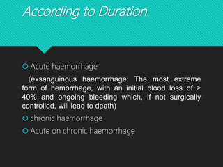
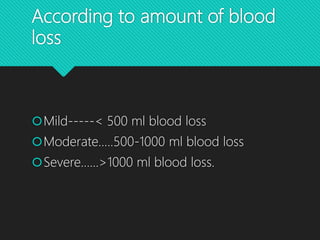
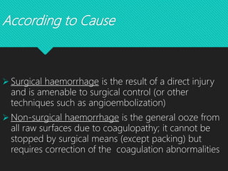
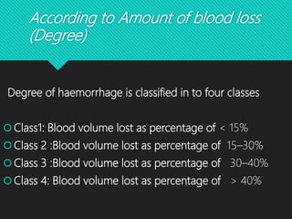
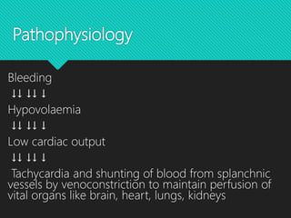
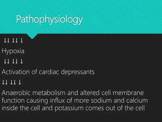
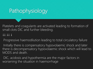
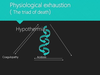
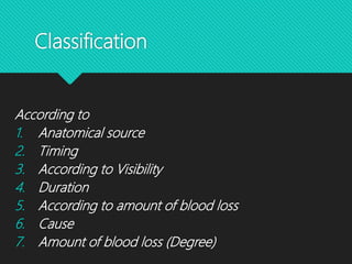
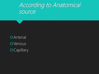
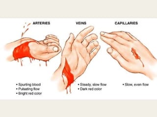
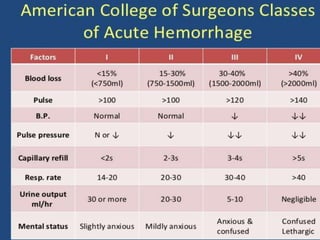
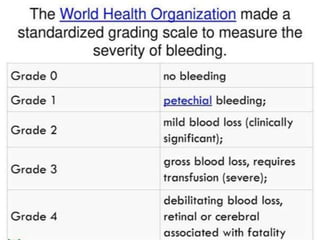
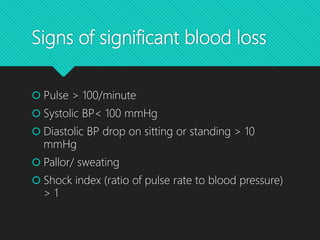
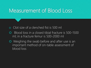
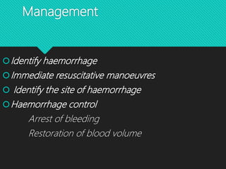
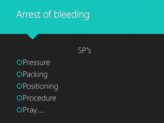
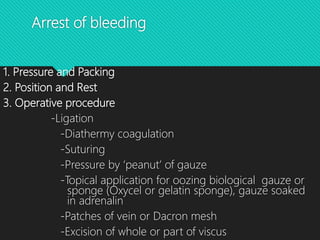
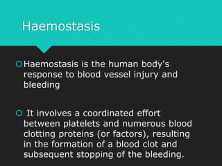
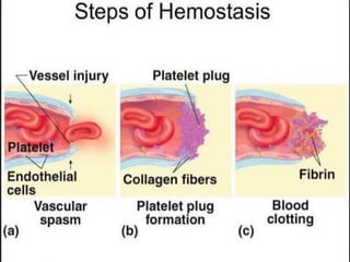
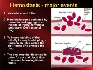
This document discusses haemorrhage or bleeding from blood vessels. It defines haemorrhage and describes the pathophysiology where bleeding leads to hypovolaemic shock and complications like acidosis. It then classifies haemorrhage based on anatomical source, timing, visibility, duration and amount of blood loss. Clinical features and investigations for haemorrhage are provided. Management of haemorrhage focuses on arresting bleeding through pressure, packing and procedures before restoring blood volume.
Fluids and Electrolytes Imbalance and ManagementNUMED SCIENCE
www.numedscience.blogspot.com
Electrolytes are minerals in your body that have an electric charge. They are in your blood, urine, tissues, and other body fluids. Electrolytes are important because they help
Balance the amount of water in your body
Balance your body's acid/base (pH) level
Move nutrients into your cells
Move wastes out of your cells
Make sure that your nerves, muscles, the heart, and the brain work the way they should
Sodium, calcium, potassium, chloride, phosphate, and magnesium are all electrolytes. You get them from the foods you eat and the fluids you drink.
Hemorrhoids are swollen veins in the lowest part of your rectum and anus. Sometimes, the walls of these blood vessels stretch so thin that the veins bulge and get irritated, especially when you poop. Hemorrhoids are also called piles.
intravenous fluid and electrolytes are important topics in medical science. potassium is one of the vital electrolytes of the human body. this presentation has a discussion on several iv fluids and potassium balance and also how to manage the potassium imbalance.
Disseminated Intravascular Coagulopathy is a serious disease which needs awareness among health personnel as well as common people. this presentation helps them to understand about DIC & its management.
Hypovolemic shock is a life-threatening emergency in which severe blood or other fluid loss makes the heart unable to pump enough blood to the body. This type of shock can cause many organs to stop working.
Hypovolemic shock is a dangerous condition that happens when suddenly lose a lot of blood or fluids from body. This drops blood volume, the amount of blood circulating in body. That’s why it’s also known as low-volume shock.
Simple medical student presentation about distributive shock, type and pathophysiology of each septic shock, anaphylactic shock, neurogenic shock
including management, prognosis and disposition of patient..
brief info of type of inotropes and when to start.
here give the knowledge that you should possess to manage acute and chronic urine retention. the lecture is more concerned about practical patient care and ward setting management. you should minimally be aware about following facts regarding urine retention. the multiple causes of retention will be discussed later in detailed manner. Direction of the lecture seems more toward BPH and acute retention management. beware there are many aspects of a patient present with an AUR. do no harm and always try to keep patient satisfaction. Let me know about your comments an Ideas. try to improve the quality. good luck.
Dr. Ch VT- The topic describe about the general as well the the dental aspect too. The classification, clinical features and management. The dental conditions associated with haemorrhage and shock has been highlighted with their management
Haemorrhage and Shock: Relevance in Periodontal SurgeryNavneet Randhawa
Haemorrhage types and definition, shock types and definition, relevance of shock and haemorrhage in Periodontics, Methods to cope with haemorrhage and shock in Periodontal Surgery
Cardiac tamponade
Synonyms Pericardial tamponade
Hemorragic effusion.jpg
A very large pericardial effusion resulting in tamponade as a result of bleeding from cancer as seen on ultrasound. Closed arrow - the heart; open arrow - the effusion
Specialty Cardiac surgery
Symptoms Shortness of breath, weakness, lightheadedness, cough[1]
Usual onset Rapid or more gradual[2]
Causes Cancer, kidney failure, chest trauma, pericarditis, tuberculosis[2][1]
Diagnostic method Symptoms and ultrasound of the heart[2]
Treatment Drainage (pericardiocentesis, pericardial window, pericardiectomy)[2]
Frequency 2 per 10,000 per year (US)[3]
Cardiac tamponade, also known as pericardial tamponade, is when fluid in the pericardium (the sac around the heart) builds up, resulting in compression of the heart.
Shock is a life-threatening condition that occurs when the body is not getting enough blood flow. Lack of blood flow means the cells and organs do not get enough oxygen and nutrients to function properly. Many organs can be damaged as a result.
A very narrative discussion over Shock & Haemorrhage, Blood Transfusion, Blood Products which is presented in seminers. A concise guideline of a vast chapter.
Knee anatomy and clinical tests 2024.pdfvimalpl1234
This includes all relevant anatomy and clinical tests compiled from standard textbooks, Campbell,netter etc..It is comprehensive and best suited for orthopaedicians and orthopaedic residents.
These lecture slides, by Dr Sidra Arshad, offer a quick overview of the physiological basis of a normal electrocardiogram.
Learning objectives:
1. Define an electrocardiogram (ECG) and electrocardiography
2. Describe how dipoles generated by the heart produce the waveforms of the ECG
3. Describe the components of a normal electrocardiogram of a typical bipolar lead (limb II)
4. Differentiate between intervals and segments
5. Enlist some common indications for obtaining an ECG
6. Describe the flow of current around the heart during the cardiac cycle
7. Discuss the placement and polarity of the leads of electrocardiograph
8. Describe the normal electrocardiograms recorded from the limb leads and explain the physiological basis of the different records that are obtained
9. Define mean electrical vector (axis) of the heart and give the normal range
10. Define the mean QRS vector
11. Describe the axes of leads (hexagonal reference system)
12. Comprehend the vectorial analysis of the normal ECG
13. Determine the mean electrical axis of the ventricular QRS and appreciate the mean axis deviation
14. Explain the concepts of current of injury, J point, and their significance
Study Resources:
1. Chapter 11, Guyton and Hall Textbook of Medical Physiology, 14th edition
2. Chapter 9, Human Physiology - From Cells to Systems, Lauralee Sherwood, 9th edition
3. Chapter 29, Ganong’s Review of Medical Physiology, 26th edition
4. Electrocardiogram, StatPearls - https://www.ncbi.nlm.nih.gov/books/NBK549803/
5. ECG in Medical Practice by ABM Abdullah, 4th edition
6. Chapter 3, Cardiology Explained, https://www.ncbi.nlm.nih.gov/books/NBK2214/
7. ECG Basics, http://www.nataliescasebook.com/tag/e-c-g-basics
Integrating Ayurveda into Parkinson’s Management: A Holistic ApproachAyurveda ForAll
Explore the benefits of combining Ayurveda with conventional Parkinson's treatments. Learn how a holistic approach can manage symptoms, enhance well-being, and balance body energies. Discover the steps to safely integrate Ayurvedic practices into your Parkinson’s care plan, including expert guidance on diet, herbal remedies, and lifestyle modifications.
ABDOMINAL TRAUMA in pediatrics part one.drhasanrajab
Abdominal trauma in pediatrics refers to injuries or damage to the abdominal organs in children. It can occur due to various causes such as falls, motor vehicle accidents, sports-related injuries, and physical abuse. Children are more vulnerable to abdominal trauma due to their unique anatomical and physiological characteristics. Signs and symptoms include abdominal pain, tenderness, distension, vomiting, and signs of shock. Diagnosis involves physical examination, imaging studies, and laboratory tests. Management depends on the severity and may involve conservative treatment or surgical intervention. Prevention is crucial in reducing the incidence of abdominal trauma in children.
- Video recording of this lecture in English language: https://youtu.be/kqbnxVAZs-0
- Video recording of this lecture in Arabic language: https://youtu.be/SINlygW1Mpc
- Link to download the book free: https://nephrotube.blogspot.com/p/nephrotube-nephrology-books.html
- Link to NephroTube website: www.NephroTube.com
- Link to NephroTube social media accounts: https://nephrotube.blogspot.com/p/join-nephrotube-on-social-media.html
Basavarajeeyam is a Sreshta Sangraha grantha (Compiled book ), written by Neelkanta kotturu Basavaraja Virachita. It contains 25 Prakaranas, First 24 Chapters related to Rogas& 25th to Rasadravyas.
Best Ayurvedic medicine for Gas and IndigestionSwastikAyurveda
Here is the updated list of Top Best Ayurvedic medicine for Gas and Indigestion and those are Gas-O-Go Syp for Dyspepsia | Lavizyme Syrup for Acidity | Yumzyme Hepatoprotective Capsules etc
Local Advanced Lung Cancer: Artificial Intelligence, Synergetics, Complex Sys...Oleg Kshivets
Overall life span (LS) was 1671.7±1721.6 days and cumulative 5YS reached 62.4%, 10 years – 50.4%, 20 years – 44.6%. 94 LCP lived more than 5 years without cancer (LS=2958.6±1723.6 days), 22 – more than 10 years (LS=5571±1841.8 days). 67 LCP died because of LC (LS=471.9±344 days). AT significantly improved 5YS (68% vs. 53.7%) (P=0.028 by log-rank test). Cox modeling displayed that 5YS of LCP significantly depended on: N0-N12, T3-4, blood cell circuit, cell ratio factors (ratio between cancer cells-CC and blood cells subpopulations), LC cell dynamics, recalcification time, heparin tolerance, prothrombin index, protein, AT, procedure type (P=0.000-0.031). Neural networks, genetic algorithm selection and bootstrap simulation revealed relationships between 5YS and N0-12 (rank=1), thrombocytes/CC (rank=2), segmented neutrophils/CC (3), eosinophils/CC (4), erythrocytes/CC (5), healthy cells/CC (6), lymphocytes/CC (7), stick neutrophils/CC (8), leucocytes/CC (9), monocytes/CC (10). Correct prediction of 5YS was 100% by neural networks computing (error=0.000; area under ROC curve=1.0).
5. Pathophysiology
Bleeding
↓↓ ↓↓ ↓
Hypovolaemia
↓↓ ↓↓ ↓
Low cardiac output
↓↓ ↓↓ ↓
Tachycardia and shunting of blood from splanchnic
vessels by venoconstriction to maintain perfusion of
vital organs like brain, heart, lungs, kidneys
6. Pathophysiology
↓↓ ↓↓ ↓
Hypoxia
↓↓ ↓↓ ↓
Activation of cardiac depressants
↓↓ ↓↓ ↓
Anaerobic metabolism and altered cell membrane
function causing influx of more sodium and calcium
inside the cell and potassium comes out of the cell
7. Pathophysiology
↓↓ ↓↓ ↓
Hyponatraemic, hyperkalaemic, hypocalcaemic
metabolic acidosis
↓↓ ↓↓ ↓
Lysosomes of cell get lysed releasing powerful
enzymes which is lethal to cell itself
↓↓ ↓↓ ↓
SICK CELL SYNDROME
8. Pathophysiology
Platelets and coagulants are activated leading to formation of
small clots DIC and further bleeding.
↓↓ ↓↓ ↓
Progressive haemodilution leading to total circulatory failure
Initially there is compensatory hypovolaemic shock and later
there is decompensatory hypovolaemic shock which will lead to
MODS and death.
DIC, acidosis and hypothermia are the major factors in
worsening the situation in haemorrhage
10. Classification
According to
1. Anatomical source
2. Timing
3. According to Visibility
4. Duration
5. According to amount of blood loss
6. Cause
7. Amount of blood loss (Degree)
13. According to Timing
Primary haemorrhage
-- occurring
immediately as a result
of an injury (or
surgery)
Reactionary haemorrhage is
delayed haemorrhage (within 24
hours) caused by
Dislodgement of clot by
resuscitation
Normalization of blood pressure
Vasodilatation
Slippage of a ligature
14. According to Timing
Secondary haemorrhage
usually occurs 7–14
days after injury
caused by sloughing
of the wall of a vessel
precipitated by
infection,
pressure necrosis
(such as from a drain)
or
malignancy.
15. According to Visibility
*Revealed haemorrhage
obvious external
haemorrhage, such as
exsanguination from an
open arterial wound
*Concealed haemorrhage
contained within the body
cavity
In trauma within the chest,
abdomen, pelvis or
retroperitoneum or in the
limbs, with contained
vascular injury or associated
with long-bone fractures
non-traumatic concealed
haemorrhage include occult
gastrointestinal bleeding or
ruptured aortic aneurysm.
18. Initially concealed but later
revealed
Any bleeding from
natural orifices
Haemoptysis
Haematemesis
Epistaxis
Haematuria
Haematocazia
Melaena
P/V bleeding
Bleeding through Ear
19. According to Duration
Acute haemorrhage
(exsanguinous haemorrhage: The most extreme
form of hemorrhage, with an initial blood loss of >
40% and ongoing bleeding which, if not surgically
controlled, will lead to death)
chronic haemorrhage
Acute on chronic haemorrhage
20. According to amount of blood
loss
Mild-----< 500 ml blood loss
Moderate…..500-1000 ml blood loss
Severe……>1000 ml blood loss.
22. According to Cause
Surgical haemorrhage is the result of a direct injury
and is amenable to surgical control (or other
techniques such as angioembolization)
Non-surgical haemorrhage is the general ooze from
all raw surfaces due to coagulopathy; it cannot be
stopped by surgical means (except packing) but
requires correction of the coagulation abnormalities
23. According to Amount of blood loss
(Degree)
Degree of haemorrhage is classified in to four classes
Class1: Blood volume lost as percentage of < 15%
Class 2 :Blood volume lost as percentage of 15–30%
Class 3 :Blood volume lost as percentage of 30–40%
Class 4: Blood volume lost as percentage of > 40%
24.
25.
26. Clinical Features of
Haemorrhage
• Pallor, thirsty
• Cyanosis
• Tachycardia
• Tachypnoea
• Air hunger.
• Cold clammy skin due to vasoconstriction
• Dry face, dry mouth and goose skin appearance (due to
contraction of arrector pilorum)
29. Signs of significant blood loss
Pulse > 100/minute
Systolic BP< 100 mmHg
Diastolic BP drop on sitting or standing > 10
mmHg
Pallor/ sweating
Shock index (ratio of pulse rate to blood pressure)
> 1
30. Measurement of Blood Loss
Clot size of a clenched fist is 500 ml
Blood loss in a closed tibial fracture is 500-1500
ml; in a fracture femur is 500-2000 ml
Weighing the swab before and after use is an
important method of on-table assessment of
blood loss
31. Measurement of Blood Loss
Hb% and PCV estimation.
Blood volume estimation using radioiodine
technique or micro hematocrit method.
Measurement of CVP or PCWP
Investigations specific for cause: U/S abdomen,
Doppler and often angiogram in vascular injury,
chest X-ray in haemothorax, CT scan in major
injuries, CT scan head in head injuries
32. Rains Factor
Total amount or of Blood loss =
Total difference in swab weight × 1.5
Or
Total difference in swab weight × 2 (For larger
wounds and larger operations)
38. Arrest of bleeding
1. Pressure and Packing
2. Position and Rest
3. Operative procedure
-Ligation
-Diathermy coagulation
-Suturing
-Pressure by ‘peanut’ of gauze
-Topical application for oozing biological gauze or
sponge (Oxycel or gelatin sponge), gauze soaked
in adrenalin
-Patches of vein or Dacron mesh
-Excision of whole or part of viscus
39. Haemostasis
Haemostasis is the human body's
response to blood vessel injury and
bleeding
It involves a coordinated effort
between platelets and numerous blood
clotting proteins (or factors), resulting
in the formation of a blood clot and
subsequent stopping of the bleeding.