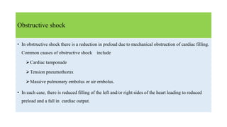
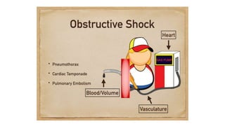
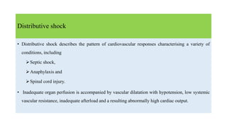
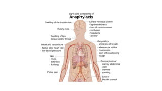
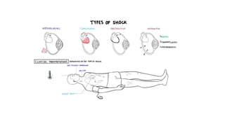
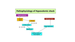
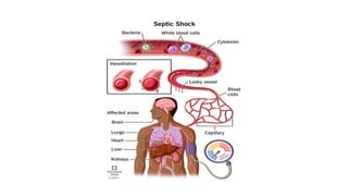
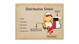
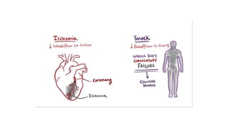
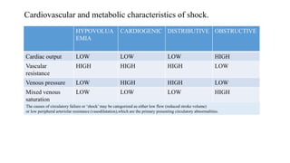
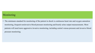
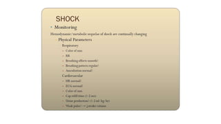
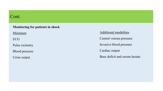
Dr. Abdullah Al Mamun
SHOCK
Level of oxygen delivery(DO2) which fails to meet
the metabolic requerment of tissue.
‘Shock’ is not synonymous with hypotension.
While hypotension is a sinister development and
requires urgent attention, it is often a late
manifestation of circulatory failure or shock, and also
the cardiac output and oxygen delivery may be
critically low even though the BP remains normal
DEFINITION:
CLASSIFICATION
1. HYPOVOLAEMIC SHOCK
2. CARDIOGENIC SHOCK
3. OBSTRUCTIVE SHOCK
4. DISTRIBITIVE SHOCK
5. ENDROCRINE SHCK
HYPOVOLAEMIC SHOCK
• Hypovolaemic shock is due to a reduced circulating volume.
• Hypovolaemia may be due to haemorrhagic or nonhaemor rhagic causes.
Nonhaemorrhagic causes include poor fluid intake (dehydration), excessive fluid loss due to
vomiting, diarrhoea urinary loss (e.g. diabetes) evaporation, or ‘thirdspacing’ where fluid is lost into
the gastrointestinal tract and interstitial spaces, as for example in bowel obstruction or pancreatitis.
CARDIOGENIC SHOCH
• Cardiogenic shock is due to primary failure of the heart to pump blood to the tissues. Causes of
cardiogenic shock include
Myocardial infarction
Cardiac dysrhythmias
Valvular heart disease
Blunt myocardial injury and cardiomyopathy.
• Cardiac insufficiency may also be due to myocardial depression caused by endogenous factors (e.g.
bacterial and humoral agents released in sepsis) or exogenous factors, such as pharmaceutical
agents or drug abuse.
Obstructive shock
• In obstructive shock there is a reduction in preload due to mechanical obstruction of cardiac filling.
Common causes of obstructive shock include
Cardiac tamponade
Tension pneumothorax
Massive pulmonary embolus or air embolus.
• In each case, there is reduced filling of the left and/or right sides of the heart leading to reduced
preload and a fall in cardiac output.
Distributive shock
• Distributive shock describes the pattern of cardiovascular responses characterising a variety of
conditions, including
Septic shock,
Anaphylaxis and
Spinal cord injury.
• Inadequate organ perfusion is accompanied by vascular dilatation with hypotension, low systemic
vascular resistance, inadequate afterload and a resulting abnormally high cardiac output.
• In anaphylaxis, vasodilatation is due to histamine release,while in high spinal cord injury there is
failure of sympatheticoutflow and adequate vascular tone (neurogenic shock).
• The cause in sepsis is less clear but is related to the release of bacterial products (endotoxin) and
the activation of cellular and humoral components of the immune system. There is maldistribution
of blood flow at a microvascular level with arteriovenous shunting and dysfunction of cellular
utilization of oxygen.
In the later phases of septic shock there is hypovolaemia from fluid loss into interstitial spaces and
there may be concomitant myocardial depression, complicating the clinical picture
Cont.
Endocrine shock
• Endocrine shock may present as a combination of hypovolaemic, cardiogenic or distributive shock.
Causes of endocrineshock include hypo and hyperthyroidism and adrenal insufficiency.
• Hypothyroidism causes a shock state similar to that of neurogenic shock due to disordered vascular
and cardiac responsiveness to circulating catecholamines. Cardiac output falls due to low inotropy
and bradycardia. There may also be an associated cardiomyopathy.
• Thyrotoxicosis may cause a high output cardiac failure.
• Adrenal insufficiency leads to shock due to hypovolaemia and a poor response to circulating
and exogenous catecholamines. Adrenal insufficiency may be due to preexisting Addison’s
disease or be a relative insufficiency due to a pathological disease state, such as systemic sepsis.
Cont.
Cardiovascular and metabolic characteristics of shock.
HYPOVOLUA
EMIA
CARDIOGENIC DISTRIBUTIVE OBSTRUCTIVE
Cardiac output LOW LOW LOW HIGH
Vascular
resistance
HIGH HIGH HIGH LOW
Venous pressure LOW HIGH HIGH LOW
Mixed venous
saturation
LOW LOW LOW HIGH
Base deficit HIGH HIGH HIGH HIGH
The causes of circulatory failure or ‘shock’ may be categorized as either low flow (reduced stroke volume)
or low peripheral arteriolar resistance (vasodilatation),which are the primary presenting circulatory abnormalities.
Low stroke volume
• Hypovolaemic: any condition provoking a major reduction in blood volume, e.g. internal or
external haemorrhage, severe burns, salt and water depletion.
• Cardiogenic: severe mechanical impairment, e.g. myocardial infarction, acute mitral regurgitation.
• Obstructive: obstruction to blood flow around the circulation, e.g. major pulmonary embolism,
cardiac tamponade, tension pneumothorax
Vasodilatation
• Septic/SIRS: infection or other causes of a systemic inflammatory response that produce wide
spread endothelial damage with vasodilatation, arteriovenous shunting, microvascular occlusion,
capillary leak and tissue oedema.
• Anaphylactic: inappropriate vasodilatation triggered by an allergen (e.g. bee sting) often associated
with endothelial disruption and capillary leak.
• Neurogenic: caused by major brain or spinal injury which disrupts brain-stem and neurogenic
vasomotor control.
Clinical assessment and complications
• Cold peripheries
• Reduced or absent peripheral pulses
• Weak central pulses and evidence of a low cardiac output.
• In contrast, septic shock and anaphylactic shock are usually associated with warm peripheries,
bounding pulses and features of a high cardiac output.
• Neurogenic shock often results in vasodilated hypotension with a paradoxically slow heart rate.
Prognosis
• If the precipitating cause and accompanying circulatory failure (hypotension and frequently severe
hypovolaemia due to venodilatation and fluid loss through the leaky vascular endothelium) are
dealt with promptly before significant organ failure occurs (‘early’ shock),the prognosis is good. If
not, there is progressive deterioration in organ function and MOF ensues (‘late’shock).
• The mortality of MOF is high and increases with the number of organs that have failed, the
duration of organ failure and the patient’s age. Failure of four or more organs is associated with a
mortality > 80%.
TREATMENT
• the timing and nature of resuscitation will depend on the type of
Shock and the timing and severity of the insult.
• Rapid clinical examination will provide adequate clues to make an appropriate first determination,
even if a source of bleeding or sepsis
is not immediately identifiable
• If there is initial doubt aboutthe cause of shock, it is safer to assume the cause is hypovolaemia
and begin with fluid resuscitation, and then assess the response.
• In patients who are actively bleeding (major trauma, aortic aneurysm rupture, gastrointestinal
haemorrhage), it is counter productive to institute high volume fluid therapy without controlling
the site of haemorrhage.
• Increasing blood pressure merely increases bleeding from the site while fluid therapy cools the
patient and dilutes available coagulation factors.Thus operative haemorrhage control should
not be delayed and resuscitation should proceed in parallel with surgery.
Cont.
• a patient with bowel obstruction and hypovolaemic shock must be adequately resuscitated
before under going surgery otherwise the additional surgical injury and hypovolaemia induced
during the procedure will exacerbate the inflammatory activation and increase the incidence
and severity of endorgan insult.
Fluid therapy:
• In all cases of shock, regardless of classification, hypovolaemia and inadequate preload must be
addressed before other therapy is instituted. Patients will enter the unresuscitatable stage of
shock as the myocardium becomes progressively more ischaemic and unresponsive to
resuscitative attempts.
Cont.
• Firstline therapy, therefore, is intravenous access and administration of intravenous fluids.
Access should be through short, widebore catheters that allow rapid infusion of fluids as
necessary. Long, narrow lines, such as central venous catheters, have too high a resistance to
allow rapid infusion and are more appropriate for monitoring than fluid replacement therapy.
Cont.
Dynamic fluid response
• The shock status can be determined dynamically by the cardiovascular response to the rapid
administration of a fluid bolus. In total, 250–500 mL of fluid is rapidly given (over 5–10minutes)
and the cardiovascular responses in terms of heart rate, blood pressure and central venous pressure
are observed. Patients can be divided into ‘responders’, ‘transient responders’ and ‘non-
responders’.
Responders have an improvement in their cardiovascular status that is sustained. These patients are
not actively losing fluid but require filling to a normal volume status.
Cont.
Transient responders have an improvement, but this then reverts to the previous state over the next
10–20 minutes. These patients have moderate ongoing fluid losses(either overt haemorrhage or
further fluid shifts reducing intravascular volume).
Non-responders are severely volume depleted and are likely to have major ongoing loss of
intravascular volume, usually through persistent uncontrolled haemorrhage .
Cont.
Vasopressor and inotropic support
• Vasopressor or inotropic therapy is not indicated as firstline therapy in hypovolaemia.
• Vasopressor agents (phenylephrine, noradrenaline) are indicated in distributive shock states (sepsis,
neurogenicshock ) where there is peripheral vasodilatation, and a low systemic vascular resistance, leading to
hypotension despite a high cardiac output. Where the vasodilatation is resistant to catecholamines (e.g.
absolute or relative steroid deficiency)vasopressin may be used as an alternative vasopressor.
• In cardiogenic shock, or where myocardial depression has complicated a shock state (e.g. severe septic shock
with low cardiac output), inotropic therapy may be required to increase cardiac output and therefore oxygen
delivery. The inodilator dobutamine is the agent of choice.
Monitoring
• The minimum standard for monitoring of the patient in shock is continuous heart rate and oxygen saturation
monitoring, frequent noninvasive blood pressure monitoring and hourly urine output measurements. Most
patients will need more aggressive invasive monitoring, including central venous pressure and invasive blood
pressure monitoring.
Additional modalities
Central venous pressure
Invasive blood pressure
Cardiac output
Base deficit and serum lactate
Cont.
Monitoring for patients in shock
Minimum
ECG
Pulse oximetry
Blood pressure
Urine output
Thank You

SHOCK

  • 1.
    Dr. Abdullah AlMamun SHOCK
  • 2.
    Level of oxygendelivery(DO2) which fails to meet the metabolic requerment of tissue. ‘Shock’ is not synonymous with hypotension. While hypotension is a sinister development and requires urgent attention, it is often a late manifestation of circulatory failure or shock, and also the cardiac output and oxygen delivery may be critically low even though the BP remains normal DEFINITION:
  • 3.
    CLASSIFICATION 1. HYPOVOLAEMIC SHOCK 2.CARDIOGENIC SHOCK 3. OBSTRUCTIVE SHOCK 4. DISTRIBITIVE SHOCK 5. ENDROCRINE SHCK
  • 5.
    HYPOVOLAEMIC SHOCK • Hypovolaemicshock is due to a reduced circulating volume. • Hypovolaemia may be due to haemorrhagic or nonhaemor rhagic causes. Nonhaemorrhagic causes include poor fluid intake (dehydration), excessive fluid loss due to vomiting, diarrhoea urinary loss (e.g. diabetes) evaporation, or ‘thirdspacing’ where fluid is lost into the gastrointestinal tract and interstitial spaces, as for example in bowel obstruction or pancreatitis.
  • 8.
    CARDIOGENIC SHOCH • Cardiogenicshock is due to primary failure of the heart to pump blood to the tissues. Causes of cardiogenic shock include Myocardial infarction Cardiac dysrhythmias Valvular heart disease Blunt myocardial injury and cardiomyopathy. • Cardiac insufficiency may also be due to myocardial depression caused by endogenous factors (e.g. bacterial and humoral agents released in sepsis) or exogenous factors, such as pharmaceutical agents or drug abuse.
  • 10.
    Obstructive shock • Inobstructive shock there is a reduction in preload due to mechanical obstruction of cardiac filling. Common causes of obstructive shock include Cardiac tamponade Tension pneumothorax Massive pulmonary embolus or air embolus. • In each case, there is reduced filling of the left and/or right sides of the heart leading to reduced preload and a fall in cardiac output.
  • 12.
    Distributive shock • Distributiveshock describes the pattern of cardiovascular responses characterising a variety of conditions, including Septic shock, Anaphylaxis and Spinal cord injury. • Inadequate organ perfusion is accompanied by vascular dilatation with hypotension, low systemic vascular resistance, inadequate afterload and a resulting abnormally high cardiac output.
  • 17.
    • In anaphylaxis,vasodilatation is due to histamine release,while in high spinal cord injury there is failure of sympatheticoutflow and adequate vascular tone (neurogenic shock). • The cause in sepsis is less clear but is related to the release of bacterial products (endotoxin) and the activation of cellular and humoral components of the immune system. There is maldistribution of blood flow at a microvascular level with arteriovenous shunting and dysfunction of cellular utilization of oxygen. In the later phases of septic shock there is hypovolaemia from fluid loss into interstitial spaces and there may be concomitant myocardial depression, complicating the clinical picture Cont.
  • 19.
    Endocrine shock • Endocrineshock may present as a combination of hypovolaemic, cardiogenic or distributive shock. Causes of endocrineshock include hypo and hyperthyroidism and adrenal insufficiency. • Hypothyroidism causes a shock state similar to that of neurogenic shock due to disordered vascular and cardiac responsiveness to circulating catecholamines. Cardiac output falls due to low inotropy and bradycardia. There may also be an associated cardiomyopathy.
  • 21.
    • Thyrotoxicosis maycause a high output cardiac failure. • Adrenal insufficiency leads to shock due to hypovolaemia and a poor response to circulating and exogenous catecholamines. Adrenal insufficiency may be due to preexisting Addison’s disease or be a relative insufficiency due to a pathological disease state, such as systemic sepsis. Cont.
  • 23.
    Cardiovascular and metaboliccharacteristics of shock. HYPOVOLUA EMIA CARDIOGENIC DISTRIBUTIVE OBSTRUCTIVE Cardiac output LOW LOW LOW HIGH Vascular resistance HIGH HIGH HIGH LOW Venous pressure LOW HIGH HIGH LOW Mixed venous saturation LOW LOW LOW HIGH Base deficit HIGH HIGH HIGH HIGH The causes of circulatory failure or ‘shock’ may be categorized as either low flow (reduced stroke volume) or low peripheral arteriolar resistance (vasodilatation),which are the primary presenting circulatory abnormalities.
  • 24.
    Low stroke volume •Hypovolaemic: any condition provoking a major reduction in blood volume, e.g. internal or external haemorrhage, severe burns, salt and water depletion. • Cardiogenic: severe mechanical impairment, e.g. myocardial infarction, acute mitral regurgitation. • Obstructive: obstruction to blood flow around the circulation, e.g. major pulmonary embolism, cardiac tamponade, tension pneumothorax
  • 25.
    Vasodilatation • Septic/SIRS: infectionor other causes of a systemic inflammatory response that produce wide spread endothelial damage with vasodilatation, arteriovenous shunting, microvascular occlusion, capillary leak and tissue oedema. • Anaphylactic: inappropriate vasodilatation triggered by an allergen (e.g. bee sting) often associated with endothelial disruption and capillary leak. • Neurogenic: caused by major brain or spinal injury which disrupts brain-stem and neurogenic vasomotor control.
  • 27.
    Clinical assessment andcomplications • Cold peripheries • Reduced or absent peripheral pulses • Weak central pulses and evidence of a low cardiac output. • In contrast, septic shock and anaphylactic shock are usually associated with warm peripheries, bounding pulses and features of a high cardiac output. • Neurogenic shock often results in vasodilated hypotension with a paradoxically slow heart rate.
  • 29.
    Prognosis • If theprecipitating cause and accompanying circulatory failure (hypotension and frequently severe hypovolaemia due to venodilatation and fluid loss through the leaky vascular endothelium) are dealt with promptly before significant organ failure occurs (‘early’ shock),the prognosis is good. If not, there is progressive deterioration in organ function and MOF ensues (‘late’shock). • The mortality of MOF is high and increases with the number of organs that have failed, the duration of organ failure and the patient’s age. Failure of four or more organs is associated with a mortality > 80%.
  • 30.
    TREATMENT • the timingand nature of resuscitation will depend on the type of Shock and the timing and severity of the insult. • Rapid clinical examination will provide adequate clues to make an appropriate first determination, even if a source of bleeding or sepsis is not immediately identifiable • If there is initial doubt aboutthe cause of shock, it is safer to assume the cause is hypovolaemia and begin with fluid resuscitation, and then assess the response.
  • 32.
    • In patientswho are actively bleeding (major trauma, aortic aneurysm rupture, gastrointestinal haemorrhage), it is counter productive to institute high volume fluid therapy without controlling the site of haemorrhage. • Increasing blood pressure merely increases bleeding from the site while fluid therapy cools the patient and dilutes available coagulation factors.Thus operative haemorrhage control should not be delayed and resuscitation should proceed in parallel with surgery. Cont.
  • 33.
    • a patientwith bowel obstruction and hypovolaemic shock must be adequately resuscitated before under going surgery otherwise the additional surgical injury and hypovolaemia induced during the procedure will exacerbate the inflammatory activation and increase the incidence and severity of endorgan insult. Fluid therapy: • In all cases of shock, regardless of classification, hypovolaemia and inadequate preload must be addressed before other therapy is instituted. Patients will enter the unresuscitatable stage of shock as the myocardium becomes progressively more ischaemic and unresponsive to resuscitative attempts. Cont.
  • 34.
    • Firstline therapy,therefore, is intravenous access and administration of intravenous fluids. Access should be through short, widebore catheters that allow rapid infusion of fluids as necessary. Long, narrow lines, such as central venous catheters, have too high a resistance to allow rapid infusion and are more appropriate for monitoring than fluid replacement therapy. Cont.
  • 35.
    Dynamic fluid response •The shock status can be determined dynamically by the cardiovascular response to the rapid administration of a fluid bolus. In total, 250–500 mL of fluid is rapidly given (over 5–10minutes) and the cardiovascular responses in terms of heart rate, blood pressure and central venous pressure are observed. Patients can be divided into ‘responders’, ‘transient responders’ and ‘non- responders’. Responders have an improvement in their cardiovascular status that is sustained. These patients are not actively losing fluid but require filling to a normal volume status. Cont.
  • 36.
    Transient responders havean improvement, but this then reverts to the previous state over the next 10–20 minutes. These patients have moderate ongoing fluid losses(either overt haemorrhage or further fluid shifts reducing intravascular volume). Non-responders are severely volume depleted and are likely to have major ongoing loss of intravascular volume, usually through persistent uncontrolled haemorrhage . Cont.
  • 37.
    Vasopressor and inotropicsupport • Vasopressor or inotropic therapy is not indicated as firstline therapy in hypovolaemia. • Vasopressor agents (phenylephrine, noradrenaline) are indicated in distributive shock states (sepsis, neurogenicshock ) where there is peripheral vasodilatation, and a low systemic vascular resistance, leading to hypotension despite a high cardiac output. Where the vasodilatation is resistant to catecholamines (e.g. absolute or relative steroid deficiency)vasopressin may be used as an alternative vasopressor. • In cardiogenic shock, or where myocardial depression has complicated a shock state (e.g. severe septic shock with low cardiac output), inotropic therapy may be required to increase cardiac output and therefore oxygen delivery. The inodilator dobutamine is the agent of choice.
  • 38.
    Monitoring • The minimumstandard for monitoring of the patient in shock is continuous heart rate and oxygen saturation monitoring, frequent noninvasive blood pressure monitoring and hourly urine output measurements. Most patients will need more aggressive invasive monitoring, including central venous pressure and invasive blood pressure monitoring.
  • 40.
    Additional modalities Central venouspressure Invasive blood pressure Cardiac output Base deficit and serum lactate Cont. Monitoring for patients in shock Minimum ECG Pulse oximetry Blood pressure Urine output
  • 41.