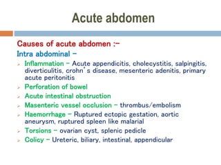
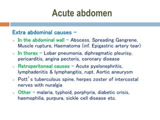
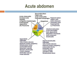
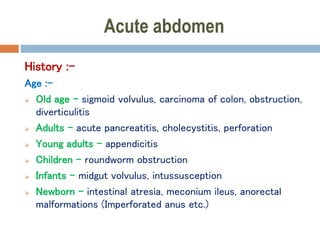
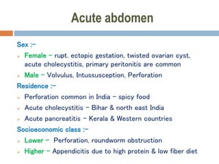
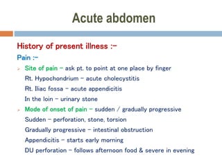
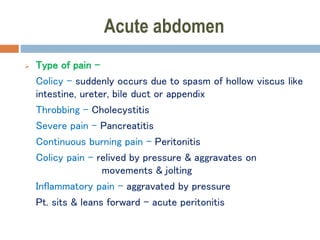
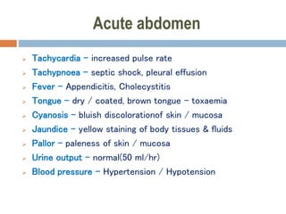
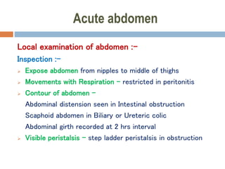
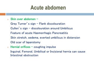
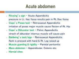
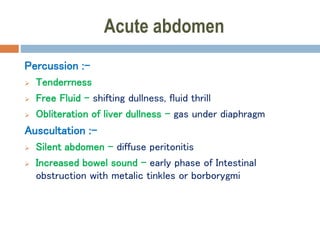
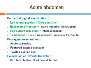
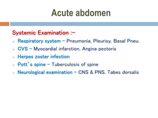
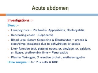
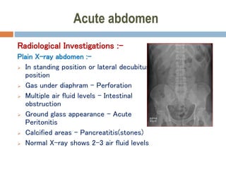
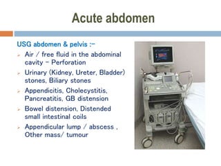
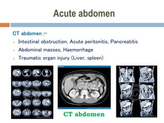
Dr. Yogesh S. Borase
M.S. Shalyatantra
Professor & HOD
Department of Shalyatantra,
MES Ayurved Mahavidyalaya, Ghanekhunt- Lote
Acute abdomen
Acute abdomen
 Sudden severe attack of abdominal pain to such an extent
that pt. is in severe agony & often shock
 It may be life threatening like Intestinal perforation
Acute abdomen
Causes of acute abdomen :-
Intra abdominal –
 Inflammation – Acute appendicitis, cholecystitis, salpingitis,
diverticulitis, crohn’s disease, mesenteric adenitis, primary
acute peritonitis
 Perforation of bowel
 Acute intestinal obstruction
 Masenteric vessel occlusion – thrombus/embolism
 Haemorrhage – Ruptured ectopic gestation, aortic
aneurysm, ruptured spleen like malarial
 Torsions – ovarian cyst, splenic pedicle
 Colicy - Ureteric, biliary, intestinal, appendicular
Acute abdomen
Extra abdominal causes –
 In the abdominal wall – Abscess, Spreading Gangrene,
Muscle rupture, Haematoma (inf. Epigastric artery tear)
 In thorax – Lobar pneumonia, diphragmatic pleurisy,
pericarditis, angina pectoris, coronary disease
 Retroperitoneal causes – Acute pyelonephritis,
lymphadenitis & lymphangitis, rupt. Aortic aneurysm
 Pott’s tuberculous spine, herpes zoster of intercostal
nerves with nuralgia
 Other – malaria, typhoid, porphyria, diabetic crisis,
haemophilia, purpura, sickle cell disease etc.
Acute abdomen
Acute abdomen
History :-
Age :-
 Old age – sigmoid volvulus, carcinoma of colon, obstruction,
diverticulitis
 Adults – acute pancreatitis, cholecystitis, perforation
 Young adults – appendicitis
 Children – roundworm obstruction
 Infants – midgut volvulus, intussusception
 Newborn – intestinal atresia, meconium ileus, anorectal
malformations (Imperforated anus etc.)
Acute abdomen
Sex :-
 Female – rupt. ectopic gestation, twisted ovarian cyst,
acute cholecystitis, primary peritonitis are common
 Male – Volvulus, Intussusception, Perforation
Residence :-
 Perforation common in India – spicy food
 Acute cholecystitis – Bihar & north east India
 Acute pancreatitis – Kerala & Western countries
Socioeconomic class :-
 Lower – Perforation, roundworm obstruction
 Higher – Appendicitis due to high protein & low fiber diet
Acute abdomen
History of present illness :-
Pain :-
 Site of pain – ask pt. to point at one place by finger
Rt. Hypochondrium – acute cholecystitis
Rt. Iliac fossa – acute appendicitis
In the loin – urinary stone
 Mode of onset of pain – sudden / gradually progressive
Sudden – perforation, stone, torsion
Gradually progressive – intestinal obstruction
Appendicitis – starts early morning
DU perforation – follows afternoon food & severe in evening
Acute abdomen
 Type of pain –
Colicy – suddenly occurs due to spasm of hollow viscus like
intestine, ureter, bile duct or appendix
Throbbing – Cholecystitis
Severe pain – Pancreatitis
Continuous burning pain – Peritonitis
Colicy pain – relived by pressure & aggravates on
movements & jolting
Inflammatory pain – aggravated by pressure
Pt. sits & leans forward – acute peritonitis
Acute abdomen
 Spread of pain –
Acute appendicitis – Umbilical then shift to Rt. Iliac fossa
DU perforation – Epigastrium to all over abdomen
Pancreatitis – Epigastrium & radiates to back
Ureteric colic – pain radiates from loin to groin to
scrotum & upper medial part of thigh
Gallstone colic – Rt. Hypochondrium to inf. angle of
scapula
Gastroduodenal & Jejunal – to Epigastrium
Ileum & appendix – to Umbilical region
Colonic disease – to Hypogastrium
Acute abdomen
Vomiting :-
 Projectile or regurgitant
 Frequency of vomiting – intestinal obstruction
 Quantity & nature of Vomitus – Gastric, bilious, intestinal,
faeculant or blood stained
 Early feature – Proximal intestinal obstruction
Late feature – Distal intestinal obstruction
 Murphy’s triod (Appendicitis) – pain, vomiting, fever
Acute abdomen
Bowel habits :-
 Appendicitis – diarrhoea, colonic spasm
 Acute intastinal obstruction – absolute constipation
 Mesenteric ischaemia – bloody putrid stool
 Intussusception – blood & mucus with distension
Abdominal distension :-
 Fullness of abdomen gradually progressive
 Associated with constipation & vomiting
Urinary symptoms :-
burning micturition, frequency, strangury
Acute abdomen
Other history :-
 Fever, chills & rigors
 Jaundice – Cholecystitis, Pancreatitis
 Laparotomy – Intestinal obstruction
 Gastric / Duodenal ulcer – Perforation
Personal history :-
 Missed menstrual cycle – Ectopic pregnancy
 Smoking, Alcohol intake, Diet history
Acute abdomen
General Examination :-
 Facies hippocratica – anxious look,
sunken eyes, pinched face, cold sweat
in terminal stage of peritonitis
 Signs of dehydration – sunken eyes,
dry tongue, drawn cheeks
 Sudden pallor with shock – ruptured ectopic gestation
 Pt. Stays still in bed – acute peritonitis
 Pt. rolls in bed with agony – colicy pain
Acute abdomen
 Tachycardia – increased pulse rate
 Tachypnoea – septic shock, pleural effusion
 Fever – Appendicitis, Cholecystitis
 Tongue – dry / coated, brown tongue - toxaemia
 Cyanosis – bluish discolorationof skin / mucosa
 Jaundice – yellow staining of body tissues & fluids
 Pallor – paleness of skin / mucosa
 Urine output – normal(50 ml/hr)
 Blood pressure – Hypertension / Hypotension
Acute abdomen
Local examination of abdomen :-
Inspection :-
 Expose abdomen from nipples to middle of thighs
 Movements with Respiration – restricted in peritonitis
 Contour of abdomen –
Abdominal distension seen in Intestinal obstruction
Scaphoid abdomen in Biliary or Ureteric colic
Abdominal girth recorded at 2 hrs interval
 Visible peristalsis – step ladder peristalsis in obstruction
Acute abdomen
 Skin over abdomen –
Grey Turner’s sign - Flank discolouration
Cullen’s sign – discolouration around Umbilicus
Feature of acute Haemorrhagic Pancreatitis
Skin stretch, oedema, everted umbilicus in distension
Old scar of laparotomy
 Hernial orifices – coughing impulse
Inguinal, Femoral, Umbilical or Incisional hernia can cause
Intestinal obstruction
Acute abdomen
Palpation :-
 Hyperaesthesia –
Scratch the skin or gently hold the fold of skin
Acute Gangrenous Appendicitis - Sherren’s triangle
Acute Cholecystitis - between 9th & 11th rib (Boas’s sign)
 Tenderness –
Acute Appendicitis - Mc Burney’s point
Acute Cholelithiasis – tip of 9th costal cartilage (Rt.)
 Rebound tenderness – Blumberg’s sign
due to inflammed parietal peritonium in acute Appendicitis,
aute Peritonitis, intestinal obstruction with gangrenous
bowel
Acute abdomen
 Rovsing’s sign – Acute Appendicitis
pressure in Lt. iliac fossa results pain in Rt. Iliac fossa
 Cope’s Psoas test – Retrocaecal Appendicitis
irritation of psoas major muscle cause flexion of Rt. Hip
 Cope’s Obturator test – Pelvic Appendicitis
strech of obturator internus muscle will cause pain
 Baldwing’s test/sign – Retrocaecal Appendicitis
flank is pressed with hand & Rt. Leg raised up
 Muscle guarding & rigidity – Parietal peritonitis
 Mass abdomen – Appendicular, Colonic etc.
 Hernial sites
Acute abdomen
Percussion :-
 Tenderrness
 Free Fluid – shifting dullness, fluid thrill
 Obliteration of liver dullness – gas under diaphragm
Auscultation :-
 Silent abdomen – diffuse peritonitis
 Increased bowel sound – early phase of Intestinal
obstruction with metalic tinkles or borborygmi
Acute abdomen
Per rectal digital examination :-
 Left lateral position / Dorsal position
 Ballooning of rectum – acute Intestinal obstruction
 Red currant jelly stool – Intussusception
 Tenderness – Pelvic Appendicitis, Abscess, Peritonitis
Pervaginal examination :-
 Acute salpingitis
 Ruptured ectopic gestation
 Twisted ovarian cyst
Examination of External Genitalia :-
Scrotum, Testes, Cord, Vas deferens
Acute abdomen
Systemic Examination :-
 Respiratory system – Pneumonia, Pleurisy, Basal Pneu.
 CVS – Myocardial infarction, Angina pectoris
 Herpes zoster infection
 Pott’s spine – Tuberculosis of spine
 Neurological examination – CNS & PNS, Tabes dorsalis
Acute abdomen
Investigations :-
Blood :-
 Leucocytosis – Peritonitis, Appendicitis, Cholecystitis
 Decreasing count – Septicemia
 Blood urea, Serum Creatinine & Electrolytes – uremia &
electrolyte imbalance due to dehydration or sepsis
 Liver function test, platelet count, sr. amylase, sr. calcium,
sr. lipase, prothrombin time – Pancreatitis
 Plasma fibrinogen, C reactive protein, methaemoglobin
Urine analysis :- for Pus cells & RBC
Acute abdomen
Radiological Investigations :-
Plain X-ray abdomen :-
 In standing position or lateral decubitus
position
 Gas under diaphram – Perforation
 Multiple air fluid levels – Intestinal
obstruction
 Ground glass appearance – Acute
Peritonitis
 Calcified areas – Pancreatitis(stones)
 Normal X-ray shows 2-3 air fluid levels
Acute abdomen
USG abdomen & pelvis :-
 Air / free fluid in the abdominal
cavity - Perforation
 Urinary (Kidney, Ureter, Bladder)
stones, Biliary stones
 Appendicitis, Cholecystitis,
Pancreatitis, GB distension
 Bowel distension, Distended
small intestinal coils
 Appendicular lump / abscess ,
Other mass/ tumour
Acute abdomen
CT abdomen :-
 Intestinal obstruction, Acute peritonitis, Pancreatitis
 Abdominal masses, Haemorrhage
 Traumatic organ injury (Liver, spleen)
CT abdomen
Acute abdomen
 Cholescientegraphy using HIDA – hippuric immunodiacetic
or I131 Rose Bengal radioisotope scan has got 100%
accuracy in diagnosis of Acute Cholecystitis
 Laparoscopy – new modality to identify the pathology &
also used for treatment of the disease as Laparoscopic
surgery for Appendicectomy, Cholecystectomy, closure of
Perforation, Hernia repair etc.
 GI contrast studies (Barium) or Endoscopy is not done in
acute abdomen as Barium will leak in peritonial cavity
causing chemical peritonitis

Acute abdomen

  • 1.
    Dr. Yogesh S.Borase M.S. Shalyatantra Professor & HOD Department of Shalyatantra, MES Ayurved Mahavidyalaya, Ghanekhunt- Lote Acute abdomen
  • 2.
    Acute abdomen  Suddensevere attack of abdominal pain to such an extent that pt. is in severe agony & often shock  It may be life threatening like Intestinal perforation
  • 3.
    Acute abdomen Causes ofacute abdomen :- Intra abdominal –  Inflammation – Acute appendicitis, cholecystitis, salpingitis, diverticulitis, crohn’s disease, mesenteric adenitis, primary acute peritonitis  Perforation of bowel  Acute intestinal obstruction  Masenteric vessel occlusion – thrombus/embolism  Haemorrhage – Ruptured ectopic gestation, aortic aneurysm, ruptured spleen like malarial  Torsions – ovarian cyst, splenic pedicle  Colicy - Ureteric, biliary, intestinal, appendicular
  • 4.
    Acute abdomen Extra abdominalcauses –  In the abdominal wall – Abscess, Spreading Gangrene, Muscle rupture, Haematoma (inf. Epigastric artery tear)  In thorax – Lobar pneumonia, diphragmatic pleurisy, pericarditis, angina pectoris, coronary disease  Retroperitoneal causes – Acute pyelonephritis, lymphadenitis & lymphangitis, rupt. Aortic aneurysm  Pott’s tuberculous spine, herpes zoster of intercostal nerves with nuralgia  Other – malaria, typhoid, porphyria, diabetic crisis, haemophilia, purpura, sickle cell disease etc.
  • 5.
  • 6.
    Acute abdomen History :- Age:-  Old age – sigmoid volvulus, carcinoma of colon, obstruction, diverticulitis  Adults – acute pancreatitis, cholecystitis, perforation  Young adults – appendicitis  Children – roundworm obstruction  Infants – midgut volvulus, intussusception  Newborn – intestinal atresia, meconium ileus, anorectal malformations (Imperforated anus etc.)
  • 7.
    Acute abdomen Sex :- Female – rupt. ectopic gestation, twisted ovarian cyst, acute cholecystitis, primary peritonitis are common  Male – Volvulus, Intussusception, Perforation Residence :-  Perforation common in India – spicy food  Acute cholecystitis – Bihar & north east India  Acute pancreatitis – Kerala & Western countries Socioeconomic class :-  Lower – Perforation, roundworm obstruction  Higher – Appendicitis due to high protein & low fiber diet
  • 8.
    Acute abdomen History ofpresent illness :- Pain :-  Site of pain – ask pt. to point at one place by finger Rt. Hypochondrium – acute cholecystitis Rt. Iliac fossa – acute appendicitis In the loin – urinary stone  Mode of onset of pain – sudden / gradually progressive Sudden – perforation, stone, torsion Gradually progressive – intestinal obstruction Appendicitis – starts early morning DU perforation – follows afternoon food & severe in evening
  • 9.
    Acute abdomen  Typeof pain – Colicy – suddenly occurs due to spasm of hollow viscus like intestine, ureter, bile duct or appendix Throbbing – Cholecystitis Severe pain – Pancreatitis Continuous burning pain – Peritonitis Colicy pain – relived by pressure & aggravates on movements & jolting Inflammatory pain – aggravated by pressure Pt. sits & leans forward – acute peritonitis
  • 10.
    Acute abdomen  Spreadof pain – Acute appendicitis – Umbilical then shift to Rt. Iliac fossa DU perforation – Epigastrium to all over abdomen Pancreatitis – Epigastrium & radiates to back Ureteric colic – pain radiates from loin to groin to scrotum & upper medial part of thigh Gallstone colic – Rt. Hypochondrium to inf. angle of scapula Gastroduodenal & Jejunal – to Epigastrium Ileum & appendix – to Umbilical region Colonic disease – to Hypogastrium
  • 11.
    Acute abdomen Vomiting :- Projectile or regurgitant  Frequency of vomiting – intestinal obstruction  Quantity & nature of Vomitus – Gastric, bilious, intestinal, faeculant or blood stained  Early feature – Proximal intestinal obstruction Late feature – Distal intestinal obstruction  Murphy’s triod (Appendicitis) – pain, vomiting, fever
  • 12.
    Acute abdomen Bowel habits:-  Appendicitis – diarrhoea, colonic spasm  Acute intastinal obstruction – absolute constipation  Mesenteric ischaemia – bloody putrid stool  Intussusception – blood & mucus with distension Abdominal distension :-  Fullness of abdomen gradually progressive  Associated with constipation & vomiting Urinary symptoms :- burning micturition, frequency, strangury
  • 13.
    Acute abdomen Other history:-  Fever, chills & rigors  Jaundice – Cholecystitis, Pancreatitis  Laparotomy – Intestinal obstruction  Gastric / Duodenal ulcer – Perforation Personal history :-  Missed menstrual cycle – Ectopic pregnancy  Smoking, Alcohol intake, Diet history
  • 14.
    Acute abdomen General Examination:-  Facies hippocratica – anxious look, sunken eyes, pinched face, cold sweat in terminal stage of peritonitis  Signs of dehydration – sunken eyes, dry tongue, drawn cheeks  Sudden pallor with shock – ruptured ectopic gestation  Pt. Stays still in bed – acute peritonitis  Pt. rolls in bed with agony – colicy pain
  • 15.
    Acute abdomen  Tachycardia– increased pulse rate  Tachypnoea – septic shock, pleural effusion  Fever – Appendicitis, Cholecystitis  Tongue – dry / coated, brown tongue - toxaemia  Cyanosis – bluish discolorationof skin / mucosa  Jaundice – yellow staining of body tissues & fluids  Pallor – paleness of skin / mucosa  Urine output – normal(50 ml/hr)  Blood pressure – Hypertension / Hypotension
  • 16.
    Acute abdomen Local examinationof abdomen :- Inspection :-  Expose abdomen from nipples to middle of thighs  Movements with Respiration – restricted in peritonitis  Contour of abdomen – Abdominal distension seen in Intestinal obstruction Scaphoid abdomen in Biliary or Ureteric colic Abdominal girth recorded at 2 hrs interval  Visible peristalsis – step ladder peristalsis in obstruction
  • 17.
    Acute abdomen  Skinover abdomen – Grey Turner’s sign - Flank discolouration Cullen’s sign – discolouration around Umbilicus Feature of acute Haemorrhagic Pancreatitis Skin stretch, oedema, everted umbilicus in distension Old scar of laparotomy  Hernial orifices – coughing impulse Inguinal, Femoral, Umbilical or Incisional hernia can cause Intestinal obstruction
  • 18.
    Acute abdomen Palpation :- Hyperaesthesia – Scratch the skin or gently hold the fold of skin Acute Gangrenous Appendicitis - Sherren’s triangle Acute Cholecystitis - between 9th & 11th rib (Boas’s sign)  Tenderness – Acute Appendicitis - Mc Burney’s point Acute Cholelithiasis – tip of 9th costal cartilage (Rt.)  Rebound tenderness – Blumberg’s sign due to inflammed parietal peritonium in acute Appendicitis, aute Peritonitis, intestinal obstruction with gangrenous bowel
  • 19.
    Acute abdomen  Rovsing’ssign – Acute Appendicitis pressure in Lt. iliac fossa results pain in Rt. Iliac fossa  Cope’s Psoas test – Retrocaecal Appendicitis irritation of psoas major muscle cause flexion of Rt. Hip  Cope’s Obturator test – Pelvic Appendicitis strech of obturator internus muscle will cause pain  Baldwing’s test/sign – Retrocaecal Appendicitis flank is pressed with hand & Rt. Leg raised up  Muscle guarding & rigidity – Parietal peritonitis  Mass abdomen – Appendicular, Colonic etc.  Hernial sites
  • 20.
    Acute abdomen Percussion :- Tenderrness  Free Fluid – shifting dullness, fluid thrill  Obliteration of liver dullness – gas under diaphragm Auscultation :-  Silent abdomen – diffuse peritonitis  Increased bowel sound – early phase of Intestinal obstruction with metalic tinkles or borborygmi
  • 21.
    Acute abdomen Per rectaldigital examination :-  Left lateral position / Dorsal position  Ballooning of rectum – acute Intestinal obstruction  Red currant jelly stool – Intussusception  Tenderness – Pelvic Appendicitis, Abscess, Peritonitis Pervaginal examination :-  Acute salpingitis  Ruptured ectopic gestation  Twisted ovarian cyst Examination of External Genitalia :- Scrotum, Testes, Cord, Vas deferens
  • 22.
    Acute abdomen Systemic Examination:-  Respiratory system – Pneumonia, Pleurisy, Basal Pneu.  CVS – Myocardial infarction, Angina pectoris  Herpes zoster infection  Pott’s spine – Tuberculosis of spine  Neurological examination – CNS & PNS, Tabes dorsalis
  • 23.
    Acute abdomen Investigations :- Blood:-  Leucocytosis – Peritonitis, Appendicitis, Cholecystitis  Decreasing count – Septicemia  Blood urea, Serum Creatinine & Electrolytes – uremia & electrolyte imbalance due to dehydration or sepsis  Liver function test, platelet count, sr. amylase, sr. calcium, sr. lipase, prothrombin time – Pancreatitis  Plasma fibrinogen, C reactive protein, methaemoglobin Urine analysis :- for Pus cells & RBC
  • 24.
    Acute abdomen Radiological Investigations:- Plain X-ray abdomen :-  In standing position or lateral decubitus position  Gas under diaphram – Perforation  Multiple air fluid levels – Intestinal obstruction  Ground glass appearance – Acute Peritonitis  Calcified areas – Pancreatitis(stones)  Normal X-ray shows 2-3 air fluid levels
  • 25.
    Acute abdomen USG abdomen& pelvis :-  Air / free fluid in the abdominal cavity - Perforation  Urinary (Kidney, Ureter, Bladder) stones, Biliary stones  Appendicitis, Cholecystitis, Pancreatitis, GB distension  Bowel distension, Distended small intestinal coils  Appendicular lump / abscess , Other mass/ tumour
  • 26.
    Acute abdomen CT abdomen:-  Intestinal obstruction, Acute peritonitis, Pancreatitis  Abdominal masses, Haemorrhage  Traumatic organ injury (Liver, spleen) CT abdomen
  • 27.
    Acute abdomen  Cholescientegraphyusing HIDA – hippuric immunodiacetic or I131 Rose Bengal radioisotope scan has got 100% accuracy in diagnosis of Acute Cholecystitis  Laparoscopy – new modality to identify the pathology & also used for treatment of the disease as Laparoscopic surgery for Appendicectomy, Cholecystectomy, closure of Perforation, Hernia repair etc.  GI contrast studies (Barium) or Endoscopy is not done in acute abdomen as Barium will leak in peritonial cavity causing chemical peritonitis